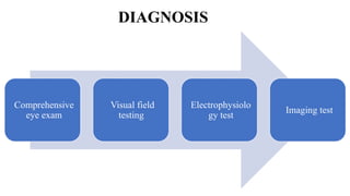
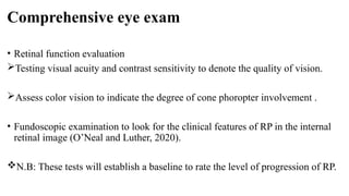
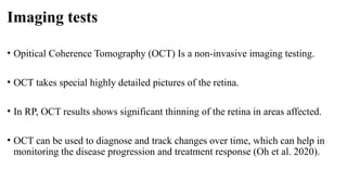
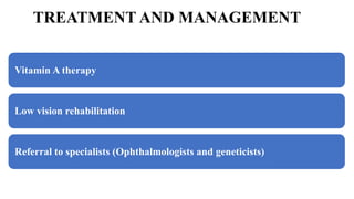
RETINITIS PIGMENTOSA
BY
DR. REJOICE EMEGHARA
AUGUST 16, 2024
INTRODUCTION
• Retinitis pigmentosa (RP) is a group
of genetic eye disorder that affects
the retina (Pennesi et al 2010).
• It is characterized by the progressive
degeneration of photoreceptor cells
in the retina, leading to vision loss.
• It typically begins with night
blindness and gradually narrows the
visual field, often resulting in tunnel
vision (O’Neal and Luther, 2020).
Fig 1: Image from a normal vision and vision with RP
(Levine, 2021).
PREVALENCE
• RP is very rare genetic eye disorder.
• Globally, it affects about 1 in 4,000 people.
• It slightly affects male more than female.
• The age of onset can vary.
• It usually diagnosed in young adulthood (Verbakel et al. 2018).
CAUSES
• RP is caused by genetic mutation. These genes carry the instructions for making
proteins that are needed in cells within the retina, called photoreceptors.
• RP can cause mutation of at least 12 different genes in Autosomal dominant.
• Autosomal dominant is usually the mildest form of RP with later age of onset.
• RP can trigger mutation of 22 different genes in Autosomal recessive.
• RP can cause 2 genes mutation in X linked.
• In X linked, RP tends to present early (Daiger et al. 2014).
VISUAL SYMPTOMS
• Loss of night vision due to early
damage of rod cells
• Gradual loss of peripheral vision
(tunnel vision) due to gradual damage
of rod cells.
• Loss of central vision due to damage of
cone cells (late stage) (Kamde and
Anjankar, 2023).
N.B: RP can start unilaterally and move
bilaterally (Moini & Piran, 2020).
Fig 2: Image of normal vision and tunnel vision in RP
(Mandal, 2023).
CLINICAL FEATURES
• Retinal pigmentary changes.
• Bone spicule formation.
• Attenuated vessels.
• Optic disc pallor (Jimenez-
Davila et al. 2022).
Fig 3: Image of clinical features of retinitis pigmentosa
(Sofi et al. 2021).
DIAGNOSIS
Comprehensive
eye exam
Visual field
testing
Electrophysiolo
gy test
Imaging test
Comprehensive eye exam
• Retinal function evaluation
Testing visual acuity and contrast sensitivity to denote the quality of vision.
Assess color vision to indicate the degree of cone phoropter involvement .
• Fundoscopic examination to look for the clinical features of RP in the internal
retinal image (O’Neal and Luther, 2020).
N.B: These tests will establish a baseline to rate the level of progression of RP.
Comprehensive eye exam cont’d
Fig 4: Image of normal eye fundus and fundus with RP (Hansen, 2024).
Visual field testing
• Visual field helps to measure your side vision and find any blind spots that may
be developing.
• Visual field testing can be used to assess the affected peripheral (side) vision
caused by RP (Boyd, 2019).
• Visual field assessment with Kinetic perimetry is considered the most effective
way to evaluate the loss of peripheral vision (O’Neal and Luther, 2020).
Visual field testing cont’d
Fig 5: Image of Humphrey kinetic Visual field testing result of RP (Sayo et al. 2017).
Electrophysiology test
• Electroretinogram (ERG) testing is used to measure the electrical activity in the
retina (how well the retina responds to light).
• In RP, ERG responses are found to be reduced from normal (indicating impaired
rod and cone function).
• ERG responses usually correlates with visual function and age.
• ERG could detect RP even before clinical features starts appearing in the fundus
(Menghini et al. 2020).
Electrophysiology test cont’d
Fig 6: Image of Electroretinogram (ERG) Result from normal eye and eye with RP (Creel, 2012).
Imaging tests
• Opitical Coherence Tomography (OCT) Is a non-invasive imaging testing.
• OCT takes special highly detailed pictures of the retina.
• In RP, OCT results shows significant thinning of the retina in areas affected.
• OCT can be used to diagnose and track changes over time, which can help in
monitoring the disease progression and treatment response (Oh et al. 2020).
Imaging test cont’d
Fig 7: Image of Electroretinogram (ERG) Result from normal eye and eye with RP (Tan et al. 2021).
TREATMENT AND MANAGEMENT
Vitamin A therapy
Low vision rehabilitation
Referral to specialists (Ophthalmologists and geneticists)
Vitamin A therapy
• Vit. A is a precursor of 11-cis-retinal.
• By maintaining adequate levels of 11-cis-retinal, Vit. A helps preserve the function of
photoreceptors, potentially slowing the progression of RP (Sajovic et al. 2022).
• A study show that high-dose Vit. A (15,000 IU/day) could slow the rate of retinal
degeneration in some RP patients.
• High-dose Vit. A (15,000 IU/day) is generally safe for most adults.
• High-dose Vit. A should be taking under medical supervision to avoid excessive amount
of Vit. A in the blood that is harmful to the body system (Shechtman, & Karpecki, 2010).
Low vision rehabilitation
• Low vision rehabilitation is essential for patients with advanced RP to maintain
independence and improve their quality of life.
• The primary goals are:
Enhancing visual function using optical and electronic aids (e.g using magnifiers or
wearable electronic glasses).
Improving mobility by training patients to navigate safely and confidently (e.g using
cane, guide dogs or electronic navigation aids).
Providing emotional and psychological support to cope with vision loss (Kumari,
2022).
PROGNOSIS
• The prognosis for patients with RP depends on the age of onset and pattern of
inheritance.
• RP caused by autosomal recessive form has early onset symptoms, severe
vision-loss and night blindness.
• RP Caused by autosomal dominant form is the least severe and more gradual
onset of symptoms later in adulthood.
• RP caused by X-linked form has the most severe vision loss (Desai & Alibhai,
2024).
PROGNOSIS CONT’D
• Tunnel vision is expected late in all forms of RP.
• Almost all patients will be legally blind at some point of the disease
progression.
• Fortunately, total vision loss is uncommon, as macular function will generally
allow light perception, even after acuity is loss.
• Many patient retains central vision into their 40s or 50s (O’Neal and Luther,
2020).
Emerging treatments
Genetic therapy
Stem cell therapy
Genetic therapy
• Genetic therapy remains a promising approach for treatment of RP.
• Genetic therapies can be delivered to the subretinal space.
• To provide functional copy of a gene or to correct a mutation (Wu et al. 2023).
Stem cell therapy
• Several types of stem cells (e.g embryonic and mesemchymal) are being studied
as a potential treatment modality in retinal dystrophies.
• Pre-clinical studies have shown that stem cell treatment can:
Replace damage cells
Adding nutritional support to the remaining functional cells,
Protect retinal vascularity and promote synaptic connection (Jimenez-Davila et
al. 2022).
CONCLUSION
Retinitis pigmentosa (RP) is a group of inherited retinal disorder that lead to
progressive vision loss. Although there is currently no cure, advancements in
genetic research and emerging therapies offer hope for future treatments. Early
diagnosis and supportive care, including the use of assistive devices and lifestyle
adaptations, can improve the quality of life for individuals affected by RP.
Ongoing research and clinic trials are essential in the quest to find effective
treatments and, ultimately, a cure for this debilitating condition.
REFERENCES
Abdelkader, E., Enani, L., & Kozak, I. (2017). A case of unilateral retinitis
pigmentosa associated with full thickness macular hole. Middle East African
Journal of Ophthalmology, 24(2), 113. https://doi.org/10.4103/meajo.meajo_97_17
Boyd, K. (2019). What Is Retinitis Pigmentosa? American Academy of
Ophthalmology.
https://www.aao.org/eye-health/diseases/what-is-retinitis-pigmentosa
Creel, D. (2012). The Electroretinogram and Electro-oculogram: Clinical
Applications by Donnell J. Creel – Webvision. Utah.edu.
https://webvision.med.utah.edu/book/electrophysiology/the-electroretinogram-
clinical-applications/
REFERENCES CONT’D
Daiger, S. P., Bowne, S. J., & Sullivan, L. S. (2014). Genes and Mutations Causing
Autosomal Dominant Retinitis Pigmentosa. Cold Spring Harbor Perspectives in
Medicine, 5(10), a017129. https://doi.org/10.1101/cshperspect.a017129
Desai, S. J., & Alibhai, Y. A. (2024). Retinitis Pigmentosa. Elsevier EBooks, 195–
196. https://doi.org/10.1016/b978-0-323-93043-7.00075-3
Hansen, J. (2024). ALG6 acts as a modifier gene in the inherited genetic eye
disease retinitis pigmentosa 59. UAB News.
https://www.uab.edu/news/research/item/14024-alg6-acts-as-a-modifier-gene-in-
the-inherited-genetic-eye-disease-retinitis-pigmentosa-59
REFERENCES CONT’D
Jimenez-Davila, J., Procopio, A. & Klufas, A. (2022). A Review of Retinitis
Pigmentosa. Www.reviewofophthalmology.com.
https://www.reviewofophthalmology.com/article/a-review-of-retinitis-pigmento
sa
Kamde, S. P. & Anjankar, A. (2023). Retinitis Pigmentosa: Pathogenesis,
Diagnostic Findings, and Treatment. Cureus.
https://doi.org/10.7759/cureus.48006
Kumari, K. (2022). Retinitis Pigmentosa – Challenging Visual Rehabilitation.
Vision Science Academy. https://visionscienceacademy.org/retinitis-pigmentosa-
challenging-visual-rehabilitation/
REFERENCES CONT’D
Levine, H. (2021, October 11). How Your Own DNA Could Someday Save Your
Vision. Content Lab U.S.
https://www.jnj.com/innovation/about-gene-therapy-for-retinitis-pigmentosa-an
d-eye-conditions
Mandal, A. (2023). What is Retinitis Pigmentosa? News-Medical.net.
https://www.news-medical.net/health/What-is-Retinitis-Pigmentosa.aspx
Menghini, M., Cehajic-Kapetanovic, J., & MacLaren, R. E. (2020). Monitoring
progression of retinitis pigmentosa: current recommendations and recent
advances. Expert Opinion on Orphan Drugs, 8(2-3), 67–78.
https://doi.org/10.1080/21678707.2020.1735352
REFERENCES CONT’D
Moini, J., & Piran, P. (2020). Visual system. Functional and Clinical Neuroanatomy,
417–466. https://doi.org/10.1016/b978-0-12-817424-1.00014-8
Oh, J., Nuzbrokh, Y., Lima, R., Ryu, J., & Tsang, S. H. (2020). Optical coherence
tomography in the evaluation of retinitis pigmentosa. Ophthalmic Genetics, 41(5), 413–
419. https://doi.org/10.1080/13816810.2020.1780619
O’Neal, T. B., & Luther, E. E. (2020). Retinitis Pigmentosa. PubMed; StatPearls
Publishing. https://www.ncbi.nlm.nih.gov/books/NBK519518/
Pennesi, M.E., Francis, P. J., & Weleber, R.G. (2010). Primary Photoreceptor
Degenerations: Terminology. Elsevier EBooks, 516–525.
https://doi.org/10.1016/b978-0-12-374203-2.00209-8
REFERENCES CONT’D
Sajovic, J., Meglič, A., Glavač, D., Markelj, Š., Hawlina, M., & Fakin, A. (2022). The
Role of Vitamin A in Retinal Diseases. International Journal of Molecular Sciences,
23(3), 1014. https://doi.org/10.3390/ijms23031014
Sayo, A., Ueno, S., Kominami, T., Nishida, K., Inooka, D., Nakanishi, A., Yasuda, S.,
Okado, S., Takahashi, K., Matsui, S., & Terasaki, H. s41598-017-16640-7 (2017).
Longitudinal study of visual field changes determined by Humphrey Field Analyzer 10-
2 in patients with Retinitis Pigmentosa. Scientific Reports, 7(1). https://doi.org/10.1038/
Shechtman, D., & Karpecki, P. (2010). Nutritional Supplements for RP.
Www.reviewofoptometry.com. https://www.reviewofoptometry.com/article/nutritional-
supplements-for-rp
REFERENCES CONT’D
Sofi, F., Sodi, A., Franco, F., Murro, V., Biagini, D., Miele, A., Abbruzzese, G., Mucciolo,
D. P., Virgili, G., Menchini, U., Casini, A., & Rizzo, S. (2016). Dietary profile of patients
with Stargardt’s disease and Retinitis Pigmentosa: is there a role for a nutritional
approach? BMC Ophthalmology, 16. https://doi.org/10.1186/s12886-016-0187-3
Tan, L., Long, Y., Li, Z., Xiao, Y., Ren, J., Sun, C., Meng, X., & Li, S. (2021). Ocular
abnormalities in a large patient cohort with retinitis pigmentosa in Western China. 21(1).
https://doi.org/10.1186/s12886-020-01797-z
Verbakel, S. K., van Huet, R. A. C., Boon, C. J. F., den Hollander, A. I., Collin, R. W. J.,
Klaver, C. C. W., Hoyng, C. B., Roepman, R., & Klevering, B. J. (2018). Non-syndromic
retinitis pigmentosa. Progress in Retinal and Eye Research, 66, 157–186.
https://doi.org/10.1016/j.preteyeres.2018.03.005
REFERENCES CONT’D
Wu, K. Y., Kulbay, M., Toameh, D., Xu, A. Q., Kalevar, A., & Tran, S. D.
(2023). Retinitis Pigmentosa: Novel Therapeutic Targets and Drug
Development. Pharmaceutics, 15(2), 685.
https://doi.org/10.3390/pharmaceutics15020685

RETINITIS PIGMENTOSA Seminar Presentation

  • 1.
    RETINITIS PIGMENTOSA BY DR. REJOICEEMEGHARA AUGUST 16, 2024
  • 2.
    INTRODUCTION • Retinitis pigmentosa(RP) is a group of genetic eye disorder that affects the retina (Pennesi et al 2010). • It is characterized by the progressive degeneration of photoreceptor cells in the retina, leading to vision loss. • It typically begins with night blindness and gradually narrows the visual field, often resulting in tunnel vision (O’Neal and Luther, 2020). Fig 1: Image from a normal vision and vision with RP (Levine, 2021).
  • 3.
    PREVALENCE • RP isvery rare genetic eye disorder. • Globally, it affects about 1 in 4,000 people. • It slightly affects male more than female. • The age of onset can vary. • It usually diagnosed in young adulthood (Verbakel et al. 2018).
  • 4.
    CAUSES • RP iscaused by genetic mutation. These genes carry the instructions for making proteins that are needed in cells within the retina, called photoreceptors. • RP can cause mutation of at least 12 different genes in Autosomal dominant. • Autosomal dominant is usually the mildest form of RP with later age of onset. • RP can trigger mutation of 22 different genes in Autosomal recessive. • RP can cause 2 genes mutation in X linked. • In X linked, RP tends to present early (Daiger et al. 2014).
  • 5.
    VISUAL SYMPTOMS • Lossof night vision due to early damage of rod cells • Gradual loss of peripheral vision (tunnel vision) due to gradual damage of rod cells. • Loss of central vision due to damage of cone cells (late stage) (Kamde and Anjankar, 2023). N.B: RP can start unilaterally and move bilaterally (Moini & Piran, 2020). Fig 2: Image of normal vision and tunnel vision in RP (Mandal, 2023).
  • 6.
    CLINICAL FEATURES • Retinalpigmentary changes. • Bone spicule formation. • Attenuated vessels. • Optic disc pallor (Jimenez- Davila et al. 2022). Fig 3: Image of clinical features of retinitis pigmentosa (Sofi et al. 2021).
  • 7.
  • 8.
    Comprehensive eye exam •Retinal function evaluation Testing visual acuity and contrast sensitivity to denote the quality of vision. Assess color vision to indicate the degree of cone phoropter involvement . • Fundoscopic examination to look for the clinical features of RP in the internal retinal image (O’Neal and Luther, 2020). N.B: These tests will establish a baseline to rate the level of progression of RP.
  • 9.
    Comprehensive eye examcont’d Fig 4: Image of normal eye fundus and fundus with RP (Hansen, 2024).
  • 10.
    Visual field testing •Visual field helps to measure your side vision and find any blind spots that may be developing. • Visual field testing can be used to assess the affected peripheral (side) vision caused by RP (Boyd, 2019). • Visual field assessment with Kinetic perimetry is considered the most effective way to evaluate the loss of peripheral vision (O’Neal and Luther, 2020).
  • 11.
    Visual field testingcont’d Fig 5: Image of Humphrey kinetic Visual field testing result of RP (Sayo et al. 2017).
  • 12.
    Electrophysiology test • Electroretinogram(ERG) testing is used to measure the electrical activity in the retina (how well the retina responds to light). • In RP, ERG responses are found to be reduced from normal (indicating impaired rod and cone function). • ERG responses usually correlates with visual function and age. • ERG could detect RP even before clinical features starts appearing in the fundus (Menghini et al. 2020).
  • 13.
    Electrophysiology test cont’d Fig6: Image of Electroretinogram (ERG) Result from normal eye and eye with RP (Creel, 2012).
  • 14.
    Imaging tests • OpiticalCoherence Tomography (OCT) Is a non-invasive imaging testing. • OCT takes special highly detailed pictures of the retina. • In RP, OCT results shows significant thinning of the retina in areas affected. • OCT can be used to diagnose and track changes over time, which can help in monitoring the disease progression and treatment response (Oh et al. 2020).
  • 15.
    Imaging test cont’d Fig7: Image of Electroretinogram (ERG) Result from normal eye and eye with RP (Tan et al. 2021).
  • 16.
    TREATMENT AND MANAGEMENT VitaminA therapy Low vision rehabilitation Referral to specialists (Ophthalmologists and geneticists)
  • 17.
    Vitamin A therapy •Vit. A is a precursor of 11-cis-retinal. • By maintaining adequate levels of 11-cis-retinal, Vit. A helps preserve the function of photoreceptors, potentially slowing the progression of RP (Sajovic et al. 2022). • A study show that high-dose Vit. A (15,000 IU/day) could slow the rate of retinal degeneration in some RP patients. • High-dose Vit. A (15,000 IU/day) is generally safe for most adults. • High-dose Vit. A should be taking under medical supervision to avoid excessive amount of Vit. A in the blood that is harmful to the body system (Shechtman, & Karpecki, 2010).
  • 18.
    Low vision rehabilitation •Low vision rehabilitation is essential for patients with advanced RP to maintain independence and improve their quality of life. • The primary goals are: Enhancing visual function using optical and electronic aids (e.g using magnifiers or wearable electronic glasses). Improving mobility by training patients to navigate safely and confidently (e.g using cane, guide dogs or electronic navigation aids). Providing emotional and psychological support to cope with vision loss (Kumari, 2022).
  • 19.
    PROGNOSIS • The prognosisfor patients with RP depends on the age of onset and pattern of inheritance. • RP caused by autosomal recessive form has early onset symptoms, severe vision-loss and night blindness. • RP Caused by autosomal dominant form is the least severe and more gradual onset of symptoms later in adulthood. • RP caused by X-linked form has the most severe vision loss (Desai & Alibhai, 2024).
  • 20.
    PROGNOSIS CONT’D • Tunnelvision is expected late in all forms of RP. • Almost all patients will be legally blind at some point of the disease progression. • Fortunately, total vision loss is uncommon, as macular function will generally allow light perception, even after acuity is loss. • Many patient retains central vision into their 40s or 50s (O’Neal and Luther, 2020).
  • 21.
  • 22.
    Genetic therapy • Genetictherapy remains a promising approach for treatment of RP. • Genetic therapies can be delivered to the subretinal space. • To provide functional copy of a gene or to correct a mutation (Wu et al. 2023).
  • 23.
    Stem cell therapy •Several types of stem cells (e.g embryonic and mesemchymal) are being studied as a potential treatment modality in retinal dystrophies. • Pre-clinical studies have shown that stem cell treatment can: Replace damage cells Adding nutritional support to the remaining functional cells, Protect retinal vascularity and promote synaptic connection (Jimenez-Davila et al. 2022).
  • 24.
    CONCLUSION Retinitis pigmentosa (RP)is a group of inherited retinal disorder that lead to progressive vision loss. Although there is currently no cure, advancements in genetic research and emerging therapies offer hope for future treatments. Early diagnosis and supportive care, including the use of assistive devices and lifestyle adaptations, can improve the quality of life for individuals affected by RP. Ongoing research and clinic trials are essential in the quest to find effective treatments and, ultimately, a cure for this debilitating condition.
  • 25.
    REFERENCES Abdelkader, E., Enani,L., & Kozak, I. (2017). A case of unilateral retinitis pigmentosa associated with full thickness macular hole. Middle East African Journal of Ophthalmology, 24(2), 113. https://doi.org/10.4103/meajo.meajo_97_17 Boyd, K. (2019). What Is Retinitis Pigmentosa? American Academy of Ophthalmology. https://www.aao.org/eye-health/diseases/what-is-retinitis-pigmentosa Creel, D. (2012). The Electroretinogram and Electro-oculogram: Clinical Applications by Donnell J. Creel – Webvision. Utah.edu. https://webvision.med.utah.edu/book/electrophysiology/the-electroretinogram- clinical-applications/
  • 26.
    REFERENCES CONT’D Daiger, S.P., Bowne, S. J., & Sullivan, L. S. (2014). Genes and Mutations Causing Autosomal Dominant Retinitis Pigmentosa. Cold Spring Harbor Perspectives in Medicine, 5(10), a017129. https://doi.org/10.1101/cshperspect.a017129 Desai, S. J., & Alibhai, Y. A. (2024). Retinitis Pigmentosa. Elsevier EBooks, 195– 196. https://doi.org/10.1016/b978-0-323-93043-7.00075-3 Hansen, J. (2024). ALG6 acts as a modifier gene in the inherited genetic eye disease retinitis pigmentosa 59. UAB News. https://www.uab.edu/news/research/item/14024-alg6-acts-as-a-modifier-gene-in- the-inherited-genetic-eye-disease-retinitis-pigmentosa-59
  • 27.
    REFERENCES CONT’D Jimenez-Davila, J.,Procopio, A. & Klufas, A. (2022). A Review of Retinitis Pigmentosa. Www.reviewofophthalmology.com. https://www.reviewofophthalmology.com/article/a-review-of-retinitis-pigmento sa Kamde, S. P. & Anjankar, A. (2023). Retinitis Pigmentosa: Pathogenesis, Diagnostic Findings, and Treatment. Cureus. https://doi.org/10.7759/cureus.48006 Kumari, K. (2022). Retinitis Pigmentosa – Challenging Visual Rehabilitation. Vision Science Academy. https://visionscienceacademy.org/retinitis-pigmentosa- challenging-visual-rehabilitation/
  • 28.
    REFERENCES CONT’D Levine, H.(2021, October 11). How Your Own DNA Could Someday Save Your Vision. Content Lab U.S. https://www.jnj.com/innovation/about-gene-therapy-for-retinitis-pigmentosa-an d-eye-conditions Mandal, A. (2023). What is Retinitis Pigmentosa? News-Medical.net. https://www.news-medical.net/health/What-is-Retinitis-Pigmentosa.aspx Menghini, M., Cehajic-Kapetanovic, J., & MacLaren, R. E. (2020). Monitoring progression of retinitis pigmentosa: current recommendations and recent advances. Expert Opinion on Orphan Drugs, 8(2-3), 67–78. https://doi.org/10.1080/21678707.2020.1735352
  • 29.
    REFERENCES CONT’D Moini, J.,& Piran, P. (2020). Visual system. Functional and Clinical Neuroanatomy, 417–466. https://doi.org/10.1016/b978-0-12-817424-1.00014-8 Oh, J., Nuzbrokh, Y., Lima, R., Ryu, J., & Tsang, S. H. (2020). Optical coherence tomography in the evaluation of retinitis pigmentosa. Ophthalmic Genetics, 41(5), 413– 419. https://doi.org/10.1080/13816810.2020.1780619 O’Neal, T. B., & Luther, E. E. (2020). Retinitis Pigmentosa. PubMed; StatPearls Publishing. https://www.ncbi.nlm.nih.gov/books/NBK519518/ Pennesi, M.E., Francis, P. J., & Weleber, R.G. (2010). Primary Photoreceptor Degenerations: Terminology. Elsevier EBooks, 516–525. https://doi.org/10.1016/b978-0-12-374203-2.00209-8
  • 30.
    REFERENCES CONT’D Sajovic, J.,Meglič, A., Glavač, D., Markelj, Š., Hawlina, M., & Fakin, A. (2022). The Role of Vitamin A in Retinal Diseases. International Journal of Molecular Sciences, 23(3), 1014. https://doi.org/10.3390/ijms23031014 Sayo, A., Ueno, S., Kominami, T., Nishida, K., Inooka, D., Nakanishi, A., Yasuda, S., Okado, S., Takahashi, K., Matsui, S., & Terasaki, H. s41598-017-16640-7 (2017). Longitudinal study of visual field changes determined by Humphrey Field Analyzer 10- 2 in patients with Retinitis Pigmentosa. Scientific Reports, 7(1). https://doi.org/10.1038/ Shechtman, D., & Karpecki, P. (2010). Nutritional Supplements for RP. Www.reviewofoptometry.com. https://www.reviewofoptometry.com/article/nutritional- supplements-for-rp
  • 31.
    REFERENCES CONT’D Sofi, F.,Sodi, A., Franco, F., Murro, V., Biagini, D., Miele, A., Abbruzzese, G., Mucciolo, D. P., Virgili, G., Menchini, U., Casini, A., & Rizzo, S. (2016). Dietary profile of patients with Stargardt’s disease and Retinitis Pigmentosa: is there a role for a nutritional approach? BMC Ophthalmology, 16. https://doi.org/10.1186/s12886-016-0187-3 Tan, L., Long, Y., Li, Z., Xiao, Y., Ren, J., Sun, C., Meng, X., & Li, S. (2021). Ocular abnormalities in a large patient cohort with retinitis pigmentosa in Western China. 21(1). https://doi.org/10.1186/s12886-020-01797-z Verbakel, S. K., van Huet, R. A. C., Boon, C. J. F., den Hollander, A. I., Collin, R. W. J., Klaver, C. C. W., Hoyng, C. B., Roepman, R., & Klevering, B. J. (2018). Non-syndromic retinitis pigmentosa. Progress in Retinal and Eye Research, 66, 157–186. https://doi.org/10.1016/j.preteyeres.2018.03.005
  • 32.
    REFERENCES CONT’D Wu, K.Y., Kulbay, M., Toameh, D., Xu, A. Q., Kalevar, A., & Tran, S. D. (2023). Retinitis Pigmentosa: Novel Therapeutic Targets and Drug Development. Pharmaceutics, 15(2), 685. https://doi.org/10.3390/pharmaceutics15020685