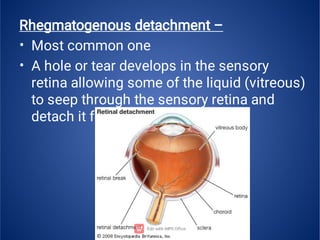
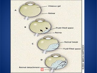
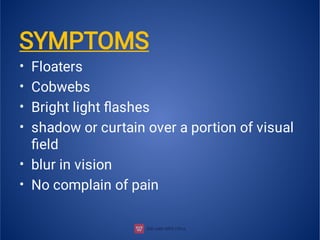
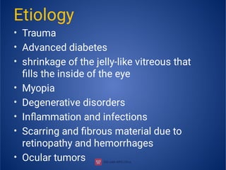
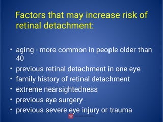
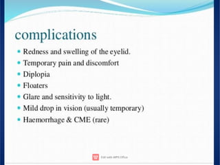
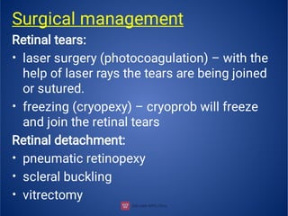
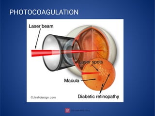
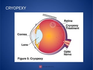
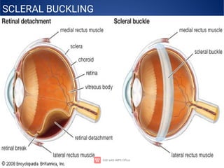
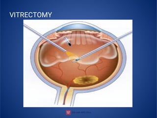
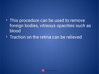
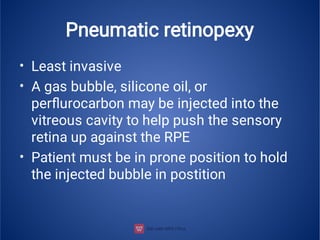
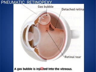
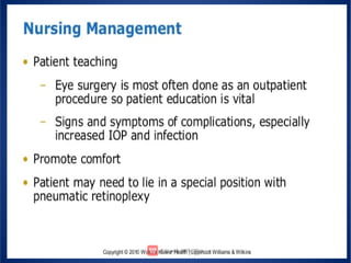
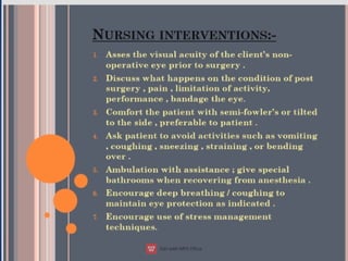
RETINAL DETACHMENT
SUBMITTED TO :
MRS. MAMTA TOPPO ASSOCIATED PROFESSOR
COLLEGE OF NURSING
RIMS RANCHI
SUBMITTED TO:
NITU KUMARI
ROLL NO-15
BASIC BSC NURSING 3RD YEAR
CONTENT
•
•
•
•
•
•
•
•
•
•
•
•
•
•
•
•
•
RETINA
RETINAL DETACHMENT
TYPE OF RETINAL DETACHMENT
PATHOPHYSIOLOGY
SYMPTOMS
ETIOLOGY
DIAGNOSIS
COMPLICATION
SURGICAL MANAGEMENT
NURSING MANAGEMENT
PRE OPERATIVE CARE
POST OPERATIVE CARE
HOME CARE
NURSIND DIAGNOSIS
NURSING INTERVENTION
NEW REASEARCH
BIBLIOGRAPHY
•
•
•
•
RETINA
light-sensitive tissue layer
sends visual messages
through the optic nerve
Retina have 2 layers
Sensory retina – rods and
cones are present
Retinal pigment
epithelium (RPE)
RETINAL DETACHMENT
•
•
•
•
Retinal detachment is the separation of
the retinal layers (sensory layer from RPE)
due to a break or tear over the retinal
layers
Four types
Rhegmatogenous
Traction
Combined form of rhegmatogenous and
traction
•
•
Rhegmatogenous detachment –
Most common one
A hole or tear develops in the sensory
retina allowing some of the liquid (vitreous)
to seep through the sensory retina and
detach it from the RPE
•
•
Traction – a pulling force is responsible
Traction can be occur due to any scars or
bands of fibrous material providing
traction to the retina
Vitreous hemorrhage, retinopathy can
cause traction effect
Exudative – due to production of serous
fluid under the retina. (uveitis,
degenerative disorders)
Pathophysiology
•
•
•
•
•
•
Due to etiological factors (a torn or break
in retina)
Vitreous fluid or serous fluid leaks in
between the layers of retina or behind the
retinal layers
Detachment of retinal layer
Retina can peel away from the underlying
layer of blood vessels
Lack of oxygenation in tissues of retina
Vision disturbances
•
•
•
•
•
•
SYMPTOMS
Floaters
Cobwebs
Bright light flashes
shadow or curtain over a portion of visual
field
blur in vision
No complain of pain
• floaters - bits of debris in field of vision
that look like spots, hairs or strings
•
•
•
•
•
•
•
•
Etiology
Trauma
Advanced diabetes
shrinkage of the jelly-like vitreous that
fills the inside of the eye
Myopia
Degenerative disorders
Inflammation and infections
Scarring and fibrous material due to
retinopathy and hemorrhages
Ocular tumors
•
•
•
•
•
•
Factors that may increase risk of
retinal detachment:
aging - more common in people older than
40
previous retinal detachment in one eye
family history of retinal detachment
extreme nearsightedness
previous eye surgery
previous severe eye injury or trauma
Diagnostic findings
•
•
•
•
Visual acuity
Ophthalmoscopy
Slit lamp microscopy
Unltrasound
•
•
•
•
•
Surgical management
Retinal tears:
laser surgery (photocoagulation) – with the
help of laser rays the tears are being joined
or sutured.
freezing (cryopexy) – cryoprob will freeze
and join the retinal tears
Retinal detachment:
pneumatic retinopexy
scleral buckling
vitrectomy
PHOTOCOAGULATION
CRYOPEXY
Scleral buckling
•
•
•
•
Compression of sclera to indent the
scleral wall from the outside of the eye
and bring the two retinal layers in contact
with each other
High success rate
It uses silicone sponge, rubber, or semi-
hard plastic for buckling
It will provide a traction on the retina and
allow the retinal tear to settle against the
wall of the eyes
SCLERAL BUCKLING
Vitrectomy
•
•
•
•
•
•
It is the surgical removal of the vitreous gel
from the eye. It can also remove blood in the
vitreous gel
Three incisions are made
One incision allows the introduction of light
source
Other incision for vitrectomy instrument
The third incision for the instrument to refill
the vitreous substitute (silicone oil)
Duration is 2 to 3 hours
VITRECTOMY
•
•
This procedure can be used to remove
foreign bodies, vitreous opacities such as
blood
Traction on the retina can be relieved
Pneumatic retinopexy
•
•
•
Least invasive
A gas bubble, silicone oil, or
perflurocarbon may be injected into the
vitreous cavity to help push the sensory
retina up against the RPE
Patient must be in prone position to hold
the injected bubble in postition
PNEUMATIC RETINOPEXY
NEW RESEARCH IN INDIA
SUMMARY
• Retinal detachment is a disorder of the
eye in which the retina separates from the
layer underneath . Symptoms include an
increase in the number of floaters , flashes
of light , and worsening of the outer part of
the visual field. This may be describe as a
curtain over part of the field of vision.
EVALUATION
1)
2)
3)
4)
5)
6)
What is retinal detachment?
What is floater symptoms?
What are the risk factor of retinal
detachment?
What is vitrectomy?
How many type of retinal detachment?
Explain the nursing management for
retinal detachment?
BIBLIOGRAPHY
•
•
•
Ansari javed , a text book of medical
surgical
nursing –II publication page no- 204
-207
Brunner and suddarth s text book of
medical
surgical nursing voll- south Asian
edition,
wolters Kluwer, page no-204-208
www.Wikipedia.com
Retinal detachment  presentation

Retinal detachment presentation