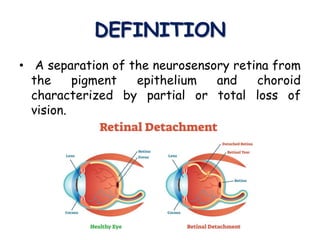
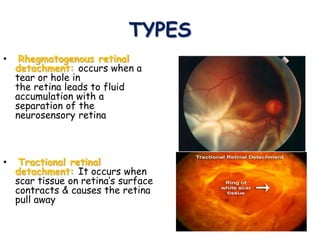
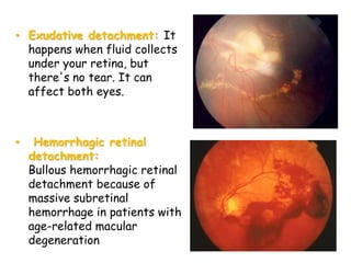
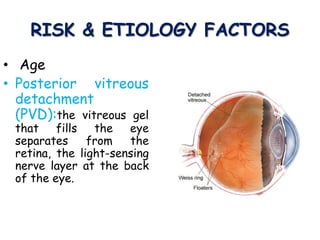
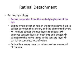
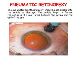
2. RETINA
• The retina is light-sensitive membrane located at the
back of the eye
• The purpose of the retina is to receive light that the
lens has focused, convert the light into neural signals,
and send these signals on to the brain for visual
recognition
3. DEFINITION
• A separation of the neurosensory retina from
the pigment epithelium and choroid
characterized by partial or total loss of
vision.
4.
5. TYPES
• Rhegmatogenous retinal
detachment: occurs when a
tear or hole in
the retina leads to fluid
accumulation with a
separation of the
neurosensory retina
• Tractional retinal
detachment: It occurs when
scar tissue on retina’s surface
contracts & causes the retina
pull away
6. • Exudative detachment: It
happens when fluid collects
under your retina, but
there's no tear. It can
affect both eyes.
• Hemorrhagic retinal
detachment:
Bullous hemorrhagic retinal
detachment because of
massive subretinal
hemorrhage in patients with
age-related macular
degeneration
7. RISK & ETIOLOGY FACTORS
• Age
• Posterior vitreous
detachment
(PVD):the vitreous gel
that fills the eye
separates from the
retina, the light-sensing
nerve layer at the back
of the eye.
8. • Family history
• Diabetes
• Cataract surgery complication
• Severe myopia
• Orbital trauma
9. CLINICAL MANIFESTATIONS
• Photospia ( is the presence of
perceived flashes of light in the field
of vision)
• No pain associated with
retinal tear
• Blurred vision
10. • Gradually reduced side
(peripheral) vision
• Black spots, floaters,
blind spot
• Frequently tears in a
horse shoe shape
• A curtain-like shadow
over your visual field
15. SCLERA BUCKLING PROCEDURE
It is a surgical procedure in which a piece of silicone plastic or
sponge is sewn onto the sclera at the site of a retinal tear to
push the sclera toward the retinal tear. The buckle holds the
retina against the sclera until scarring seals the tear
16. PNEUMATIC RETINOPEXY
The eye doctor (ophthalmologist) injects a gas bubble into
the middle of the eye. The bubble helps to flatten
the retina until a seal forms between the retina and the
wall of the eye
20. BENIGN PROSTATIC
HYPERTROPHY
• MR. MOUNESH. BADIGER
• SENIOR TUTOR
• MEDICAL SURGICAL DEPARTMENT
• KAHER INSTITUTE OF NURSING SCIENCES,
• BELAGAVI,
THANK YOU