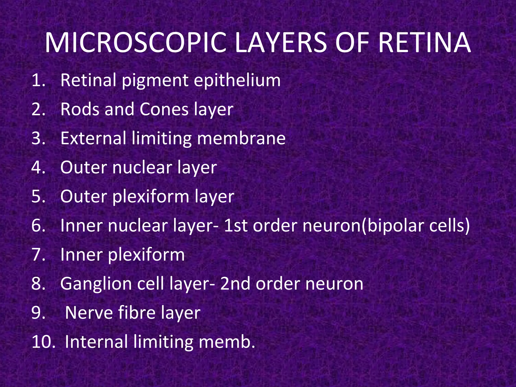
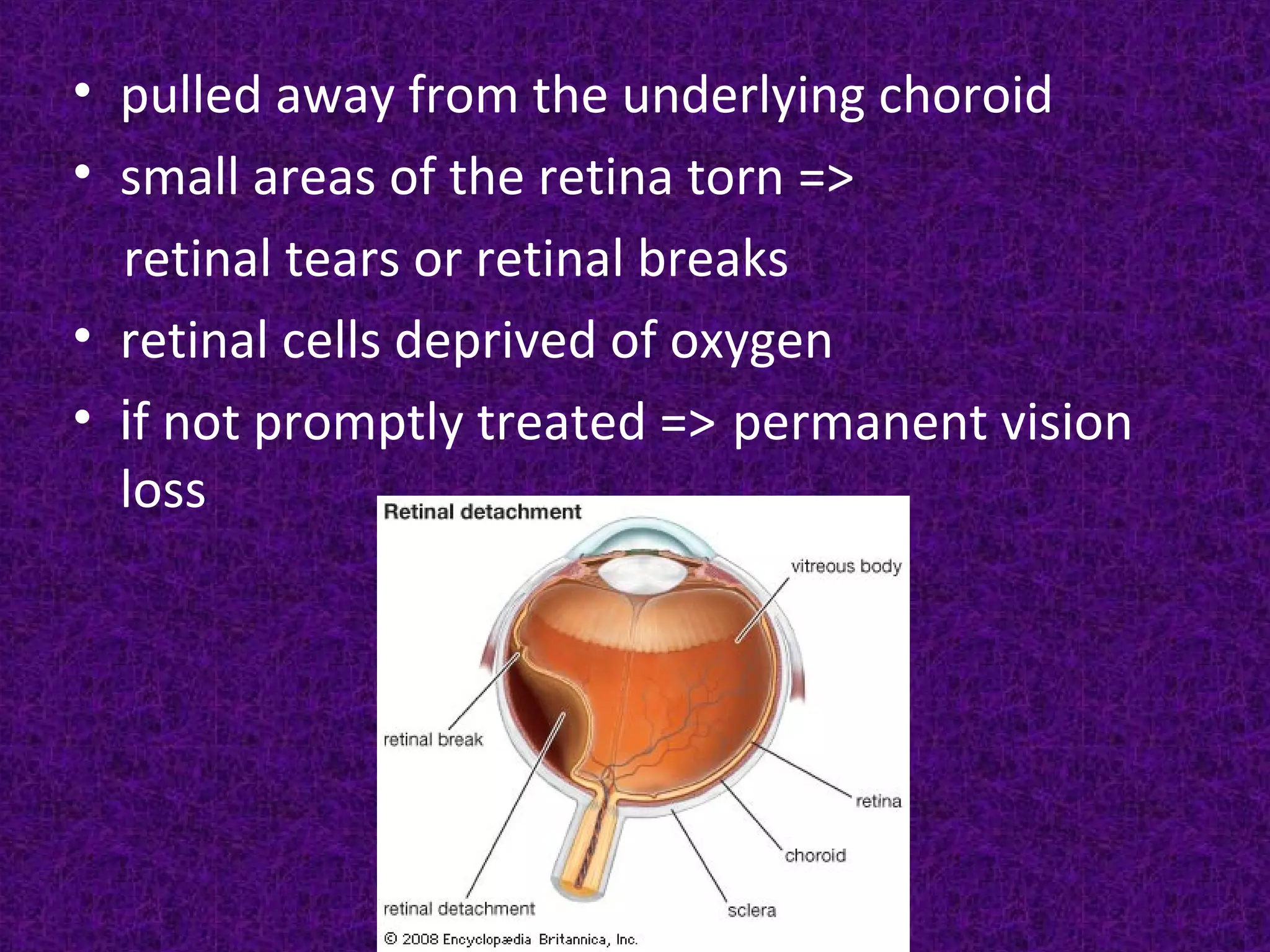
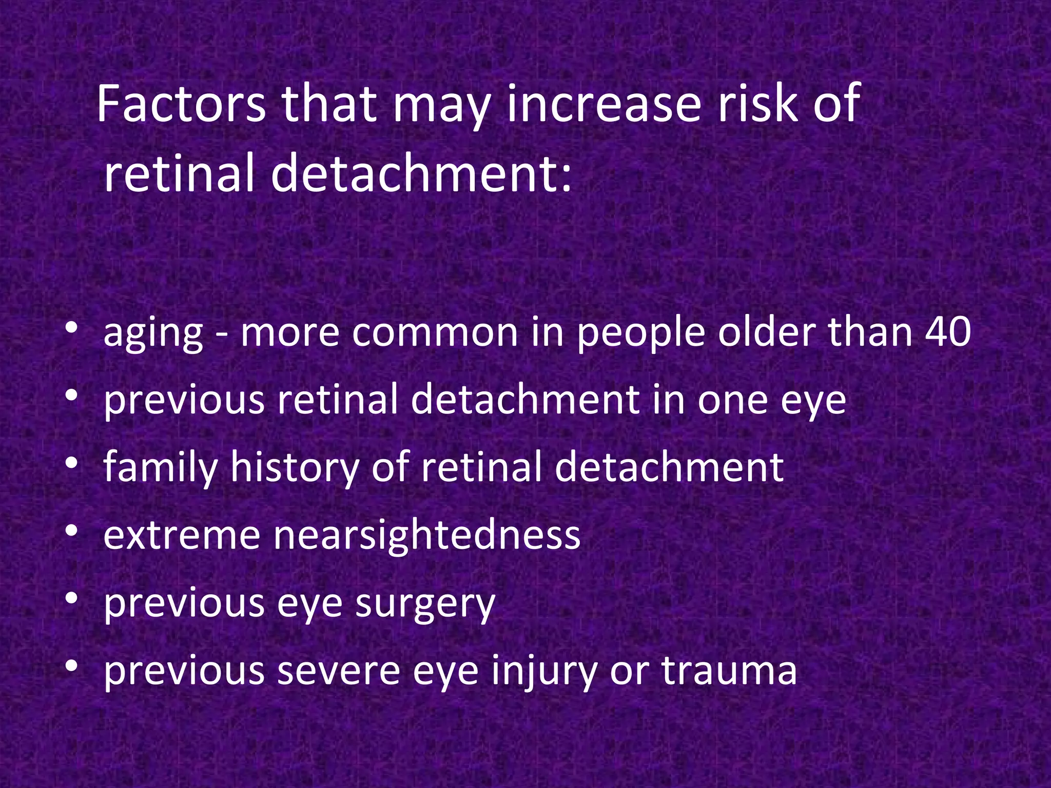
RETINAL DETACHMENT
GLADIS K MATHEW
LECTURER
RETINA
• light-sensitive layer
of tissue
• sends visual
messages through
the optic nerve
MICROSCOPIC LAYERS OF RETINA
1. Retinal pigment epithelium
2. Rods and Cones layer
3. External limiting membrane
4. Outer nuclear layer
5. Outer plexiform layer
6. Inner nuclear layer- 1st order neuron(bipolar cells)
7. Inner plexiform
8. Ganglion cell layer- 2nd order neuron
9. Nerve fibre layer
10. Internal limiting memb.
Retinal Detachment
• A Retinal Detachment (RD) describes the
separation of the neurosensory retina (NSR)
from the retinal pigment epithelium (RPE).
This results in the accumulation of subretinal
fluid (SRF) in the potential space between the
NSR and RPE.
• pulled away from the underlying choroid
• small areas of the retina torn =>
retinal tears or retinal breaks
• retinal cells deprived of oxygen
• if not promptly treated => permanent vision
loss
Types
1. Rhegmatogenous (rhegma – break), occurs
secondarily to a full-thickness defect in the
sensory retina, which permits fluid derived
from vitreous to gain access to the subretinal
space.
2. Tractional in which the NSR is pulled away
from the RPE by contracting vitreoretinal
membranes in the absence of a retinal break.
3. Exudative (serous, secondary) RD is caused
neither by a break nor traction; the SRF is
derived from fluid in the vessels of the NSR or
choroid, or both.
4. Combined tractional-rhegmatogenous, is the
result of a combination of a retinal break and
retinal traction. The retinal break is caused by
traction from an adjacent area of fibrovascular
proliferation and is most commonly seen in
advanced proliferative diabetic retinopathy.
Can occur as a result of:
• trauma
• advanced diabetes
• an inflammatory disorder, such as
sarcoidosis
• shrinkage of the jelly-like vitreous that fills
the inside of the eye
Retinal detachment can occur as a result of;
• Obesity
• Trauma
• Nearsightedness
Causes in non rhegmatogenous
detachment
1. The retina being pushed away from its bed
• Accumulation of fluid. Eg- blood (choroidal
haemorrhage) or exudates
• Neoplasm
2. The retina being pulled away from its bed
• The contraction of fibrous tissue bands in the
vitreous
Factors that may increase risk of
retinal detachment:
• aging - more common in people older than 40
• previous retinal detachment in one eye
• family history of retinal detachment
• extreme nearsightedness
• previous eye surgery
• previous severe eye injury or trauma
• vitreous liquid leaks through retinal tear and
accumulates underneath retina
• retina can peel away from underlying layer of
blood vessels
SYMPTOMS
• floaters
• light flashes (photopsia)
• shadow or curtain over a portion of visual
field
• blur in vision
SYMPTOMS
• floaters - bits of debris in field of vision that
look like spots, hairs or strings
Signs
• Plane mirror examination- defective or no red
glow seen
Fundus examination
• The detached retina looks greyish- white and
raised above the surface
• The retinal vessels are dark with no central
light reflex
• Detached retina is thrown into multiple folds
which oscillate with the movement of the eye
• Holes or tear can seen
• Visual fields – scotomas are present
• Electroretinography- it is subnormal
• Ultrasonography confirms the diagnosis
Complications
• Total detachment of the retina
• Complicated cataract is seen in the posterior
cortex
• Chronic uveitis and phthisis
TREATMENTS
Retinal tears:
• laser surgery (photocoagulation)
• freezing (cryopexy)
Retinal detachment:
• pneumatic retinopexy
• scleral buckling
• vitrectomy
PHOTOCOAGULATION
A triple row of burns is placed around the break
to coagulate the area
CRYOPEXY- is done to seal the retinal breaks by
causing tissue necrosis
PNEUMATIC RETINOPEXY
It is another method of repairing a retinal
detachment in which a gas bubble is injected
into the eye after which laser or freezing
treatment is applied to the retinal hole.
PNEUMATIC RETINOPEXY
Scleral buckling
• Scleral buckle surgery is an established
treatment in which the eye surgeon sews one
or more silicone bands to the sclera. The
bands push the wall of the eye inward against
the retinal hole, closing the break or reducing
fluid flow through it and reducing the effect of
vitreous traction thereby allowing the retina
to re-attach.
SCLERAL BUCKLING
vitrectomy
• It breaks the tractional band in the vitreous
thus releasing the pull on the retina in cases of
tractional retinal detachment
• Drainage of subretinal fluid is required in long
standing cases
VITRECTOMY
Nursing diagnoses for Retinal
Detachment
• Disturbed sensory perception (visual).
• Anxiety.
• Risk for injury.
Implementation
• Asses visual status and functional vision in the
unaffected eye to determine self care needs.
• Prepare the client for surgery by explaining
possible surgical interventions and technique
to alleviate some of the client's anxiety.
• Discourage straining during defecation,
bending down and hard coughing, sneezing or
vomiting to avoid activities that increase
intraocular pressure.
• Assist with ambulation, as needed, to help the
client remain independent.
• Approach the clients from the unaffected side
to avoid startling him.
• Provide assistance with activities of daily living
to minimize frustation and strain.
• Orient the client to his environment to reduce
the risk of injury.
• Postoperatively instruct the client to lie on his
back or on his unoperated side to reduce
intraocular pressure in the affected area.

Retinal detachment

  • 1.
  • 2.
    RETINA • light-sensitive layer oftissue • sends visual messages through the optic nerve
  • 3.
    MICROSCOPIC LAYERS OFRETINA 1. Retinal pigment epithelium 2. Rods and Cones layer 3. External limiting membrane 4. Outer nuclear layer 5. Outer plexiform layer 6. Inner nuclear layer- 1st order neuron(bipolar cells) 7. Inner plexiform 8. Ganglion cell layer- 2nd order neuron 9. Nerve fibre layer 10. Internal limiting memb.
  • 4.
    Retinal Detachment • ARetinal Detachment (RD) describes the separation of the neurosensory retina (NSR) from the retinal pigment epithelium (RPE). This results in the accumulation of subretinal fluid (SRF) in the potential space between the NSR and RPE.
  • 5.
    • pulled awayfrom the underlying choroid • small areas of the retina torn => retinal tears or retinal breaks • retinal cells deprived of oxygen • if not promptly treated => permanent vision loss
  • 6.
    Types 1. Rhegmatogenous (rhegma– break), occurs secondarily to a full-thickness defect in the sensory retina, which permits fluid derived from vitreous to gain access to the subretinal space. 2. Tractional in which the NSR is pulled away from the RPE by contracting vitreoretinal membranes in the absence of a retinal break.
  • 7.
    3. Exudative (serous,secondary) RD is caused neither by a break nor traction; the SRF is derived from fluid in the vessels of the NSR or choroid, or both. 4. Combined tractional-rhegmatogenous, is the result of a combination of a retinal break and retinal traction. The retinal break is caused by traction from an adjacent area of fibrovascular proliferation and is most commonly seen in advanced proliferative diabetic retinopathy.
  • 8.
    Can occur asa result of: • trauma • advanced diabetes • an inflammatory disorder, such as sarcoidosis • shrinkage of the jelly-like vitreous that fills the inside of the eye
  • 9.
    Retinal detachment canoccur as a result of; • Obesity • Trauma • Nearsightedness
  • 10.
    Causes in nonrhegmatogenous detachment 1. The retina being pushed away from its bed • Accumulation of fluid. Eg- blood (choroidal haemorrhage) or exudates • Neoplasm 2. The retina being pulled away from its bed • The contraction of fibrous tissue bands in the vitreous
  • 11.
    Factors that mayincrease risk of retinal detachment: • aging - more common in people older than 40 • previous retinal detachment in one eye • family history of retinal detachment • extreme nearsightedness • previous eye surgery • previous severe eye injury or trauma
  • 12.
    • vitreous liquidleaks through retinal tear and accumulates underneath retina • retina can peel away from underlying layer of blood vessels
  • 13.
    SYMPTOMS • floaters • lightflashes (photopsia) • shadow or curtain over a portion of visual field • blur in vision
  • 14.
    SYMPTOMS • floaters -bits of debris in field of vision that look like spots, hairs or strings
  • 16.
    Signs • Plane mirrorexamination- defective or no red glow seen Fundus examination • The detached retina looks greyish- white and raised above the surface • The retinal vessels are dark with no central light reflex • Detached retina is thrown into multiple folds which oscillate with the movement of the eye • Holes or tear can seen
  • 17.
    • Visual fields– scotomas are present • Electroretinography- it is subnormal • Ultrasonography confirms the diagnosis
  • 18.
    Complications • Total detachmentof the retina • Complicated cataract is seen in the posterior cortex • Chronic uveitis and phthisis
  • 19.
    TREATMENTS Retinal tears: • lasersurgery (photocoagulation) • freezing (cryopexy) Retinal detachment: • pneumatic retinopexy • scleral buckling • vitrectomy
  • 20.
    PHOTOCOAGULATION A triple rowof burns is placed around the break to coagulate the area
  • 21.
    CRYOPEXY- is doneto seal the retinal breaks by causing tissue necrosis
  • 22.
    PNEUMATIC RETINOPEXY It isanother method of repairing a retinal detachment in which a gas bubble is injected into the eye after which laser or freezing treatment is applied to the retinal hole.
  • 23.
  • 24.
    Scleral buckling • Scleralbuckle surgery is an established treatment in which the eye surgeon sews one or more silicone bands to the sclera. The bands push the wall of the eye inward against the retinal hole, closing the break or reducing fluid flow through it and reducing the effect of vitreous traction thereby allowing the retina to re-attach.
  • 25.
  • 26.
    vitrectomy • It breaksthe tractional band in the vitreous thus releasing the pull on the retina in cases of tractional retinal detachment • Drainage of subretinal fluid is required in long standing cases
  • 27.
  • 28.
    Nursing diagnoses forRetinal Detachment • Disturbed sensory perception (visual). • Anxiety. • Risk for injury.
  • 29.
    Implementation • Asses visualstatus and functional vision in the unaffected eye to determine self care needs. • Prepare the client for surgery by explaining possible surgical interventions and technique to alleviate some of the client's anxiety. • Discourage straining during defecation, bending down and hard coughing, sneezing or vomiting to avoid activities that increase intraocular pressure.
  • 30.
    • Assist withambulation, as needed, to help the client remain independent. • Approach the clients from the unaffected side to avoid startling him. • Provide assistance with activities of daily living to minimize frustation and strain. • Orient the client to his environment to reduce the risk of injury. • Postoperatively instruct the client to lie on his back or on his unoperated side to reduce intraocular pressure in the affected area.