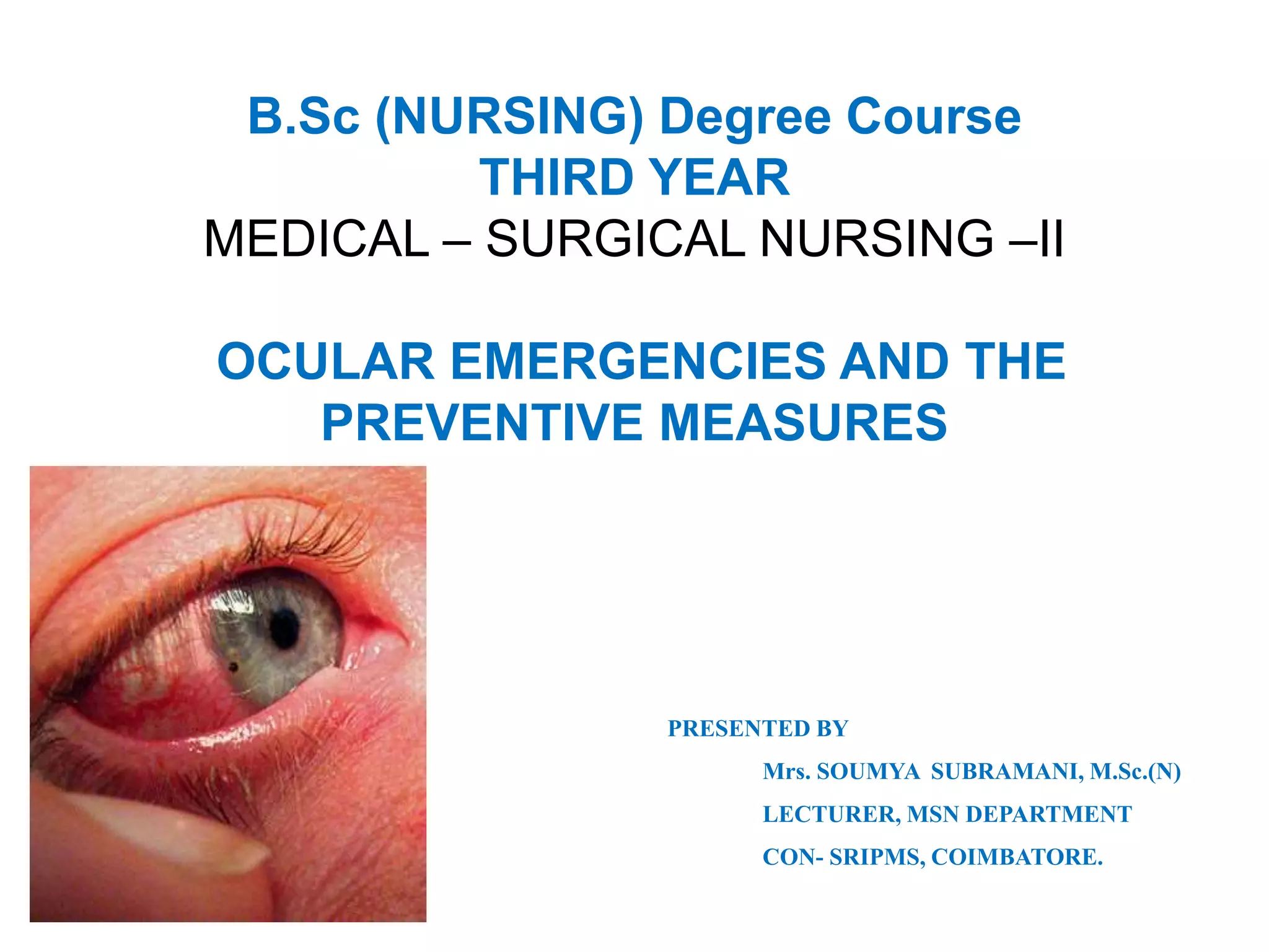
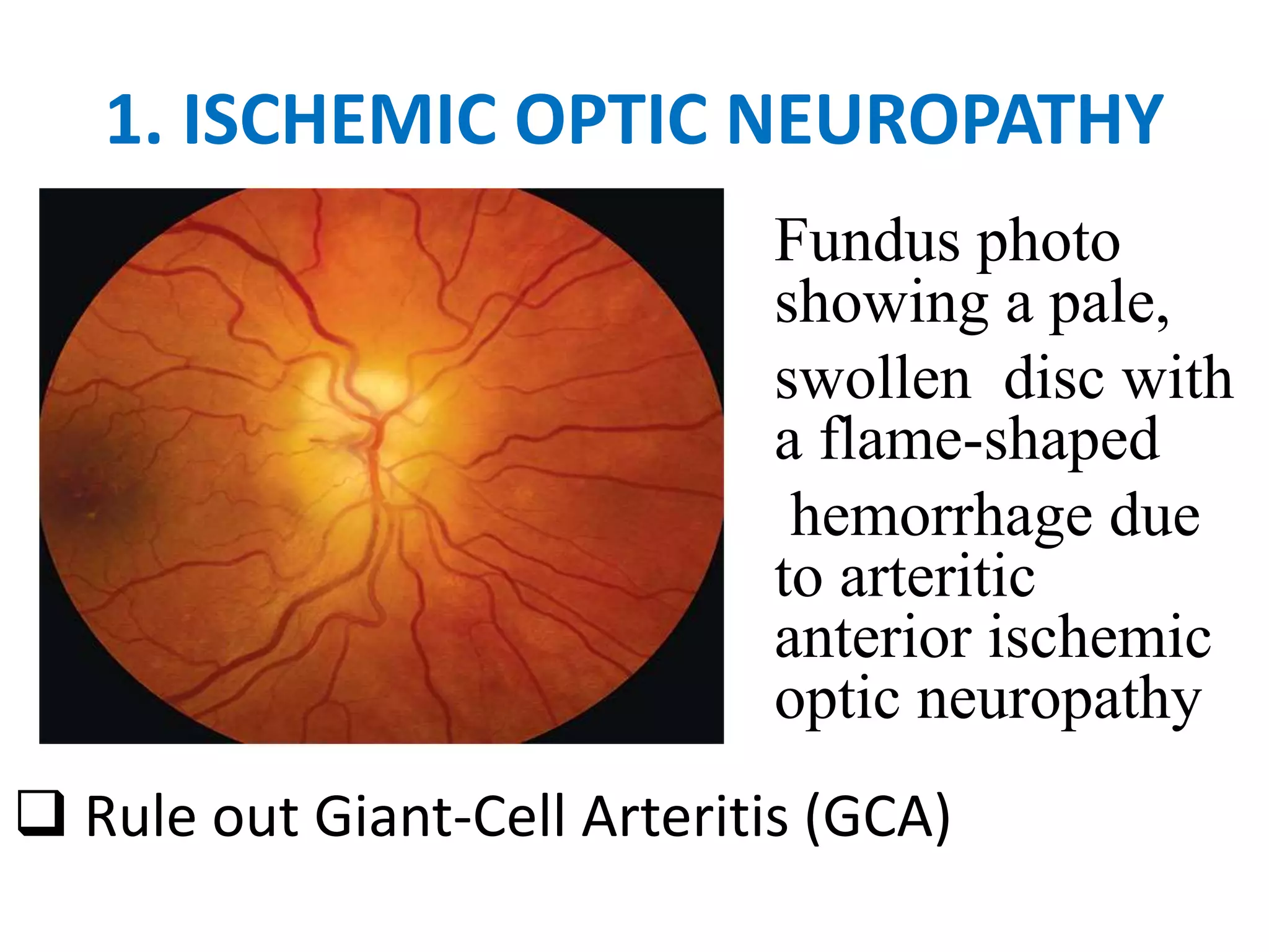
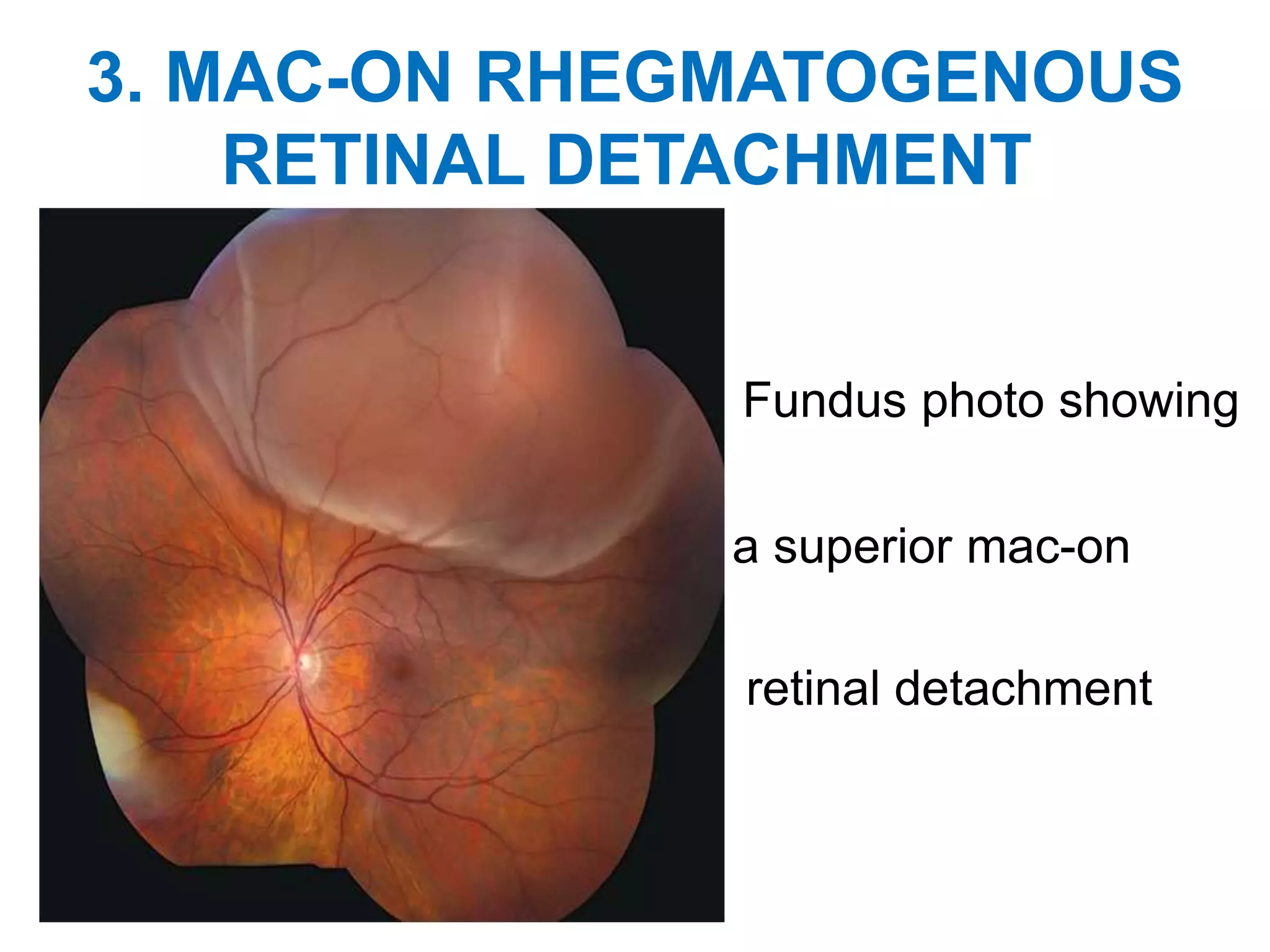
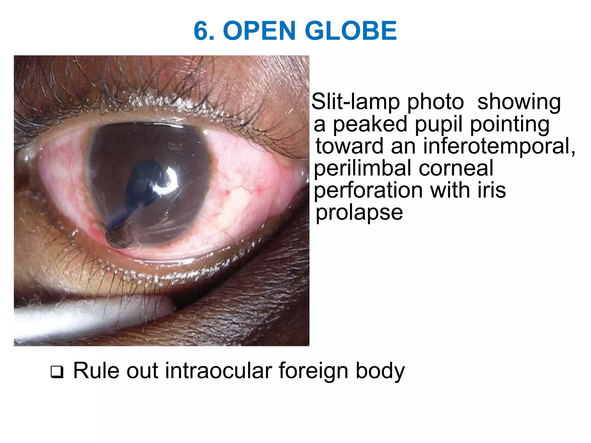
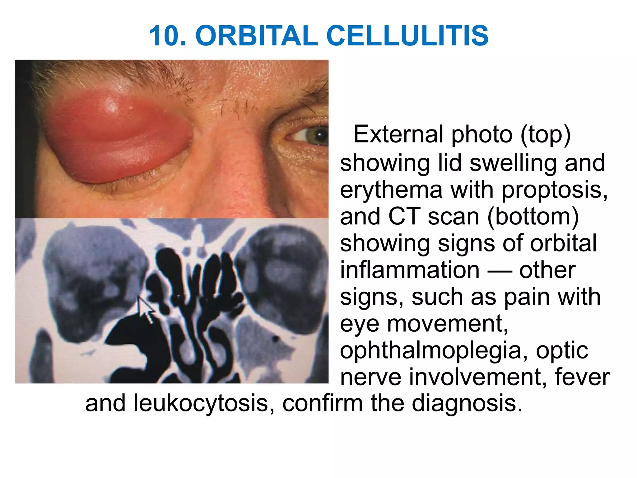
B.Sc (NURSING) Degree Course
THIRD YEAR
MEDICAL – SURGICAL NURSING –II
OCULAR EMERGENCIES AND THE
PREVENTIVE MEASURES
PRESENTED BY
Mrs. SOUMYA SUBRAMANI, M.Sc.(N)
LECTURER, MSN DEPARTMENT
CON- SRIPMS, COIMBATORE.
OBJECTIVES
At the end of this presentation the Student
Nurses will be able to
• Define Ocular Emergencies
• State the classification of Ocular Emergencies
• Describe the Top Ten Ocular Emergencies
• List out the symptoms of Ocular Emergencies
• Explain about Chemical injuries, Trauma and Foreign
body in the eyes and their management
• Mention about common Assessment and Diagnostic
findings
• Enlist the complications
• Instruct what not to do if we have eye injury
• Briefly explain about the preventive measures
INTRODUCTION
“Beautiful eyes look good in others, The heart
smiles through the eyes”. All these phrases tells the
significance of our EYES. Sometimes physical or
chemical injuries and patho physiological changes of
the eyes may end up in loss of vision if not treated in
time.
There have been numerous individual reports on Ocular
Emergencies. W.H.O has reported 55 Million eye
emergencies causing restriction of daily activities of which
1.6 million go blind everyday. The prevalence of Ocular
trauma to be 2.4% of population in an urban city in India.
Let us view some of the Ocular Emergencies to
treat and prevent vision loss in us and in others too..
WHAT IS AN OCULAR EMERGENCY?
An ocular emergency is a condition that
can cause a sudden loss of, or decrease in a
person’s vision that could lead to a permanent
condition.
This may occur at any time when a person
have foreign object or chemicals in his eye(s),
or when an injury or burn affects the eye area.
CLASSIFICATION OF OCULAR
EMERGENCIES
 Emergency
 Urgent
 Semi Urgent
EMERGENCY
Must be treated within minutes
Example:
Chemical Burns of …
 Conjunctiva
 Cornea
URGENT
Must be treated within hours.
Examples……
• Penetrating globe injuries
• Corneal Abrasions
• Corneal foreign bodies;
• Hyphema
• Eyelid lacerations that are deep
• Radiant energy burns such as Arc eye (welder's
burn, Snow blindness)
• Traumatic Optic neuropathy
SEMI-URGENT
Must be managed within 1–2 days.
Examples….
• Orbital fractures
• Sub Conjunctival Haemorrhages
ASSESSMENT and DIAGNOSTIC FINDINGS
• Ocular History – Type of injury
• Past ocular surgery
• Nature of ocular injury
• Nature of cause and force
• Vision loss-sudden/slow/progressive
• Identify the chemical agent in burns and test for its
pH level
• Examine the corneal surface for foreign body,
wounds and abrasions
• Examine the pupillary size,shape,reaction to light
and compare with the unaffected eye if possible
• Assess ocular mobility
Contin...
 Usual Eye examination to be done and may
require a Topical anesthetic in order to be
tolerable.
 Proxymetacaine has been found to have the best
tolerance.
 Depending on the Medical history and preliminary
examination, the diagnosis is made and classified
as an emergency, urgent or semi-urgent
TOP 10 OCULAR EMERGENCIES
Since the eyes are easily damaged any of
these conditions can lead to vision loss if not
treated in time.
Here follows Top 10 Ocular emergencies for
better understanding
1. ISCHEMIC OPTIC NEUROPATHY
Fundus photo
showing a pale,
swollen disc with
a flame-shaped
hemorrhage due
to arteritic
anterior ischemic
optic neuropathy
 Rule out Giant-Cell Arteritis (GCA)
2.CENTRAL RETINAL ARTERY
OCCLUSION (Amaurosis Fugax)
Fundus photo showing
diffuse Retinal whitening
and a foveal cherry-red
spot
Rule out GCA and causes of emboli/thrombus
3. MAC-ON RHEGMATOGENOUS
RETINAL DETACHMENT
Fundus photo showing
a superior mac-on
retinal detachment
4. ACUTE THIRD NERVE PALSY
Extraocular motility showing
complete ptosis, the right eye
down and out, inability to
adduct, infraduct and
supraduct the eye and a
dilated pupil
Rule out intracranial aneurysm
5. CORNEAL MICROBIAL KERATITIS
Slit-lamp photo showing
conjunctival infection and
focal White infiltrates with
hypopyon.
Culture and treat with empiric antibiotics and follow
closely
6. OPEN GLOBE
Slit-lamp photo showing
a peaked pupil pointing
toward an inferotemporal,
perilimbal corneal
perforation with iris
prolapse
 Rule out intraocular foreign body
7. ACUTE ANGLE CLOSURE GLAUCOMA
Slit-lamp photo showing
conjunctival infection,
corneal haze with
microcystic edema, a
fixed, mid-dilated pupil
and a shallow anterior
chamber
8. ENDOPHTHALMITIS
Slit-lamp photo showing
conjunctival infection,
mild corneal edema and
haze and anterior
chamber hypopyon
9. ALKALI INJURY
Slit-lamp photo showing
perilimbal conjunctival
blanching, conjunctival
infection and diffuse
corneal haze
Requires urgent and copious irrigation
10. ORBITAL CELLULITIS
External photo (top)
showing lid swelling and
erythema with proptosis,
and CT scan (bottom)
showing signs of orbital
inflammation — other
signs, such as pain with
eye movement,
ophthalmoplegia, optic
nerve involvement, fever
and leukocytosis, confirm the diagnosis.
SYMPTOMS OF OCULAR EMERGENCIES
The following symptoms may indicate an
emergency requiring immediate medical attention..
 Black spots or flashes of light
 Curtain-like disappearance of vision
 Eye injury or pain
 Seeing halos or rainbows around light
 Loss of peripheral (side) vision
 Sudden hazy or blurred vision
 Sudden vision loss in one eye
 Red, crusty or swollen eyelids
Symptoms Conti…..
 Bruising around the eye
 Bleeding from the eye
 Blood in the white part of the eye
 Discharge from the eye
 Severe itching
 Pupils that are not the same size
 One eye is not moving like the other
 One eye is Sticking out or bulging
Symptoms Conti….
The following symptoms may not be
emergencies but still require examination from
the eye doctor:
 Blurred or double vision
 Excessive tearing or watering of the eyes
 Itchy, burning, or dry eyes
 Difficulty seeing in dark environments
 Seeing spots or ghost-like images
 Light sensitivity
 Eye strain and/or frequent mild headaches
CHEMICAL INJURIES TO THE EYE
Agents causing chemical injuries are…..
 Cleaning products
 Garden chemicals
 Industrial chemicals
 Aerosols and
 Fumes
 If get acid in the eye, early treatment generally
results in a good prognosis. However, alkaline
products like drain cleaners, sodium hydroxide,
Dye, or lime can permanently damage the
cornea.
CHEMICAL BURNS OF EYES
First Aid measures for Chemical injuries
of Eyes
 Wash hands with soap and water to remove any
chemicals that may have adhere on hands.
 Turn the head so the injured eye is down and to the
side.
 Hold the eyelid open and flush with clean cool tap
water for 15 minutes. This can also be done in the
shower.
 If wearing contact lenses and they’re still in eyes after
flushing, try to remove them.
 Get to an emergency room or urgent care center as
quickly as possible. If possible, continue to flush the
eyes with clean water while waiting for an ambulance
or traveling to the medical center.
MANAGEMENT OF CHEMICAL OCULAR BURNS
 Appears as Superficial Punctate Keratopathy
 Copious irrigation with Tap water or Normal
saline or with Neutral solution to be done
immediately
 Apply local anaesthetic and eye lid speculum
 Remove the particulate matters by moistened
cotton tip applicators
 Check the pH of the corneal surface with strip
 Instill Antibiotics and patch the eye or use
therapeutic lenses
Contin.....
 Needs thorough inspection, cleansing and repair of
the wound
 Cold compress are to be used in early phase
followed by warm compress
 Surgical draining of hematoma may be required
 Corticosteroid therapy or optic nerve
decompression surgery is indicated to reduce optic
nerve swelling
 Long term treatment or rehabilitation -Grafting
procedures and Surgical restoration of corneal
integrity and optical clarity
 Scarring of the eye lids may require oculoplastic
surgery and corneal scarring may require corneal
surgery
SMALL FOREIGN BODIES IN THE EYE
 If something gets in to the eyes it can cause eye
damage or a loss of vision. Even something as small
as sand or dust can cause irritation.
 Make the person to try blinking to see if it clears eye.
Don’t rub the eye.
 Wash the hands before touching the eye. Look into
the eye to try to locate the object.
 If necessary, make him to look behind the lower lid
by pulling it down gently. Make him to look under the
upper lid by placing a cotton swab on the lid and
flipping the lid over it.
Small foreign objects in the eye
continues…..
 Use artificial tear eye drops to help rinse out the
foreign body.
 If the foreign object is stuck on one of the
eyelids, flush it with water. If the object is in the
eye, flush the eye with cool water.
 If cannot remove the object or if the irritation
continues, immediately seek medical aid.
LARGE FOREIGN OBJECTS STUCK IN EYE
 Glass, metal, or objects that enter the eye at high
speed can cause serious damage. If something is
stuck in the eye, leave it where it is.
 Do not touch it, do not apply pressure, and do
not attempt to remove it.
 This is a medical emergency and should seek
help immediately.
 Make him try to move the eye as little as possible
while waiting for medical care.
 If the object is small and help to cover both eyes
with a clean piece of cloth. This will reduce the
eye movement until the doctor examines.
FOREIGN BODIES IN THE EYES
MANAGEMENT OF FOREINBODY IN THE EYES
 Copper, Iron and vegetative materials may cause
purulent infection
 X-ray and C.T. scan to be taken
 Careful history taking
 Prohibit MRI in case of metallic foreign body
 Remove superficial foreign body
 Apply Antibiotic ointment and patch the eye
 Examine daily for evidence of infection
 Immobilize the lids if needed by patch
 Culture and sensitivity test if infection suspects
 I.V. Antibiotics followed by Oral Antibiotics to be
administered
 Surgical intervention may be done if needed
OCULAR TRAUMA
 Leading cause of blindness among children and
young adults.
 Causes may be Occupational injuries, Sports,
Weapons,Assault,Motor vehicle crashes, Blast
fragments during wars etc,.
 For chemical burn irrigate with normal saline or
with tap water
 Don’t attempt to remove pierced foreign body at
the occurrence site
 PROTECT the eye by using metal shield or by stiff
paper cup
CUTS AND SCRATCHES
 If there is a cut or scratch to the eyeball or
eyelid, that person need urgent medical care.
 Apply a loose bandage while waiting for medical
treatment, but be careful not to apply pressure
PENETRATING INJURIES OF THE EYES
May results in loss of vision and exhibit...
 Hemorrhagic Chemosis
 Conjunctival laceration
 Shallow anterior chamber
 With or without ecentrically placed pupil
 Hyphema
 Vitreous Hemorrhage
PENETRATING INJURY
Management Of Penetrating Injuries Of
The Eyes
HYPHEMA
 Hospitalize the patient with moderate activity restriction
 Apply Eye Shield
 Apply topical Corticosteroids to reduce inflammation
RUPTURED GLOBE
 Vitrectomy for traumatic retinal detachment
 Primary Enucleation (with in two weeks) if needed to
prevent Sympathetic ophthalmia in the unaffected eye.
INTRA OCULAR FOREIGNBODIES
 Diagnose and localize the foreignbody by Slit Lamp
Biomicroscopy and Direct ophthalmoscopy, C.T.Scan or
by Ultrasound
 Determine the compression,size,locationand affected
eye structures
 If Cornea is perforated administer Tetanus Toxoid
 Surgical incision and extraction of foreignbody is
decided based on its location,composition and
associated ocular injuries
 Any damaged area f the Retina is treated to prevent
retinal detachment
ASSESSMENT and DIAGNOSTIC FINDINGS
• Ocular History – Type of injury
• Past ocular surgery
• Nature of ocular injury
• Nature of cause and force
• Vision loss-sudden/slow/progressive
• Identify the chemical agent in burns and test for its
pH level
• Examine the corneal surface for foreignbody,
wounds and abrasions
• Examine the pupillary size,shape,reaction to light
and compare with the unaffected eye if possible
• Assess ocular mobility
Contin...
 Usual Eye examination to be done and may
require a Topical anesthetic in order to be
tolerable. Many topical agents cause burning upon
instillation.
 Proxymetacaine has been found to have the best
tolerance.
 Depending on the Medical history and preliminary
examination, the diagnosis is made and classified
as an emergency, urgent or semi-urgent
COMPLICATIONS
 CornealScarring
 Hyphema
 Iridodialysis
 Post traumatic glaucoma
 Uveitis cataract
 Vitreous Hemorrhage
 Retinal detachment
 The complications risk is high with retinal tears,
penetrating injuries and severe blunt trauma.
WHAT NOT TO DO IF HAVE AN EYE INJURY
Rub or apply pressure to the eye
Try to remove foreign objects that are stuck in
any part of your eye
Use tweezers or any other tools in the eye (cotton
swabs can be used, but only on the eyelid)
Put medications or ointments in the eye
If wearing Contact lenses , don’t take them out, if
suspects eye injury. Attempting to remove the
contact lenses can make injury worse.
The only exceptions to this rule are in situations
where a chemical injury and the lenses didn’t
flush out with water, or where cannot receive
immediate medical help.
The best thing we can do in an eye emergency is
to get to the doctor as soon as possible.
PREVENTING OPHTHALMIC MERGENCIES
 Eye injuries can happen anywhere, including at
home, work, athletic events, or on the playground.
Accidents can happen during high-risk activities,
but also in places where we least expect them.
 Wearing protective eyewear when use power
tools or engage in high-risk sporting events.
 Follow the directions carefully when working with
chemicals or cleaning supplies.
 Keep scissors, knives, and other sharp
instruments and keep away from the reach of
young children.
 Teach older children how to use them safely and
supervise them when they do.
PREVENTION Contin.....
Don’t let the children play with projectile toys,
such as darts or pellet guns.
Childproof the home by either removing or
cushioning items with sharp edges.
Use caution when cooking with grease and oil.
Keep heated hair appliances, like curling irons
and straightening tools, away from the eyes.
Keep distance from amateur fireworks.
To decrease the chances of developing
permanent eye damage, always see an eye
doctor after experience an eye injury
DIAGNOSIS
 Usual Eye examination to be done and may
require a Topical anesthetic in order to be
tolerable. Many topical agents cause burning upon
instillation.
 Proxymetacaine has been found to have the best
tolerance.
 Depending on the Medical history and preliminary
examination, the diagnosis is made and classified
as an emergency, urgent or semi-urgent
SUMMARY
• “The process of sorting people based on their
need for immediate medical care as compared to
their chance of benefiting from such”is the core
concept to understand about OCULAR
EMERGENCIES.
• There are unexpected situations or factors which
may injure our eyes. These may vary as
chemicals,foreign objects,trauma,eye diseases
itself ,etc. Expecting the unexpected and
practicing the preventive measures will help a lot.
Application of this knowledge is prime in the
clinical settings and in day today life also.
THANK YOU

11. ocular emergencies and their prevention

  • 2.
    B.Sc (NURSING) DegreeCourse THIRD YEAR MEDICAL – SURGICAL NURSING –II OCULAR EMERGENCIES AND THE PREVENTIVE MEASURES PRESENTED BY Mrs. SOUMYA SUBRAMANI, M.Sc.(N) LECTURER, MSN DEPARTMENT CON- SRIPMS, COIMBATORE.
  • 3.
    OBJECTIVES At the endof this presentation the Student Nurses will be able to • Define Ocular Emergencies • State the classification of Ocular Emergencies • Describe the Top Ten Ocular Emergencies • List out the symptoms of Ocular Emergencies • Explain about Chemical injuries, Trauma and Foreign body in the eyes and their management • Mention about common Assessment and Diagnostic findings • Enlist the complications • Instruct what not to do if we have eye injury • Briefly explain about the preventive measures
  • 4.
    INTRODUCTION “Beautiful eyes lookgood in others, The heart smiles through the eyes”. All these phrases tells the significance of our EYES. Sometimes physical or chemical injuries and patho physiological changes of the eyes may end up in loss of vision if not treated in time. There have been numerous individual reports on Ocular Emergencies. W.H.O has reported 55 Million eye emergencies causing restriction of daily activities of which 1.6 million go blind everyday. The prevalence of Ocular trauma to be 2.4% of population in an urban city in India. Let us view some of the Ocular Emergencies to treat and prevent vision loss in us and in others too..
  • 5.
    WHAT IS ANOCULAR EMERGENCY? An ocular emergency is a condition that can cause a sudden loss of, or decrease in a person’s vision that could lead to a permanent condition. This may occur at any time when a person have foreign object or chemicals in his eye(s), or when an injury or burn affects the eye area.
  • 6.
    CLASSIFICATION OF OCULAR EMERGENCIES Emergency  Urgent  Semi Urgent
  • 7.
    EMERGENCY Must be treatedwithin minutes Example: Chemical Burns of …  Conjunctiva  Cornea
  • 8.
    URGENT Must be treatedwithin hours. Examples…… • Penetrating globe injuries • Corneal Abrasions • Corneal foreign bodies; • Hyphema • Eyelid lacerations that are deep • Radiant energy burns such as Arc eye (welder's burn, Snow blindness) • Traumatic Optic neuropathy
  • 9.
    SEMI-URGENT Must be managedwithin 1–2 days. Examples…. • Orbital fractures • Sub Conjunctival Haemorrhages
  • 10.
    ASSESSMENT and DIAGNOSTICFINDINGS • Ocular History – Type of injury • Past ocular surgery • Nature of ocular injury • Nature of cause and force • Vision loss-sudden/slow/progressive • Identify the chemical agent in burns and test for its pH level • Examine the corneal surface for foreign body, wounds and abrasions • Examine the pupillary size,shape,reaction to light and compare with the unaffected eye if possible • Assess ocular mobility
  • 11.
    Contin...  Usual Eyeexamination to be done and may require a Topical anesthetic in order to be tolerable.  Proxymetacaine has been found to have the best tolerance.  Depending on the Medical history and preliminary examination, the diagnosis is made and classified as an emergency, urgent or semi-urgent
  • 12.
    TOP 10 OCULAREMERGENCIES Since the eyes are easily damaged any of these conditions can lead to vision loss if not treated in time. Here follows Top 10 Ocular emergencies for better understanding
  • 13.
    1. ISCHEMIC OPTICNEUROPATHY Fundus photo showing a pale, swollen disc with a flame-shaped hemorrhage due to arteritic anterior ischemic optic neuropathy  Rule out Giant-Cell Arteritis (GCA)
  • 14.
    2.CENTRAL RETINAL ARTERY OCCLUSION(Amaurosis Fugax) Fundus photo showing diffuse Retinal whitening and a foveal cherry-red spot Rule out GCA and causes of emboli/thrombus
  • 15.
    3. MAC-ON RHEGMATOGENOUS RETINALDETACHMENT Fundus photo showing a superior mac-on retinal detachment
  • 16.
    4. ACUTE THIRDNERVE PALSY Extraocular motility showing complete ptosis, the right eye down and out, inability to adduct, infraduct and supraduct the eye and a dilated pupil Rule out intracranial aneurysm
  • 17.
    5. CORNEAL MICROBIALKERATITIS Slit-lamp photo showing conjunctival infection and focal White infiltrates with hypopyon. Culture and treat with empiric antibiotics and follow closely
  • 18.
    6. OPEN GLOBE Slit-lampphoto showing a peaked pupil pointing toward an inferotemporal, perilimbal corneal perforation with iris prolapse  Rule out intraocular foreign body
  • 19.
    7. ACUTE ANGLECLOSURE GLAUCOMA Slit-lamp photo showing conjunctival infection, corneal haze with microcystic edema, a fixed, mid-dilated pupil and a shallow anterior chamber
  • 20.
    8. ENDOPHTHALMITIS Slit-lamp photoshowing conjunctival infection, mild corneal edema and haze and anterior chamber hypopyon
  • 21.
    9. ALKALI INJURY Slit-lampphoto showing perilimbal conjunctival blanching, conjunctival infection and diffuse corneal haze Requires urgent and copious irrigation
  • 22.
    10. ORBITAL CELLULITIS Externalphoto (top) showing lid swelling and erythema with proptosis, and CT scan (bottom) showing signs of orbital inflammation — other signs, such as pain with eye movement, ophthalmoplegia, optic nerve involvement, fever and leukocytosis, confirm the diagnosis.
  • 23.
    SYMPTOMS OF OCULAREMERGENCIES The following symptoms may indicate an emergency requiring immediate medical attention..  Black spots or flashes of light  Curtain-like disappearance of vision  Eye injury or pain  Seeing halos or rainbows around light  Loss of peripheral (side) vision  Sudden hazy or blurred vision  Sudden vision loss in one eye  Red, crusty or swollen eyelids
  • 24.
    Symptoms Conti…..  Bruisingaround the eye  Bleeding from the eye  Blood in the white part of the eye  Discharge from the eye  Severe itching  Pupils that are not the same size  One eye is not moving like the other  One eye is Sticking out or bulging
  • 25.
    Symptoms Conti…. The followingsymptoms may not be emergencies but still require examination from the eye doctor:  Blurred or double vision  Excessive tearing or watering of the eyes  Itchy, burning, or dry eyes  Difficulty seeing in dark environments  Seeing spots or ghost-like images  Light sensitivity  Eye strain and/or frequent mild headaches
  • 26.
    CHEMICAL INJURIES TOTHE EYE Agents causing chemical injuries are…..  Cleaning products  Garden chemicals  Industrial chemicals  Aerosols and  Fumes  If get acid in the eye, early treatment generally results in a good prognosis. However, alkaline products like drain cleaners, sodium hydroxide, Dye, or lime can permanently damage the cornea.
  • 27.
  • 28.
    First Aid measuresfor Chemical injuries of Eyes  Wash hands with soap and water to remove any chemicals that may have adhere on hands.  Turn the head so the injured eye is down and to the side.  Hold the eyelid open and flush with clean cool tap water for 15 minutes. This can also be done in the shower.  If wearing contact lenses and they’re still in eyes after flushing, try to remove them.  Get to an emergency room or urgent care center as quickly as possible. If possible, continue to flush the eyes with clean water while waiting for an ambulance or traveling to the medical center.
  • 29.
    MANAGEMENT OF CHEMICALOCULAR BURNS  Appears as Superficial Punctate Keratopathy  Copious irrigation with Tap water or Normal saline or with Neutral solution to be done immediately  Apply local anaesthetic and eye lid speculum  Remove the particulate matters by moistened cotton tip applicators  Check the pH of the corneal surface with strip  Instill Antibiotics and patch the eye or use therapeutic lenses
  • 30.
    Contin.....  Needs thoroughinspection, cleansing and repair of the wound  Cold compress are to be used in early phase followed by warm compress  Surgical draining of hematoma may be required  Corticosteroid therapy or optic nerve decompression surgery is indicated to reduce optic nerve swelling  Long term treatment or rehabilitation -Grafting procedures and Surgical restoration of corneal integrity and optical clarity  Scarring of the eye lids may require oculoplastic surgery and corneal scarring may require corneal surgery
  • 31.
    SMALL FOREIGN BODIESIN THE EYE  If something gets in to the eyes it can cause eye damage or a loss of vision. Even something as small as sand or dust can cause irritation.  Make the person to try blinking to see if it clears eye. Don’t rub the eye.  Wash the hands before touching the eye. Look into the eye to try to locate the object.  If necessary, make him to look behind the lower lid by pulling it down gently. Make him to look under the upper lid by placing a cotton swab on the lid and flipping the lid over it.
  • 32.
    Small foreign objectsin the eye continues…..  Use artificial tear eye drops to help rinse out the foreign body.  If the foreign object is stuck on one of the eyelids, flush it with water. If the object is in the eye, flush the eye with cool water.  If cannot remove the object or if the irritation continues, immediately seek medical aid.
  • 33.
    LARGE FOREIGN OBJECTSSTUCK IN EYE  Glass, metal, or objects that enter the eye at high speed can cause serious damage. If something is stuck in the eye, leave it where it is.  Do not touch it, do not apply pressure, and do not attempt to remove it.  This is a medical emergency and should seek help immediately.  Make him try to move the eye as little as possible while waiting for medical care.  If the object is small and help to cover both eyes with a clean piece of cloth. This will reduce the eye movement until the doctor examines.
  • 34.
  • 35.
    MANAGEMENT OF FOREINBODYIN THE EYES  Copper, Iron and vegetative materials may cause purulent infection  X-ray and C.T. scan to be taken  Careful history taking  Prohibit MRI in case of metallic foreign body  Remove superficial foreign body  Apply Antibiotic ointment and patch the eye  Examine daily for evidence of infection  Immobilize the lids if needed by patch  Culture and sensitivity test if infection suspects  I.V. Antibiotics followed by Oral Antibiotics to be administered  Surgical intervention may be done if needed
  • 36.
    OCULAR TRAUMA  Leadingcause of blindness among children and young adults.  Causes may be Occupational injuries, Sports, Weapons,Assault,Motor vehicle crashes, Blast fragments during wars etc,.  For chemical burn irrigate with normal saline or with tap water  Don’t attempt to remove pierced foreign body at the occurrence site  PROTECT the eye by using metal shield or by stiff paper cup
  • 37.
    CUTS AND SCRATCHES If there is a cut or scratch to the eyeball or eyelid, that person need urgent medical care.  Apply a loose bandage while waiting for medical treatment, but be careful not to apply pressure
  • 38.
    PENETRATING INJURIES OFTHE EYES May results in loss of vision and exhibit...  Hemorrhagic Chemosis  Conjunctival laceration  Shallow anterior chamber  With or without ecentrically placed pupil  Hyphema  Vitreous Hemorrhage
  • 39.
  • 40.
    Management Of PenetratingInjuries Of The Eyes HYPHEMA  Hospitalize the patient with moderate activity restriction  Apply Eye Shield  Apply topical Corticosteroids to reduce inflammation RUPTURED GLOBE  Vitrectomy for traumatic retinal detachment  Primary Enucleation (with in two weeks) if needed to prevent Sympathetic ophthalmia in the unaffected eye.
  • 41.
    INTRA OCULAR FOREIGNBODIES Diagnose and localize the foreignbody by Slit Lamp Biomicroscopy and Direct ophthalmoscopy, C.T.Scan or by Ultrasound  Determine the compression,size,locationand affected eye structures  If Cornea is perforated administer Tetanus Toxoid  Surgical incision and extraction of foreignbody is decided based on its location,composition and associated ocular injuries  Any damaged area f the Retina is treated to prevent retinal detachment
  • 42.
    ASSESSMENT and DIAGNOSTICFINDINGS • Ocular History – Type of injury • Past ocular surgery • Nature of ocular injury • Nature of cause and force • Vision loss-sudden/slow/progressive • Identify the chemical agent in burns and test for its pH level • Examine the corneal surface for foreignbody, wounds and abrasions • Examine the pupillary size,shape,reaction to light and compare with the unaffected eye if possible • Assess ocular mobility
  • 43.
    Contin...  Usual Eyeexamination to be done and may require a Topical anesthetic in order to be tolerable. Many topical agents cause burning upon instillation.  Proxymetacaine has been found to have the best tolerance.  Depending on the Medical history and preliminary examination, the diagnosis is made and classified as an emergency, urgent or semi-urgent
  • 44.
    COMPLICATIONS  CornealScarring  Hyphema Iridodialysis  Post traumatic glaucoma  Uveitis cataract  Vitreous Hemorrhage  Retinal detachment  The complications risk is high with retinal tears, penetrating injuries and severe blunt trauma.
  • 45.
    WHAT NOT TODO IF HAVE AN EYE INJURY Rub or apply pressure to the eye Try to remove foreign objects that are stuck in any part of your eye Use tweezers or any other tools in the eye (cotton swabs can be used, but only on the eyelid) Put medications or ointments in the eye If wearing Contact lenses , don’t take them out, if suspects eye injury. Attempting to remove the contact lenses can make injury worse. The only exceptions to this rule are in situations where a chemical injury and the lenses didn’t flush out with water, or where cannot receive immediate medical help. The best thing we can do in an eye emergency is to get to the doctor as soon as possible.
  • 46.
    PREVENTING OPHTHALMIC MERGENCIES Eye injuries can happen anywhere, including at home, work, athletic events, or on the playground. Accidents can happen during high-risk activities, but also in places where we least expect them.  Wearing protective eyewear when use power tools or engage in high-risk sporting events.  Follow the directions carefully when working with chemicals or cleaning supplies.  Keep scissors, knives, and other sharp instruments and keep away from the reach of young children.  Teach older children how to use them safely and supervise them when they do.
  • 47.
    PREVENTION Contin..... Don’t letthe children play with projectile toys, such as darts or pellet guns. Childproof the home by either removing or cushioning items with sharp edges. Use caution when cooking with grease and oil. Keep heated hair appliances, like curling irons and straightening tools, away from the eyes. Keep distance from amateur fireworks. To decrease the chances of developing permanent eye damage, always see an eye doctor after experience an eye injury
  • 48.
    DIAGNOSIS  Usual Eyeexamination to be done and may require a Topical anesthetic in order to be tolerable. Many topical agents cause burning upon instillation.  Proxymetacaine has been found to have the best tolerance.  Depending on the Medical history and preliminary examination, the diagnosis is made and classified as an emergency, urgent or semi-urgent
  • 49.
    SUMMARY • “The processof sorting people based on their need for immediate medical care as compared to their chance of benefiting from such”is the core concept to understand about OCULAR EMERGENCIES. • There are unexpected situations or factors which may injure our eyes. These may vary as chemicals,foreign objects,trauma,eye diseases itself ,etc. Expecting the unexpected and practicing the preventive measures will help a lot. Application of this knowledge is prime in the clinical settings and in day today life also.
  • 50.