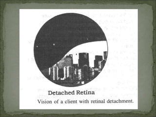
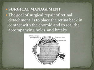
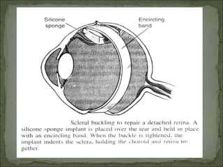
RETINAL
DETACHMENT
 RETINAL DETACHMENT
 DEFINITION AND MEANING:
 Retinal detachment occurs when there is a
separation of the neurosensory retina from
the under lying pigment epithelium layer of
the retina .
 Because the neurosensory retina the part
of the retina containing rods and cones is
detached from the nourishing retinal
pigment epithelium , these photosensitive
cells can not perform there visual function
and loss of sight results.
 CAUSES:
 Congenital malformations
 Metabolic disorders
 Vascular disease
 Intraocular inflammation
 Neoplasm
 Trauma
 Degenerative changes in in the vitreous or
retina
 Most commonly they are caused by the
mechanical forces associated with posterior
vitreous detachment and retinal tears.
 Tear-induced (rhegmatogenous) detachment
are the most common detachment
 PREDISPOSING CONDITIONS
 -Cataract extraction
 -Mostly occurs between the age of 50and 70
 -The over all incidence is 1 in 15000 people per
year
 -High Myopia
 -Lattice degeneration
 - Aphakia (surgical removal of part or all of the
crystalline lens)
 -Trauma
 -Degenerative changes (Liquefaction)
 -associated with aging
 PATHOPHISIOLOGY:
 Due to any causes or predisposing factor the
retina is separated from its choroidal blood
supply.
 It will die and Small gap is developed in between
retina and choroidal part .
 Exudative serous fluid is collected in this gap.
 The retinal tissues are at the high risk of
vascular necrosis because they are delicate
structures and have a high metabolic rate.
 That part of retina is detached and accordingly
visual defect is seen in the patient .
 E.G. If a tear in the temporal region , which
is affected more frequently , create a visual
defect in the nasal area..
 So visual field is impaired in opposite
quadrant of the actual detachment.
 CLINICAL MANIFESTATIONS and
 DIAGNOSTIC FINDING
 A patient usually reports a history of floaters or
flashing lights or both.
 The floaters may be perceived as tiny dark spot s or
cobwebs
 Later the patient may notice a spreading shadow or
curtain moving across the field of vision, resulting
in blurred vision and loss of visual field as the retina
separates from the pigmented epithelium.
 Dimness of vision gradually increased without
pain
 The onset is usually sudden
 Decreased central acuity or loss of central vision
indicates that the macula area is involved
 Examination (ophthalmoscopy)
1.Examination with direct and indirect
ophthalmoscope reveals the portion of
the retina involved and the extent of the
detachment .
2.A scleral depressor also may be used
externally on the lid or conjunctiva to assist
in rotating the eyeball and to indent the
retina for increased viewing ability .
3.Area of detachment appear bluish-gray as
opposed to the normal red pink color.
4.Tears are most often horseshoe-shaped but
may be round.
 INVESTIGATION:
 1.Haemoglobin
 2.Total count and Differential count
 3.Blood sugar
 4.ESR
 5.Blood urea
 6.S.Creatinine
 7.S.HIV
 8.HBsAG
 9.Urine routine and microscopic exam.
 10.X ray chest
 MANAGEMENT:
 EMERGENCY CARE
 If not treated promptly, a retinal
detachment may progress to involve the
macula ; this greatly compromises visual
acuity. A retinal detachment is an
ophthalmic emergency and even more so if
visual acuity is still normal.
 There is no known medical treatment for a
retinal detachment
 SURGICAL MANAGEMENT
 The goal of surgical repair of retinal
detachment is to place the retina back in
contact with the choroid and to seal the
accompanying holes and breaks.
 LASOR PHOTOCOAGULATION
 CRYOPEXY
 Cryopexy means by using of a freesing
probe seal the hole if it has not progressed
to detachment .
 Both methods create inflammation around
the area which scars and seal the hole.
 Some exudative or serous retinal
detachment due to tumor or inflammatory
that produces sub retinal fluid without a
retinal break respond to laser
photocoagulation
 Laser procedures form scar tissue on the
retina, sealing it to the pigmented
epithelium.
 Diabetic retinopathy or trauma with
vitreous hemorrhage may require vitreous
surgery to relieve the tractional forces to the
retina that they cause.
 RADIATION
 Radiation therapy may be useful in treating
retinal detachments associated with
intraocular tumors.
 SCLERAL BUCKLING :
 It is the primary surgical procedure performed
to reattach the retina.
 Transscleral cryotherapy is applied around
each retinal tear, producing a chorioretinal
adhesion that seals the break so that liquid
vitreous can no longer pass through in to the
sub retinal space .
 A piece or pieces of silicone are sutured and
infolded in to the sclera , physically indenting ,
of buckling , the sclera , choroids , and
photosensitive layers up to the pigmented
epithelium , supporting the breaks .
 When the retina thus comes in to contact
with the underlying, supportive tissue,
normal physiological function is restored.
 Often, external syringe drainage of sub
retinal fluid is necessary to bring the
detached retina closer to the buckled area so
that the retina can be reattached.
 During surgery it may be necessary to inject
inert gas (e.g. sulphahexalfuoride SF6,
octofluroptopane C3F8, or air bubble) into
the vitreous body to maintain intraocular
pressure or to assist in flattening the retina.
 Depending on which gas is used, the bubble
will be reabsorbed and replaced by aqueous
fluid in 3 days to 2 months.
 Between 90% and 95% of retinal detachments can be
reattached and good visual acuity achieved with scleral
buckling, although more than one procedure may be
needed.
 Full visual recovery may not be achieved, even with
successful reattachments, in patient with chronic
retinal detachment or in those macular involvements.
 Detachment that can not be reattached by scleral
buckling may require vitreous surgery.
 Approximately 25% of patient with complex retinal
detachments do not respond to conventional surgical
procedures.
 Instillation of perfluorocarbon liquid as an adjunt to
treatment of these patients has improve ed visual
outcome .
 PREOPERATIVE CARE:
 Written consent
 Vaccination , Inj. Tetanus toxoid 0.5ml
intramuscular state
 NBM after 10.00 pm previous night
 Laxative previous night
 Covering of head by cloth and give O.T.
dress to the patient .
 Dilate the pupil on the day of operation.
 Preoperative nursing care involves to
preparing the client for out-door surgery or
over night stay in the hospital .
 Assess the clients current level of
knowledge and understanding of the
implication of retinal detachment and the
expectations for the surgery procedure.
 Because retinal detachment repair may take
several hours, general anaesthesia is used in
many cases.
 The pupil must be widely dilated before the
operation, and client may be given a
sedative.
 POST OPERATIVE MANAGEMENT:
1.Post operatively, observe the eye patches for
any drainage.
2.Blood loss in retinal detachment surgery is
minimal, and only serous drainage is
expected on
the post operative dressing.
3 Activity restriction may be necessary if an
air or gas bubble has been injected.
4.The client will need to be positioned so that
the bubble can apply maximum pressure on
the retina by the force of gravity.
 The position usually head down and to one
side, it maintain for several days
 Provide suggestions for comfort and support
with the positioning.
6. Post operative segment surgery such has
schlera kuckling procedure , results in
considerably more discomfort then an
anterior segment procedures.
7. Ocular muscles are separate , and globe is
manipulated. To reach the posterior portion
of the eye ball. Narcotic may be needed
during the first 24 hours after surgery .
8 Nausea and vomiting may also require
management.
9. Intravenous Acetazolamide (diamox)
may be used to reduced increased
intraocular pressure .
10.The intraocular pressure is monitored
closely during the first 24 hours.
11.Encourage the client to resume a
regular diet and fluids as tolerated.
12.The eye patch and shield are removed
the next morning.
13.Redness and swelling of the lids and
conjunctiva should be expected from the
surgical manipulation. After several days , the
swelling and echymosis of the lids subsides ,
but conjunctiva may remain red or pink for a
few weeks.
14.Post operative eye medication generally
includes an antibiotics-steroid combination
drops to prevent infection and reduce swelling .
15.Cyclopesic agents are prescribe to dilate pupil
and relax the cilliary muscles, which decreases
discomfort and helps prevent the formation of
iris adhesions to the corneal endothelium.
16.Either warm or cold compresses may be
applied for comfort several times a day.
17.Instruct the client to clean the eye with
warm tap water using a clean wash cloth.
 Warm compress may be continues at home.
18.Either an eye shield or glasses should be
worn during the day. And shield should be
worn during naps and at night.
19.The client is usually instructed to avoid
vigorous activities and heavy lifting during
the immediate postoperative period.
20.If an air or gas bubble has been injected, it
may take several weeks to totally absorb. 21
Client is advised to avoid air travel during
this time because the gas and air expand at
high altitudes.
 .COMPLICATION;
 Increased intraocular pressure
 Glaucoma
 Infection
 Choroidal detachment
 Failure of the retina to reattached or
 Redetachment of the retina
 LATE COMPLICATION:
 Infection
 Extrusion of the buckling material through
the conjunctiva or
 Erosion through eyeball
 Proliferative vitreoretinopathy (scar tissue
involving the retina)
 Diplopia
 Refractive error or
 Astigmatism
RETINAL%20DETACHMENT.pptx

RETINAL%20DETACHMENT.pptx

  • 1.
  • 2.
     RETINAL DETACHMENT DEFINITION AND MEANING:  Retinal detachment occurs when there is a separation of the neurosensory retina from the under lying pigment epithelium layer of the retina .  Because the neurosensory retina the part of the retina containing rods and cones is detached from the nourishing retinal pigment epithelium , these photosensitive cells can not perform there visual function and loss of sight results.
  • 3.
     CAUSES:  Congenitalmalformations  Metabolic disorders  Vascular disease  Intraocular inflammation  Neoplasm  Trauma  Degenerative changes in in the vitreous or retina  Most commonly they are caused by the mechanical forces associated with posterior vitreous detachment and retinal tears.  Tear-induced (rhegmatogenous) detachment are the most common detachment
  • 4.
     PREDISPOSING CONDITIONS -Cataract extraction  -Mostly occurs between the age of 50and 70  -The over all incidence is 1 in 15000 people per year  -High Myopia  -Lattice degeneration  - Aphakia (surgical removal of part or all of the crystalline lens)  -Trauma  -Degenerative changes (Liquefaction)  -associated with aging
  • 5.
     PATHOPHISIOLOGY:  Dueto any causes or predisposing factor the retina is separated from its choroidal blood supply.  It will die and Small gap is developed in between retina and choroidal part .  Exudative serous fluid is collected in this gap.  The retinal tissues are at the high risk of vascular necrosis because they are delicate structures and have a high metabolic rate.  That part of retina is detached and accordingly visual defect is seen in the patient .
  • 6.
     E.G. Ifa tear in the temporal region , which is affected more frequently , create a visual defect in the nasal area..  So visual field is impaired in opposite quadrant of the actual detachment.
  • 7.
     CLINICAL MANIFESTATIONSand  DIAGNOSTIC FINDING  A patient usually reports a history of floaters or flashing lights or both.  The floaters may be perceived as tiny dark spot s or cobwebs  Later the patient may notice a spreading shadow or curtain moving across the field of vision, resulting in blurred vision and loss of visual field as the retina separates from the pigmented epithelium.  Dimness of vision gradually increased without pain  The onset is usually sudden  Decreased central acuity or loss of central vision indicates that the macula area is involved
  • 9.
     Examination (ophthalmoscopy) 1.Examinationwith direct and indirect ophthalmoscope reveals the portion of the retina involved and the extent of the detachment . 2.A scleral depressor also may be used externally on the lid or conjunctiva to assist in rotating the eyeball and to indent the retina for increased viewing ability . 3.Area of detachment appear bluish-gray as opposed to the normal red pink color. 4.Tears are most often horseshoe-shaped but may be round.
  • 10.
     INVESTIGATION:  1.Haemoglobin 2.Total count and Differential count  3.Blood sugar  4.ESR  5.Blood urea  6.S.Creatinine  7.S.HIV  8.HBsAG  9.Urine routine and microscopic exam.  10.X ray chest
  • 11.
     MANAGEMENT:  EMERGENCYCARE  If not treated promptly, a retinal detachment may progress to involve the macula ; this greatly compromises visual acuity. A retinal detachment is an ophthalmic emergency and even more so if visual acuity is still normal.  There is no known medical treatment for a retinal detachment
  • 12.
     SURGICAL MANAGEMENT The goal of surgical repair of retinal detachment is to place the retina back in contact with the choroid and to seal the accompanying holes and breaks.
  • 13.
     LASOR PHOTOCOAGULATION CRYOPEXY  Cryopexy means by using of a freesing probe seal the hole if it has not progressed to detachment .  Both methods create inflammation around the area which scars and seal the hole.  Some exudative or serous retinal detachment due to tumor or inflammatory that produces sub retinal fluid without a retinal break respond to laser photocoagulation
  • 14.
     Laser proceduresform scar tissue on the retina, sealing it to the pigmented epithelium.  Diabetic retinopathy or trauma with vitreous hemorrhage may require vitreous surgery to relieve the tractional forces to the retina that they cause.
  • 15.
     RADIATION  Radiationtherapy may be useful in treating retinal detachments associated with intraocular tumors.
  • 16.
     SCLERAL BUCKLING:  It is the primary surgical procedure performed to reattach the retina.  Transscleral cryotherapy is applied around each retinal tear, producing a chorioretinal adhesion that seals the break so that liquid vitreous can no longer pass through in to the sub retinal space .  A piece or pieces of silicone are sutured and infolded in to the sclera , physically indenting , of buckling , the sclera , choroids , and photosensitive layers up to the pigmented epithelium , supporting the breaks .
  • 17.
     When theretina thus comes in to contact with the underlying, supportive tissue, normal physiological function is restored.  Often, external syringe drainage of sub retinal fluid is necessary to bring the detached retina closer to the buckled area so that the retina can be reattached.
  • 18.
     During surgeryit may be necessary to inject inert gas (e.g. sulphahexalfuoride SF6, octofluroptopane C3F8, or air bubble) into the vitreous body to maintain intraocular pressure or to assist in flattening the retina.  Depending on which gas is used, the bubble will be reabsorbed and replaced by aqueous fluid in 3 days to 2 months.
  • 19.
     Between 90%and 95% of retinal detachments can be reattached and good visual acuity achieved with scleral buckling, although more than one procedure may be needed.  Full visual recovery may not be achieved, even with successful reattachments, in patient with chronic retinal detachment or in those macular involvements.  Detachment that can not be reattached by scleral buckling may require vitreous surgery.
  • 20.
     Approximately 25%of patient with complex retinal detachments do not respond to conventional surgical procedures.  Instillation of perfluorocarbon liquid as an adjunt to treatment of these patients has improve ed visual outcome .
  • 22.
     PREOPERATIVE CARE: Written consent  Vaccination , Inj. Tetanus toxoid 0.5ml intramuscular state  NBM after 10.00 pm previous night  Laxative previous night  Covering of head by cloth and give O.T. dress to the patient .  Dilate the pupil on the day of operation.
  • 23.
     Preoperative nursingcare involves to preparing the client for out-door surgery or over night stay in the hospital .  Assess the clients current level of knowledge and understanding of the implication of retinal detachment and the expectations for the surgery procedure.  Because retinal detachment repair may take several hours, general anaesthesia is used in many cases.  The pupil must be widely dilated before the operation, and client may be given a sedative.
  • 24.
     POST OPERATIVEMANAGEMENT: 1.Post operatively, observe the eye patches for any drainage. 2.Blood loss in retinal detachment surgery is minimal, and only serous drainage is expected on the post operative dressing. 3 Activity restriction may be necessary if an air or gas bubble has been injected. 4.The client will need to be positioned so that the bubble can apply maximum pressure on the retina by the force of gravity.
  • 25.
     The positionusually head down and to one side, it maintain for several days  Provide suggestions for comfort and support with the positioning. 6. Post operative segment surgery such has schlera kuckling procedure , results in considerably more discomfort then an anterior segment procedures. 7. Ocular muscles are separate , and globe is manipulated. To reach the posterior portion of the eye ball. Narcotic may be needed during the first 24 hours after surgery . 8 Nausea and vomiting may also require management.
  • 26.
    9. Intravenous Acetazolamide(diamox) may be used to reduced increased intraocular pressure . 10.The intraocular pressure is monitored closely during the first 24 hours. 11.Encourage the client to resume a regular diet and fluids as tolerated. 12.The eye patch and shield are removed the next morning.
  • 27.
    13.Redness and swellingof the lids and conjunctiva should be expected from the surgical manipulation. After several days , the swelling and echymosis of the lids subsides , but conjunctiva may remain red or pink for a few weeks. 14.Post operative eye medication generally includes an antibiotics-steroid combination drops to prevent infection and reduce swelling . 15.Cyclopesic agents are prescribe to dilate pupil and relax the cilliary muscles, which decreases discomfort and helps prevent the formation of iris adhesions to the corneal endothelium.
  • 28.
    16.Either warm orcold compresses may be applied for comfort several times a day. 17.Instruct the client to clean the eye with warm tap water using a clean wash cloth.  Warm compress may be continues at home. 18.Either an eye shield or glasses should be worn during the day. And shield should be worn during naps and at night.
  • 29.
    19.The client isusually instructed to avoid vigorous activities and heavy lifting during the immediate postoperative period. 20.If an air or gas bubble has been injected, it may take several weeks to totally absorb. 21 Client is advised to avoid air travel during this time because the gas and air expand at high altitudes.
  • 30.
     .COMPLICATION;  Increasedintraocular pressure  Glaucoma  Infection  Choroidal detachment  Failure of the retina to reattached or  Redetachment of the retina
  • 31.
     LATE COMPLICATION: Infection  Extrusion of the buckling material through the conjunctiva or  Erosion through eyeball  Proliferative vitreoretinopathy (scar tissue involving the retina)  Diplopia  Refractive error or  Astigmatism