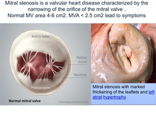
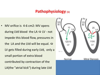
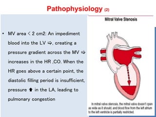
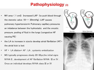
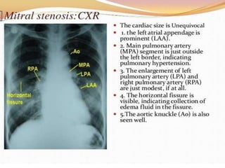
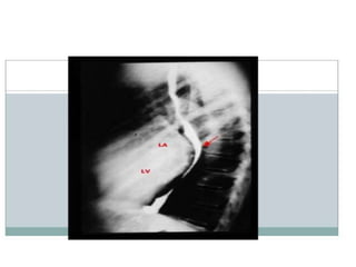
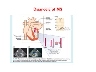
Mitral valve stenosis
Kim Chung Nguyen. MD
Head of General Cardiovascular Department
Tam Đuc Hospital
Mitral stenosis is a valvular heart disease characterized by the
narrowing of the orifice of the mitral valve .
Normal MV area 4-6 cm2. MVA < 2.5 cm2 lead to symptoms
Mitral stenosis with marked
thickening of the leaflets and left
atrial hypertrophy
Normal mitral valve
Causes of MS
• Almost all cases:
- The heart secondary to rheumatic fever # rheumatic heart disease.
• Uncommon causes:
- Calcification of the MV leaflets or annular
- Infective endocarditis where the vegetations may favor increase risk of
stenosis.
- Left atrial myxoma
- Congenital MS
• Other rare causes:
- Endomyocardial fibroelastosis
- Systemic lupus erythematosus.
Pathophysiology (1)
• MV orifice is 4-6 cm2: MV opens
during LVd blood the LA  LV - not
impede this blood flow, pressures in
the LA and the LVd will be equal. 
LV gets filled during early LVd, only a
small portion of extra blood
contributed by contraction of the
LA(the "atrial kick") during late LVd
Pathophysiology (2)
• MV area < 2 cm2: An impediment
blood into the LV , creating a
pressure gradient across the MV 
increases in the HR ,CO. When the
HR goes above a certain point, the
diastolic filling period is insufficient,
pressure  in the LA, leading to
pulmonary congestion
Pathophysiology (3)
• MV area < 1 cm2: Increased LAP (to push blood through
the stenotic valve: 10--- 20mmHg). LAP causes
pulmonary hypertension Pulmonary capillary pressures
an imbalance between the hydrostatic and the oncotic
pressure, pooling of fluid in the lungs (congestive HF
causing PE)
• the LA to increase in size:to develop atrial fibrillation (AF)-
the atrial kick is lost
• HF + LA dilation+ AF : LA , systemic embolization
• MS typically progresses slowly 20-30ys,the initial signs
NYHA II , development of AF fibrillation NYHA III or IV .
Once an individual develops NYHA class III or IV
: dyspnea on exertion, orthopnea and
paroxysmal nocturnal dyspnea (PND)
Clinical findings
• Hoarseness: compression of the left recurrent laryngeal nerve
(LRLN) by enlarged LA
• Persistent cough: Enlarged LA compress to bronchi and
pulcongestion
• Mitral facies: Pinkish purpule patches on the cheeks due to
decreased CO and vasoconstriction
• Palpation: The arterial pulses are reduced due to the decreased of
SV
• Pulses may be irregular in Afib
• A P2 may be palpable in the the 2nd left intercostal space (2nd LICS)
• Moist rales, pleural effusion
Physical examination
• S1 is usually loud because of increased force in closing the MV.
• S2 (P2) loud and late If pulmonary hypertension secondary
• Opening snap: A high-pitch additional sound after S2( severity of the MS , the
pressure in the  LA , and MV opens earlier, leaflets suddenly tense and dome into
the LVd.
• A mid-diastolic rumbling murmur with presystolic accentuation after the opening
snap. The diastolic murmur is low-pitch sound, heard best with the bell of the
stethoscope, rolling pt toward left
Imaging Tests
1. Echocardiogram:
- LA enlargement, thick and calcified MV"fish-mouth"shaped orifice
- RV Hypertrophic
- The trans-mitral gradient as measured by Doppler echocardiography is the gold
standard in the evaluation of the severity of MS
• Transesophageal echocardiogram (TEE):Wilkins scoring system to assess MV
morphology and its relationship to the success of percutaneous balloon dilation of
the MV
2
3
Diagnosis of MS
(1)
Management (2)
1. Medical management:
• Anticoagulants (blood thinners)
• Diuretics (to reduce fluid buildup through increased urine output)
• Antiarrhythmics (medications to treat abnormal heart rhythms)
• Beta-blockers (medications to slow your HR)
2.MV repair or replacement by surgery: The replacement might be biological (from a
cow, pig, or human cadaver), or mechanical. Repair is better than replacement
3.Percutaneous mitral valvuloplasty by balloon catheter :moderate or severe MS ,
nonpliable calcified valve, NYHA III–IV, and either are not candidates for surgery or
are at high risk with surgery
Mitral valve stenosis

Mitral valve stenosis

  • 1.
    Mitral valve stenosis KimChung Nguyen. MD Head of General Cardiovascular Department Tam Đuc Hospital
  • 2.
    Mitral stenosis isa valvular heart disease characterized by the narrowing of the orifice of the mitral valve . Normal MV area 4-6 cm2. MVA < 2.5 cm2 lead to symptoms Mitral stenosis with marked thickening of the leaflets and left atrial hypertrophy Normal mitral valve
  • 3.
    Causes of MS •Almost all cases: - The heart secondary to rheumatic fever # rheumatic heart disease. • Uncommon causes: - Calcification of the MV leaflets or annular - Infective endocarditis where the vegetations may favor increase risk of stenosis. - Left atrial myxoma - Congenital MS • Other rare causes: - Endomyocardial fibroelastosis - Systemic lupus erythematosus.
  • 5.
    Pathophysiology (1) • MVorifice is 4-6 cm2: MV opens during LVd blood the LA  LV - not impede this blood flow, pressures in the LA and the LVd will be equal.  LV gets filled during early LVd, only a small portion of extra blood contributed by contraction of the LA(the "atrial kick") during late LVd
  • 6.
    Pathophysiology (2) • MVarea < 2 cm2: An impediment blood into the LV , creating a pressure gradient across the MV  increases in the HR ,CO. When the HR goes above a certain point, the diastolic filling period is insufficient, pressure  in the LA, leading to pulmonary congestion
  • 7.
    Pathophysiology (3) • MVarea < 1 cm2: Increased LAP (to push blood through the stenotic valve: 10--- 20mmHg). LAP causes pulmonary hypertension Pulmonary capillary pressures an imbalance between the hydrostatic and the oncotic pressure, pooling of fluid in the lungs (congestive HF causing PE) • the LA to increase in size:to develop atrial fibrillation (AF)- the atrial kick is lost • HF + LA dilation+ AF : LA , systemic embolization • MS typically progresses slowly 20-30ys,the initial signs NYHA II , development of AF fibrillation NYHA III or IV . Once an individual develops NYHA class III or IV
  • 8.
    : dyspnea onexertion, orthopnea and paroxysmal nocturnal dyspnea (PND)
  • 9.
    Clinical findings • Hoarseness:compression of the left recurrent laryngeal nerve (LRLN) by enlarged LA • Persistent cough: Enlarged LA compress to bronchi and pulcongestion • Mitral facies: Pinkish purpule patches on the cheeks due to decreased CO and vasoconstriction • Palpation: The arterial pulses are reduced due to the decreased of SV • Pulses may be irregular in Afib • A P2 may be palpable in the the 2nd left intercostal space (2nd LICS) • Moist rales, pleural effusion
  • 10.
    Physical examination • S1is usually loud because of increased force in closing the MV. • S2 (P2) loud and late If pulmonary hypertension secondary • Opening snap: A high-pitch additional sound after S2( severity of the MS , the pressure in the  LA , and MV opens earlier, leaflets suddenly tense and dome into the LVd. • A mid-diastolic rumbling murmur with presystolic accentuation after the opening snap. The diastolic murmur is low-pitch sound, heard best with the bell of the stethoscope, rolling pt toward left
  • 12.
    Imaging Tests 1. Echocardiogram: -LA enlargement, thick and calcified MV"fish-mouth"shaped orifice - RV Hypertrophic - The trans-mitral gradient as measured by Doppler echocardiography is the gold standard in the evaluation of the severity of MS • Transesophageal echocardiogram (TEE):Wilkins scoring system to assess MV morphology and its relationship to the success of percutaneous balloon dilation of the MV
  • 14.
  • 16.
  • 17.
  • 18.
  • 19.
    Management (2) 1. Medicalmanagement: • Anticoagulants (blood thinners) • Diuretics (to reduce fluid buildup through increased urine output) • Antiarrhythmics (medications to treat abnormal heart rhythms) • Beta-blockers (medications to slow your HR) 2.MV repair or replacement by surgery: The replacement might be biological (from a cow, pig, or human cadaver), or mechanical. Repair is better than replacement 3.Percutaneous mitral valvuloplasty by balloon catheter :moderate or severe MS , nonpliable calcified valve, NYHA III–IV, and either are not candidates for surgery or are at high risk with surgery

Editor's Notes