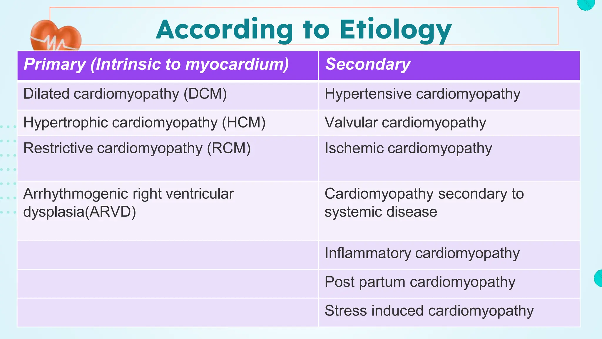
Jane Nader, MD
Lecturer of Cardiology, BUC
Unraveling
Cardiomyopathies
A Deep Dive for Medical
Students
Cardiomyopathies are diseases of heart muscle
Cardio Opathy
Myo
Definition of Cardiomyopathy
Cardiomyopathy is a myocardial disorder in which the
heart muscle is structurally and functionally abnormal in
the absence of (coronary artery disease, hypertension,
valvular disease, and congenital heart disease)
sufficient to explain the observed myocardial
abnormality
Classification of
cardiomyopathy
02 03
01
Rectrictive
Hypertrophic
Dilated
According to Pathology
According to Etiology
Primary (Intrinsic to myocardium) Secondary
Dilated cardiomyopathy (DCM) Hypertensive cardiomyopathy
Hypertrophic cardiomyopathy (HCM) Valvular cardiomyopathy
Restrictive cardiomyopathy (RCM) Ischemic cardiomyopathy
Arrhythmogenic right ventricular
dysplasia(ARVD)
Cardiomyopathy secondary to
systemic disease
Inflammatory cardiomyopathy
Post partum cardiomyopathy
Stress induced cardiomyopathy
Pathophysiology
Contraction and relaxation disorder
Ineffective energy utilization
Altered Ca ions handling
Mutation Exogenous insult
Activation of compensatory
neurohumoral mechanisms
Apoptosis
Fibrosis
Hypertrophy
Heart failure Arrhythmia, sudden death Thromboembolic complication
01
Dilated Cardiomopathy (DCM)
Decreased or lost systolic function due to
dilatation of the left ventricle
Definition of DCM
Causes
1. Genetic Factors.
2. Infections: Viral infections, such as viral myocarditis
3. Toxins: excessive alcohol consumption, cocaine use
4. Autoimmune Conditions
5. Pregnancy
6. Nutritional Deficiencies: such as thiamine (vitamin B1).
7. Ischemic Heart Disease: Coronary artery disease, which results in reduced blood flow to the
heart muscle, can lead to dilated cardiomyopathy.
8. Hypertension (High Blood Pressure): Long-term uncontrolled high blood pressure can place
strain on the heart, potentially leading to dilation and weakening.
9. Valvular Heart Disease: especially the aortic or mitral valves
10. Metabolic Disorders: hemochromatosis, and glycogen storage diseases.
Patho
physiology
• Diffuse inflammation and rapid degeneration of myocardial fibers
↓
• Depressed myocardial systolic function
↓
• LV contractile force is diminished
↓
• low cardiac output and increased end-systolic and end-diastolic
ventricular volumes
↓
• LV dilatation accompanied by compensatory hypertrophy
Patho physiology
Manifestationsofperipheralembolization
• Acuteneurologicdeficit
• Loinpain
• Hamaturia
• Cyanoticextremity
Impairedexercisetolerence
• Dyspneaonexertion
• Orhopnea
• Paroxysmalnocturnaldyspnea
• Chestpainwithfeaturesoftypicalangina
pectoris
Asthediseaseprogresses
• Anorexia,Nauseaandvomiting
• Abdominaldistension
• LLedema
Symptoms
Generalexamination:
• Weakperipheralpulsesandcoldextremities
• Hepatomegaly
• Jugularvenousdistention
• LLEdema
Signs
Auscultation:
• Abnormals3ands4gallop
• Displacedapex
• Pansystolicmurmurheartovertheapex
• Sinustachycardiaoratrialfibrillation
• Pulmonarycrepitations
1. ECG
2. Chest X ray
3. Echocardiography
- Enlarged cardiac silhouette
-Kerley B lines,
-enlarged central pulmonary arteries
-Widened right heart border
X ray
ECG
• LV hypertrophy and LA enlargement are the most
common findings
• AF & ventricular ectopy are common.
• LBBB
• Poor R-wave progression
• Low QRS voltage
Echo
• ↓ EF
• ↑ Systolic & Diastolic volumes
• Ventricular & atrial enlargement
• Tachyarrhythmias
• MR/TR
Treatment of DCM
1.Treatment of Underlying Causes
2.Lifestyle Modifications:
1. Dietary Changes: A heart-healthy diet, low in sodium and saturated
fats, can help manage symptoms and improve overall heart health.
2. Fluid Restriction: Limiting fluid intake may be recommended,
especially if there is significant fluid retention.
3. Smoking Cessation: Quitting smoking is essential for heart health.
4. Limiting Alcohol and Caffeine: These substances can exacerbate
symptoms in some individuals.
5. Exercise Program: A supervised and tailored exercise program can
be beneficial for improving cardiovascular fitness.
3. Medications:
1. Angiotensin-Converting Enzyme (ACE) Inhibitors or
Angiotensin II Receptor Blockers (ARBs): These
medications help dilate blood vessels and reduce the workload
on the heart.
2. Beta-Blockers: They can slow the heart rate, decrease blood
pressure, and improve the heart's pumping ability.
3. Diuretics: Diuretic medications help reduce fluid retention and
alleviate symptoms such as edema and dyspnea.
4. Aldosterone Antagonists: These medications can further
reduce fluid retention.
5. Dioxin: to help control rapid atrial fibrillation and improve
contraction
4. Device Therapy:
1. Implantable Cardioverter-Defibrillator (ICD): For individuals at risk of
life-threatening arrhythmias, an ICD may be implanted to monitor and
correct abnormal heart rhythms.
2. Cardiac Resynchronization Therapy (CRT): In some cases, a CRT
device is used to improve coordination in the contraction of the heart's
chambers.
5.Heart Transplantation:
In severe cases where other treatments are ineffective, heart
transplantation may be considered as a last resort.
6.Regular Monitoring:
In all cases, regular follow-up appointments with a healthcare provider are
crucial to monitor the condition, adjust medications, and address any
changes in symptoms.
2024
“As we step into the dawn of 2024, let's remember
that each day is a blank page waiting for our story
to unfold. Seize the pen, write with purpose, and
make this chapter a masterpiece of growth,
resilience, and joy.”
02
Hypertrophic Cardiomopathy (HCM)
Pathophysiology
Increased thickness of the heart muscle,
especially the left ventricular wall
Causes
Diverse morphology
Subtypes :-
i) Hypertrophic obstructive cardiomyopathy (HOCM)
ii) Idiopathic hypertrophic subaortic stenosis (IHSS)
Iii) Asymmetric septal hypertrophy
HCM Athletic Heart
LVEDD < 45 mm LVEDD > 55mm
Unusual LVH pattern Responds to detraining
Marked LA enlargement Super normal exercise capacity
Abnormal LD diastolic filling No scar tissue on MRI
Symptoms
1. Sudden cardiac death (most devastating presenting manifestation)
2. Dyspnoea on exertion (exercise-induced sinus tachy, which results in
an abrupt ↑ LV diastolic pressure & pulmonary venous HTN)
3. Syncope & pre syncope
4. Angina (imbalance between the O2 demand of the hypertrophied LV &
the available myocardial blood flow)
5. Palpitations
6. Orthopnoea & PND (early signs of CHF)
Examination
• Apical impulse is forceful and displaced laterally.
• Normal S1; S2 is usually split in severe outflow gradients; S3
and S4 are usually present.
• Systolic ejection crescendo-decrescendo murmur.
• Holosystolic murmur at the apex and axilla of MR
• Jugular venous pulse revealing a prominent ‘a’ wave
Investigations
1. CXR : Enlarged cardiac silhouette
2. Echocardiogram: LVH
Left atrial enlargement
Small ventricular cavity size
Septal hypertrophy
Abnormal systolic motion of the mitral valve MVP & MR
3. Electrocardiogram (ECG): ST-T wave abnormalities and LVH, Axis
deviation (right or left), Conduction abnormalities (BBB)
4. Cardiac MRI: Offers more detailed images of the heart, helping
to assess the extent of hypertrophy and any scarring.
5. Holter Monitor: to help capturing any intermittent arrhythmias.
6. Exercise Stress Test: Monitors the heart's response to physical
activity.
7. Genetic Testing: recommended in case of family history of
HCM or sudden cardiac death.
MANAGEMENT :
Pharmacologic therapy for HCM may include the following:-
• ᵝblockers
• CCB
• Diltiazem, Amiodarone (rate control)
• Avoid Digitalis & use diuretics with caution
Surgical & catheter-based therapeutic options include the following:-
• LV Myomectomy
• Alcohol septal ablation
• implantable cardioverter defibrillator (ICD)
• MV replacement
• Heart Transplantation
03
RESTRICTIVE CARDIOMYOPATHY :
• defined as heart muscle disease that results in "restricted"
ventricular filling, with normal or decreased diastolic volume
of either or both ventricles.
• It is among the least common of the described
Cardiomyopathies.
It may result from systemic diseases like :-
• Amyloidosis
• Sarcoidosis
• Hemochromatosis
• Progressive systemic sclerosis
• Carcinoid heart disease
• Endomyocardial fibrosis
• Hypereosinophilic syndrome
• Glycogen storage disease of the heart
• Metastatic malignancy/ Radiotherapy
• Idiopathic
Hemodynamics
i) Elevated LV and RV end-diastolic pressure
ii) Normal LV systolic function
iii) marked ↓ followed by a rapid rise & plateau in early- diastolic
ventricular pressure
Hemodynamics
The rapid rise and abrupt plateau in the early-diastolic ventricular pressure
trace produce a characteristic "square-root sign" or "dip-and-plateau" filling
pattern due to increased myocardial stiffness
CLINICAL FEATURES :-
• Gradually worsening shortness of breath
• Progressive exercise intolerance
• Fatigue (due to↓ stroke volume and cardiac output)
• Orthopnoea & PND
• Chest pain mimicking MI can be observed, primarily in patients with Amyloidosis
(possibly d/t myocardial compression of small vessels)
• Palpitations (frequently AF)
• Hepatomegaly, right upper quadrant pain, and ascites (Right-sided manifestations)
• S3 is almost always present
• Pulmonary rales
• Jugular venous distention
• Kussmaul sign (jugular venous pulse ↑ during inspiration rather than
decreasing)
• Murmurs of MR/TR may be heard
• Breath sounds are ↓ in case of pleural effusion ( present in Amyloidosis
–often bilateral & large)
• Hepatomegaly, LL edema and ascites
DIAGNOSIS :-
• MRI – the most accurate diagnostic imaging technique
• CXR – atrial dilatation and normal ventricular size
• ECG – non specific changes.
AF or conduction abnormalities like AV block
• ECHO - non-hypertrophied, non-dilated ventricle with preserved
systolic function and dilated atria
MANAGEMENT :
Medical :
• Diuretics
• Vasodilators
• ACE inhibitors
• Anticoagulation (if atrial fibrillation)
Treatment of the cause:
• Corticosteroid (Sarcoidosis, Loeffler Endocarditis)
• Chelation (Hemochromatosis)
• Chemo-Radiotherapy (Amyloidosis)
Surgical
• Endocardiectomy for Endomyocardial fibrosis
• Permanent pacing
• Heart Transplantation
R
Flash back
D H
Mercury is the closest planet to the Sun and the smallest one in
the Solar System—it’s only a bit larger than our Moon. The planet’s
name has nothing to do with the liquid metal since it was named
after the Roman messenger god, Mercury
A brief story
Cardiomyopathy Edited for medical students.pptx

Cardiomyopathy Edited for medical students.pptx

  • 1.
    Jane Nader, MD Lecturerof Cardiology, BUC Unraveling Cardiomyopathies A Deep Dive for Medical Students
  • 3.
    Cardiomyopathies are diseasesof heart muscle Cardio Opathy Myo
  • 4.
    Definition of Cardiomyopathy Cardiomyopathyis a myocardial disorder in which the heart muscle is structurally and functionally abnormal in the absence of (coronary artery disease, hypertension, valvular disease, and congenital heart disease) sufficient to explain the observed myocardial abnormality
  • 5.
  • 6.
  • 7.
    According to Etiology Primary(Intrinsic to myocardium) Secondary Dilated cardiomyopathy (DCM) Hypertensive cardiomyopathy Hypertrophic cardiomyopathy (HCM) Valvular cardiomyopathy Restrictive cardiomyopathy (RCM) Ischemic cardiomyopathy Arrhythmogenic right ventricular dysplasia(ARVD) Cardiomyopathy secondary to systemic disease Inflammatory cardiomyopathy Post partum cardiomyopathy Stress induced cardiomyopathy
  • 8.
    Pathophysiology Contraction and relaxationdisorder Ineffective energy utilization Altered Ca ions handling Mutation Exogenous insult Activation of compensatory neurohumoral mechanisms Apoptosis Fibrosis Hypertrophy Heart failure Arrhythmia, sudden death Thromboembolic complication
  • 9.
  • 10.
    Decreased or lostsystolic function due to dilatation of the left ventricle Definition of DCM
  • 11.
    Causes 1. Genetic Factors. 2.Infections: Viral infections, such as viral myocarditis 3. Toxins: excessive alcohol consumption, cocaine use 4. Autoimmune Conditions 5. Pregnancy 6. Nutritional Deficiencies: such as thiamine (vitamin B1). 7. Ischemic Heart Disease: Coronary artery disease, which results in reduced blood flow to the heart muscle, can lead to dilated cardiomyopathy. 8. Hypertension (High Blood Pressure): Long-term uncontrolled high blood pressure can place strain on the heart, potentially leading to dilation and weakening. 9. Valvular Heart Disease: especially the aortic or mitral valves 10. Metabolic Disorders: hemochromatosis, and glycogen storage diseases.
  • 12.
  • 13.
    • Diffuse inflammationand rapid degeneration of myocardial fibers ↓ • Depressed myocardial systolic function ↓ • LV contractile force is diminished ↓ • low cardiac output and increased end-systolic and end-diastolic ventricular volumes ↓ • LV dilatation accompanied by compensatory hypertrophy Patho physiology
  • 14.
    Manifestationsofperipheralembolization • Acuteneurologicdeficit • Loinpain •Hamaturia • Cyanoticextremity Impairedexercisetolerence • Dyspneaonexertion • Orhopnea • Paroxysmalnocturnaldyspnea • Chestpainwithfeaturesoftypicalangina pectoris Asthediseaseprogresses • Anorexia,Nauseaandvomiting • Abdominaldistension • LLedema Symptoms
  • 15.
    Generalexamination: • Weakperipheralpulsesandcoldextremities • Hepatomegaly •Jugularvenousdistention • LLEdema Signs Auscultation: • Abnormals3ands4gallop • Displacedapex • Pansystolicmurmurheartovertheapex • Sinustachycardiaoratrialfibrillation • Pulmonarycrepitations
  • 16.
    1. ECG 2. ChestX ray 3. Echocardiography
  • 17.
    - Enlarged cardiacsilhouette -Kerley B lines, -enlarged central pulmonary arteries -Widened right heart border X ray
  • 18.
    ECG • LV hypertrophyand LA enlargement are the most common findings • AF & ventricular ectopy are common. • LBBB • Poor R-wave progression • Low QRS voltage
  • 19.
    Echo • ↓ EF •↑ Systolic & Diastolic volumes • Ventricular & atrial enlargement • Tachyarrhythmias • MR/TR
  • 20.
  • 21.
    1.Treatment of UnderlyingCauses 2.Lifestyle Modifications: 1. Dietary Changes: A heart-healthy diet, low in sodium and saturated fats, can help manage symptoms and improve overall heart health. 2. Fluid Restriction: Limiting fluid intake may be recommended, especially if there is significant fluid retention. 3. Smoking Cessation: Quitting smoking is essential for heart health. 4. Limiting Alcohol and Caffeine: These substances can exacerbate symptoms in some individuals. 5. Exercise Program: A supervised and tailored exercise program can be beneficial for improving cardiovascular fitness.
  • 22.
    3. Medications: 1. Angiotensin-ConvertingEnzyme (ACE) Inhibitors or Angiotensin II Receptor Blockers (ARBs): These medications help dilate blood vessels and reduce the workload on the heart. 2. Beta-Blockers: They can slow the heart rate, decrease blood pressure, and improve the heart's pumping ability. 3. Diuretics: Diuretic medications help reduce fluid retention and alleviate symptoms such as edema and dyspnea. 4. Aldosterone Antagonists: These medications can further reduce fluid retention. 5. Dioxin: to help control rapid atrial fibrillation and improve contraction
  • 23.
    4. Device Therapy: 1.Implantable Cardioverter-Defibrillator (ICD): For individuals at risk of life-threatening arrhythmias, an ICD may be implanted to monitor and correct abnormal heart rhythms. 2. Cardiac Resynchronization Therapy (CRT): In some cases, a CRT device is used to improve coordination in the contraction of the heart's chambers. 5.Heart Transplantation: In severe cases where other treatments are ineffective, heart transplantation may be considered as a last resort. 6.Regular Monitoring: In all cases, regular follow-up appointments with a healthcare provider are crucial to monitor the condition, adjust medications, and address any changes in symptoms.
  • 24.
    2024 “As we stepinto the dawn of 2024, let's remember that each day is a blank page waiting for our story to unfold. Seize the pen, write with purpose, and make this chapter a masterpiece of growth, resilience, and joy.”
  • 25.
  • 26.
    Pathophysiology Increased thickness ofthe heart muscle, especially the left ventricular wall
  • 27.
  • 28.
  • 29.
    Subtypes :- i) Hypertrophicobstructive cardiomyopathy (HOCM) ii) Idiopathic hypertrophic subaortic stenosis (IHSS) Iii) Asymmetric septal hypertrophy
  • 34.
    HCM Athletic Heart LVEDD< 45 mm LVEDD > 55mm Unusual LVH pattern Responds to detraining Marked LA enlargement Super normal exercise capacity Abnormal LD diastolic filling No scar tissue on MRI
  • 35.
    Symptoms 1. Sudden cardiacdeath (most devastating presenting manifestation) 2. Dyspnoea on exertion (exercise-induced sinus tachy, which results in an abrupt ↑ LV diastolic pressure & pulmonary venous HTN) 3. Syncope & pre syncope 4. Angina (imbalance between the O2 demand of the hypertrophied LV & the available myocardial blood flow) 5. Palpitations 6. Orthopnoea & PND (early signs of CHF)
  • 36.
    Examination • Apical impulseis forceful and displaced laterally. • Normal S1; S2 is usually split in severe outflow gradients; S3 and S4 are usually present. • Systolic ejection crescendo-decrescendo murmur. • Holosystolic murmur at the apex and axilla of MR • Jugular venous pulse revealing a prominent ‘a’ wave
  • 37.
  • 38.
    1. CXR :Enlarged cardiac silhouette 2. Echocardiogram: LVH Left atrial enlargement Small ventricular cavity size Septal hypertrophy Abnormal systolic motion of the mitral valve MVP & MR 3. Electrocardiogram (ECG): ST-T wave abnormalities and LVH, Axis deviation (right or left), Conduction abnormalities (BBB)
  • 40.
    4. Cardiac MRI:Offers more detailed images of the heart, helping to assess the extent of hypertrophy and any scarring. 5. Holter Monitor: to help capturing any intermittent arrhythmias. 6. Exercise Stress Test: Monitors the heart's response to physical activity. 7. Genetic Testing: recommended in case of family history of HCM or sudden cardiac death.
  • 41.
    MANAGEMENT : Pharmacologic therapyfor HCM may include the following:- • ᵝblockers • CCB • Diltiazem, Amiodarone (rate control) • Avoid Digitalis & use diuretics with caution Surgical & catheter-based therapeutic options include the following:- • LV Myomectomy • Alcohol septal ablation • implantable cardioverter defibrillator (ICD) • MV replacement • Heart Transplantation
  • 44.
  • 45.
    RESTRICTIVE CARDIOMYOPATHY : •defined as heart muscle disease that results in "restricted" ventricular filling, with normal or decreased diastolic volume of either or both ventricles. • It is among the least common of the described Cardiomyopathies.
  • 46.
    It may resultfrom systemic diseases like :- • Amyloidosis • Sarcoidosis • Hemochromatosis • Progressive systemic sclerosis • Carcinoid heart disease • Endomyocardial fibrosis • Hypereosinophilic syndrome • Glycogen storage disease of the heart • Metastatic malignancy/ Radiotherapy • Idiopathic
  • 48.
    Hemodynamics i) Elevated LVand RV end-diastolic pressure ii) Normal LV systolic function iii) marked ↓ followed by a rapid rise & plateau in early- diastolic ventricular pressure
  • 49.
    Hemodynamics The rapid riseand abrupt plateau in the early-diastolic ventricular pressure trace produce a characteristic "square-root sign" or "dip-and-plateau" filling pattern due to increased myocardial stiffness
  • 50.
    CLINICAL FEATURES :- •Gradually worsening shortness of breath • Progressive exercise intolerance • Fatigue (due to↓ stroke volume and cardiac output) • Orthopnoea & PND • Chest pain mimicking MI can be observed, primarily in patients with Amyloidosis (possibly d/t myocardial compression of small vessels) • Palpitations (frequently AF) • Hepatomegaly, right upper quadrant pain, and ascites (Right-sided manifestations)
  • 51.
    • S3 isalmost always present • Pulmonary rales • Jugular venous distention • Kussmaul sign (jugular venous pulse ↑ during inspiration rather than decreasing) • Murmurs of MR/TR may be heard • Breath sounds are ↓ in case of pleural effusion ( present in Amyloidosis –often bilateral & large) • Hepatomegaly, LL edema and ascites
  • 52.
    DIAGNOSIS :- • MRI– the most accurate diagnostic imaging technique • CXR – atrial dilatation and normal ventricular size • ECG – non specific changes. AF or conduction abnormalities like AV block • ECHO - non-hypertrophied, non-dilated ventricle with preserved systolic function and dilated atria
  • 53.
    MANAGEMENT : Medical : •Diuretics • Vasodilators • ACE inhibitors • Anticoagulation (if atrial fibrillation) Treatment of the cause: • Corticosteroid (Sarcoidosis, Loeffler Endocarditis) • Chelation (Hemochromatosis) • Chemo-Radiotherapy (Amyloidosis)
  • 54.
    Surgical • Endocardiectomy forEndomyocardial fibrosis • Permanent pacing • Heart Transplantation
  • 55.
  • 56.
    Mercury is theclosest planet to the Sun and the smallest one in the Solar System—it’s only a bit larger than our Moon. The planet’s name has nothing to do with the liquid metal since it was named after the Roman messenger god, Mercury A brief story