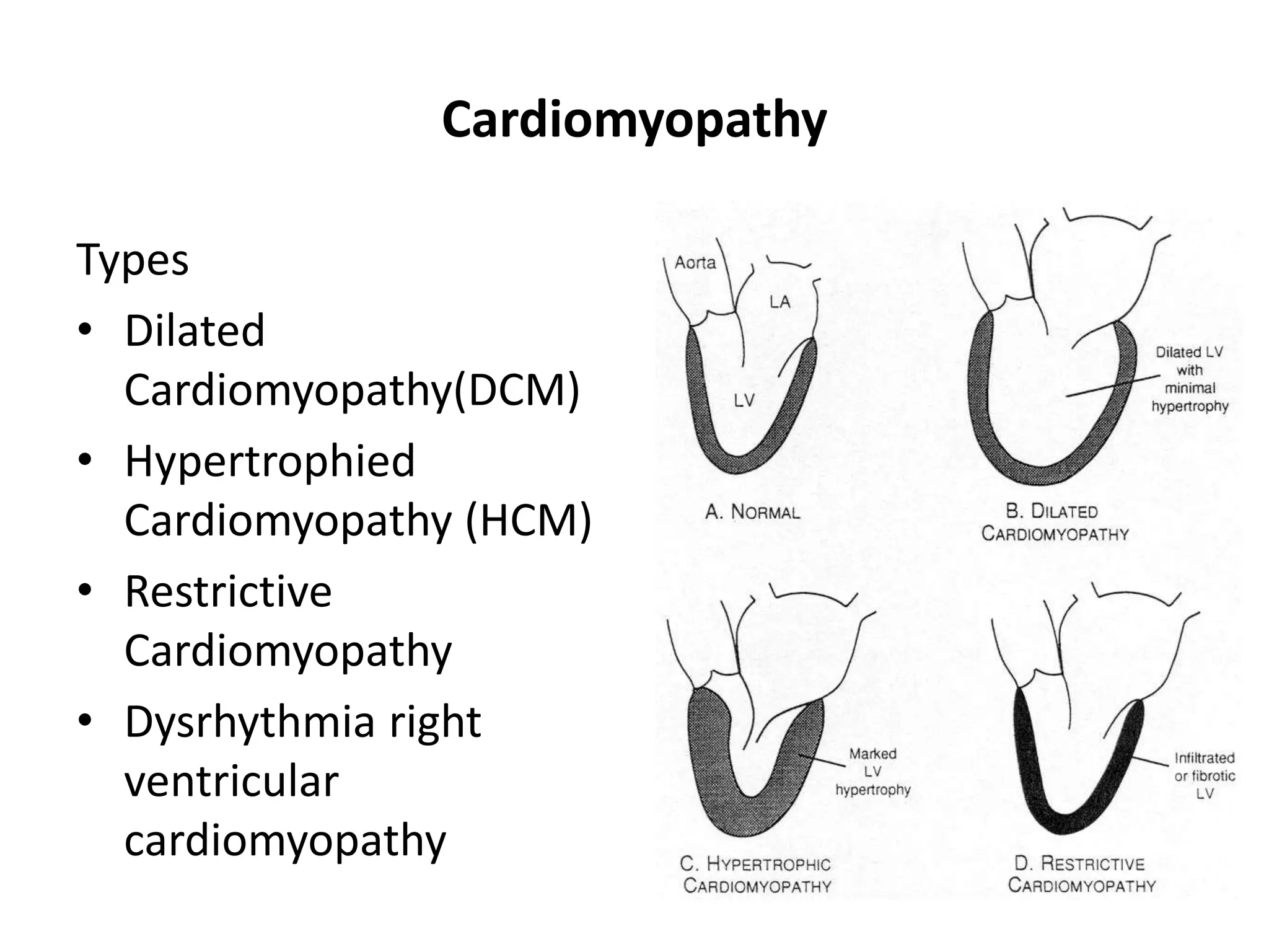
Prepared by: Nigussie Wondimu ID 188/15
Submitted To: Mr. Bikila T.(Asst.Prof.)
July,2023
Fiche, Ethiopia
SALALE UNIVERSITY COLLOGE HEALTH
SCIENCE DEPARTMENT OF NURSING
Assignment: Cardiomyopathy, Myocarditis and
Pericardial Disease
Course Outline
• Objective
• Introduction
• Cardiomyopathy
• Types of Cardiomyopathy
• Myocarditis
• Pericarditis disease
Objective
At the end of this course students will be able to:
• Define cardiomyopathy, myocarditis & Pericarditis
disease
• List types of cardiomyopathy
• Describe etiology of cardiomyopathy, myocarditis &
Pericarditis disease
• List clinical manifestation of cardiomyopathy,
myocarditis & Pericarditis disease
Introduction
• cardiomyopathies as “a heterogeneous group of
diseases of the myocardium associated with
mechanical &/or electrical dysfunction that usually (but
not invariably) exhibit inappropriate ventricular
hypertrophy or dilatation and are due to a variety of
causes that frequently are genetic.”
• Cardiomyopathies either are confined to the heart or
are a part of generalized systemic disorders
Introduction…
• As cardiomyopathy worsens, the heart becomes
weaker.
• It's less able to pump blood through the body and
maintain a normal electrical rhythm. This can lead to
heart failure or irregular heartbeats called
arrhythmias.
• In turn, heart failure can cause fluid to build up in the
lungs, ankles, feet, legs, or abdomen.
• The weakening of the heart also can cause other
complications, such as heart valve problems.
Cardiomyopathy
Definition
• cardiomyopathies as “a heterogeneous group of
diseases of the myocardium associated with
mechanical &/or electrical dysfunction that usually
(but not invariably) exhibit inappropriate ventricular
hypertrophy or dilatation and are due to a variety of
causes that frequently are genetic.”
• Cardiomyopathies either are confined to the heart or
are a part of generalized systemic disorders
Cardiomyopathy...
• 3rd most common form of heart disease in U.S.
• 2nd most common cause of adolescent sudden
death(IHSS or HOCM)
• Directly affects cardiac structure and impairs
myocardial function
Cardiomyopathy
Types
• Dilated
Cardiomyopathy(DCM)
• Hypertrophied
Cardiomyopathy (HCM)
• Restrictive
Cardiomyopathy
• Dysrhythmia right
ventricular
cardiomyopathy
Dilated Cardiomyopathy
• Dilation of one or both ventricles
• Depressed ventricular systolic function: both
ventricles or predominantly LV. Isolated RV
cardiomyopathy is rare.
• 80% of DCM cases are idiopathic
• African Americans and males have 2.5x increased risk
• Most common age of diagnosis 20-50yrs
Etiology of DCM
 Coronary artery disease
 Idiopathic
 Hypertensive heart disease
 Familial/genetic
• Viral/other infectious agents (HIV)
• Immune/autoimmune
• Alcoholic/toxic (cocaine, chemotherapeutic
drugs)
• Infiltrative (hemochromatosis, sarcoidosis,
amyloidosis)
• Post partum-;myocarditis
Dilated Cardiomyopathy
Clinical Presentation
• SSx 0f CHF
• Chest pain (due to low coronary vascular reserve)
• Mural thrombi formation
• Holosystolic regurgitant murmur or gallop
• Dependent edema, bibasilar rales
Systolic heart failure
Natural History of DCM
• Congestive heart failure
• Arrhythmias (Afib, VT)
• Chest pain
• Thromboembolism
• Sudden death
Diagnosis of DCM
• CXR- cardiomegaly and pulmonary congestion
• ECG- LVH, Left atrial enlargement, Atrial fibrillation, Q
waves, poor R wave progression
• Echo-Biventricular enlargement and global
hypokinesis.
• Cardiac cath: contrast left ventriculogram
• Radionuclide ventriculogram: RVEF, LVEF , global
hypokinesis.
Diagnosis of DCM
 Exclude other causes of contractile failure
(HTN, CAD, valvular disease).
 Test for specific etiologies
 ?Percutaneous endomyocardial biopsy
Treatment of DCM
• Goal of Rx
• Alleviate symptoms of dyspnea
• Improve exercise tolerance
• Prevent progressive cardiac dilation
(remodeling)
• Prolong survival
ED care and disposition
• Admit- newly diagnosed or symptomatic
• IV lasix and digoxin-improve symptoms
• ACE inhibitors & B-Blockers-improve survival
• Antiarrhythmics: eg. Amiodarone- for complex
ventricular ectopic
• Anticoagulation can be considered
Treatment of DCM
Hypertrophic Cardiomyopathy (HCM)
• Synonyms
 Hypertrophic Obstructive Cardiomyopathy
(HOCM)
 Idiopathic Hypertrophic Sub aortic Stenosis (IHSS)
• Asymmetric LVH and/or RVH
primarily involves septum-usually without
dilation
• Dynamic systolic obstruction of LV outflow
 Apposition of the bulging septum and the
anterior leaflet of mitral valve
Hypertrophic Cardiomyopathy
• Abnormal compliance
Impaired diastolic relaxation and filling
Normal or supernormal contractile function
• Prevalence: 1 in 500
• 50% hereditary or sporadic
 Familial (autosomal dominant with variable
penetrance)
• Myofibrillar disarray
 Mutations in sarcomeric contractile protein genes
• Mortality 4-6% in childhood/adolescence
Pathophysiology_HCM
Clinical Features_HCM
• Symptom severity progresses with age
 Dyspnea on exertion
– most common initial or presenting symptom
 Angina-like chest pain
 Palpitations
 Syncope may also be present
 Sudden death
Physical Findings _HCM
• JVP: Prominent “a” wave
• PMI: LV heave, double apical impulse (palpable “a”
wave)
• Heart sounds: Loud S4
• Systolic ejection murmur
 at apex or lower left sternal border
 Murmur increased with valsalva maneuver
Dynamic murmur of HOCM
• Smaller LV volume brings septum closer to
anterior MV leaflet: more obstruction and
louder murmur.
• Larger LV volume separates upper septum
from anterior MV leaflet: less obstruction and
softer murmur.
How to alter LV volume
• Increase LV volume
– Squatting
– Passive leg lifting
– Slow heart rate
– IV volume infusion
• Decrease LV volume
– Stand (after squatting)
– Valsalva maneuver
– Increase heart rate
– Amyl nitrate
– Volume depletion
Hypertrophic Cardiomyopathy
Diagnosis
• EKG
LVH with “strain” pattern:30%
LAE:25-50%
Large septal Q waves:25%
• CXR-usually normal
• Echo-study of choice: disproportionate septal
hypertrophy
• Radionuclide ventriculogram
• Contrast left ventriculogra
• ECG: LVH with “strain” pattern
Hypertrophic Cardiomyopathy
Management_HCM
• Enhance impaired LV diastolic function(improve filling)
 Slow heart rate
 Maintain normal sinus rhythm[Drugs to enhance
myocardial relaxation]
• Reduce obstruction caused by septal/MV apposition:
 Avoid dehydration and vasodilators
 Negative inotropic drugs (beta blockers, disopyramide)
 Surgical septal myectomy
 Dual chamber (atrial and ventricular) pacemaker
 Admit: With syncopal episode
 B-blocker: Rx of choice for HCM
 Discourage vigorous exercise(not a problem for most
patients)
Management_HCM
• Predict risk of sudden death:
-Early age at presentation
-Positive family history
-Massive hypertrophy: LV >35 mm
-Syncope
-Non-sustained VT on Holter
-Genetic typing
• Prevent sudden death
Internal cardiac defibrillator (ICD)
Management_HCM
Restrictive Cardiomyopathy
• One of least common cardiomyopathies
• Abnormally stiff myocardium:
Fibrosis, infiltration, idiopathic
• Impaired diastolic function (systolic function usually
preserved)
• Decreased or normal volume of ventricles
• Wall thickness is normal
• Mostly idiopathic- sometimes familial
• Systemic disorders-amyloidosis, sarcoidosis,
hemochromatosis, scleroderma, and carcinoid.
Pathophysiology_RCM
• Impaired biventricular filling
• Elevated right and left atrial pressures
Clinical Features_RCM
• Symptoms of CHF-dyspnea, orthopnea, pedal
edema- rarely chest pain
• Exam- rales, increased JVP, Kussmaul’s sign, S3 or S4
gallop,, hepatomegaly, pedal edema or ascites
Diagnosis_RCM
• CXR-signs of CHF without cardiomegaly
• EKG-nonspecific changes most likely
 Conduction disturbances and low-voltage QRS
complexes: with amyloidosis or sarcoidosis
• Cardiac catheterization:
Restricted filling pattern during diastole
• RV biopsy
Differential Diagnosis: RCM
• Diastolic left ventricular dysfunction(due to ischemic
or hypertensive heart disease)
• Constrictive pericarditis
 Need to differentiate RCM from constrictive
pericarditis (surgical treatment)
Treatment_RCM
 Admission: based on severity of Sxs and availability
of prompt follow up
 Symptom directed-diuretics and ACE inhibitors
 Corticosteroids for sarcoidosis
 Chelation therapy for hematochromatosis
Dysrhythmogenic Right Ventricular
Cardiomyopathy(DRVC)
• Most rare form of cardiomyopathy
• Progressive replacement of RV myocardium with
fibro fatty tissue
• Typical presentation of sudden death in young or
middle aged patient
• Exam usually normal
• EKG- RBBB may be present
• Echo-necessary for diagnosis
Myocarditis
• Is inflammation of myocardium
• Can be result of systemic disorder or infectious agent
• Viral-Coxsackie B, echovirus, influenza, parainfluenza,
Epstein-Bar, and HIV
• Bacterial-C. Diphtheria, N. meningitides, M.
pneumonia, and beta-hemolytic strep
• Frequently accompanied with pericarditis
Clinical Feature_ Myocarditis
• Fever, tachycardia out of proportion to fever,
myalgias, headache, rigors
• Chest pain due to coexisting pericarditis
• Pericardial friction rub
• Severe cases may have CHF symptoms
Diagnosis and Differential_Myocarditis
• EKG-nonspecific changes, av block, prolonged QRS
suration, or ST elevation(with pericarditis)
• CXR-Normal
• Cardiac Enzymes- may be elevated
• Differentail-ischemia or infarct, valvular disease, and
sepsis
Treatment_Myocarditis
• Supportive care
• Blood cultures
• Antibiotics for bacterial cause
• Watch for signs of progressive heart failure
Pericardial disease
• May be primary and acute
or
• Chronic
- Constrictive
- Effusive
- Or constrictive-effusive
Acute Pericarditis
• Loose visceral and dense parietal pericardium
surround heart
• Pericardial space may contain up to 50ml normally
• Etiologies: viral, bacterial, fungal, malignancy, drugs,
radiation, connective tissue disorder, uremia,
myxedema, post-MI, or idiopathic
Clinical Features_Acute Pericarditis
• Sudden or gradual onset of sharp or stabbing pain
 Radiation to back, neck, Lt shoulder or arm: most
common
 Radiation to Lt trapezial ridge: distinguishing
 Pain more severe with lying supine and relieved with
sitting
• Low grade fever, dyspnea and dysphagia
• Transient, intermittent friction rub
9/98 medslides.com 44
ECG features_Acute Pericarditis
• ST-segment elevation
– leads I, II, aVL, and V3-V6
– lead aVR usually shows ST depression
• ST concave upward
– ST in AMI concave downward like a “dome”
• PR segment depression (early stage)
• T-wave inversion
– occurs after the ST returns to baseline
ECG (acute pericarditis)
Investigations_Acute Pericarditis
• CXR-normal and can help r/o other disease
• Other tests-CBC, BUN and Cr, streptococcal serology,
viral serologies, ANA/anti-DNA abs, thyroid function,
ESR, Cardiac Enzymes
Treatment_Acute Pericarditis
• Idiopathic or presumed viral etiology: outpatient
with NSAIDs for 1-3 weeks
• Myocarditis or hemodynamic instability: admit
• Treat any identified specific causes
Cardiac Tamponade: Nontraumatic
• Pressure in pericardial sac exceeds normal filling
pressure in RV: restricts filling and COP
• Etiology: metastatic malignancy, uremia,
hemorrhage(over-anticoagulation), bacterial or
tubercular disorders, chronic pericarditis, SLE, post
radiation, myxedema
Clinical Features _Cardiac Tamponade
• Dyspnea and decreased exercise tolerance, wt loss,
pedal edema, ascites
• Tachycardia, Narrow pulse pressure
• Pulsus paradoxus
• JVD, Muffled heart sounds, Hypotension:-Triad
Diagnosis_ Cardiac Tamponade
• EKG-low voltage QRS with ST elevation and PR
depression possible
• Electrical Alternans-classic finding—P and R wave
beat to beat variability
• CXR-+/- enlarged cardiac silhoutte
• ECHO-diagnostic modality of choice
Treatment_ Cardiac Tamponade
• Admission to ICU or monitored setting
• IV Fluid Bolus: improves RV filling and improves
hemodynamics
• Pericardiocentesis-therapeutic and diagnostic
Constrictive Pericarditis
• Occurs when fibrous thickening and loss of elasticity
interfere with diastolic filling
• Etiology: Cardiac trauma, pericardiotomy,
intrapericardial hemmorhage, fungal or bacterial
pericarditis, uremic pericarditis
C/F_Constrictive Pericarditis
• Sx’s gradually develop-mimics restrictive CM-
 CHF, DOE, and decreased exercise tolerance
 Chest pain, orthopnea, PND uncommon
• Exam- JVD and Kussmaul’s sign,Pedal edema,
hepatomegaly, ascites.
• Pericardial “knock”: early diastolic sound at apex
Constrictive Pericarditis
Diagnosis
• EKG
not very helpful-may show low voltage QRS and
inverted T waves
• CXR
pericardial calcifications seen in 50% on lateral
view(not diagnostic)
• ECHO, CT, MRI are diagnostic
DDx_Constrictive Pericarditis
• Restrictive CMP
• Acute pericarditis
• Myocarditis
Treatment_Constrictive Pericarditis
• Supportive care
• Symptomatic patients: admission and
pericardiectomy
Pericardial effusion
• Is Presence of abnormal amount and/or character of
fluid in the pericardial space
• Can be caused by LOCAL/SYSTEMIC/IDIOPATHIC
• Can be ACUTE or CHRONIC (symptoms)
• Important implications for prognosis (intrathoracic
neoplasm), diagnosis (myopercarditis) or both
(dissecting of ascentding aorta)
Pericardial effusion
• Treatment directed at removal of pericardial fluid and
alleviation of the underlying cause
• Spectrum of causes of effusion is similar to acute
pericarditis
• More likely than constriction following MI and
cardiac surgery
• Can coexist with acute pericarditis and chronic
constrictive disease
PHYSIOLOGY
• Volume of fluid: 15-50 ml.
• Essentially and ultra filtrate of plasma
• Total protein generally low. Albumin conc. HIGH.
• Contribution of pericardial fluid: end-diastolic
pressure (mostly RA,RV)ensure uniform contraction of
the myocardium
• Acute (80ml) vs. Chronic (up to 2lt).
ETIOLOGY
• As a component of any pericardial disorder or 2ry to
a systemic disorder:
• Acute idiopathic or viral pericaditis
• Infectious: Viral, Purulent pericarditis, Tuberculous,
HIV
• Post MI/post cardiac surgery
• Malignancy (lung, breast, hodgkin’s, mesothelioma)
• Mediastinal radiation
• Autoimmune disease
ETIOLOGY…
• Dialysis, Ch. Renal failure
• Hypothyroidism (myxedema), ovarian hyper
stimulation synd.
• Drugs: procainamide, isoniazid, hydralazine,
anticoagulants.
• HEMORRHAGIC PERICARDIAL EFFUSION:
• Malignancy (26%)
• Trans-catheter interventions and/or pacemaker
insertion (18%)
ETIOLOGY…
• HEMORRHAGIC PERICARDIAL EFFUSION:
• Malignancy (26%)
• Trans-catheter interventions and/or pacemaker
insertion (18%)
• Post-pericardiotomy syndrome (13%)
• Complication of MI (free wall rupture, thrombolysis)
(11%)
• Idiopathic (10%)
•
ETIOLOGY…
• Uremic (7%)
• Aortic dissection (4%)
• Trauma (3%)
• Other (8%)
CLINICAL- SYMPTOMS
• CVS: chest pain, pericardial pain (relieved by sitting),
light headedness, syncope, palpitations • RESP: cough,
dyspnea, hoarsness
• GI: hiccoughs
• NEUR: anxiety, confusion
CLINICAL- SIGNS
• CVS:BECK’s triad of tamponade (hypotension, muffled
heart sounds, jugular venous distension), pulsus
paradoxus, pericardial friction rub, tachycardia,
hepatojugular reflux.
• RESP: tachypnea, decreased breath sounds, Ewart
sign
• GI: hepato-splenomegaly
• EXTREMITIES: weakened peripheral pulses, edema,
cyanosis.
DIAGNOSIS
• Suspect when:
• All cases of acute pericarditis
• Unexplained persistent fever +- source. Purulent per.
• New radiographic cardiomegaly without pul.
Congestion.
• Isolated left pleural effusion
• Hemodynamic deterioration after MI, cardiac surgery,
invasive cardiac procedures.
TREATMENT
• CONSIDER: underlying disease, hemodynamic
significance, presence of tamponade.
• Underlying disease: infectious, malignant, uremic
peric. MI, collagen vascular disease.
• Cardiac tamponade: volume resuscitation (RA
pressure 10-12mmHg).
• Pericardial fluid drainage: percutaneous/
pericadiectomy.
Summary: Cardiomyopathy
WHO Classification
anatomy & physiology of the LV
1. Dilated
• Enlarged
• Systolic dysfunction
2. Hypertrophic
• Thickened
• Diastolic dysfunction
3. Restrictive
• Myocardial stiffness
• Diastolic dysfunction
4. Arrhythmogenic RV dysplasia
• Fibrofatty replacement
PREVENTION cardiomyopathy, myocarditis &
pericarditis disease
control the underlying conditions of high blood
pressure, high blood cholesterol and diabetes:
• Get regular checkups with your health care
professional.
• Follow your health care professional's advice about
lifestyle changes.
Prevention…
• Take all your medications exactly as prescribed.
• getting prompt treatment
• following your treatment plan and
• getting ongoing medical care as needed
Reference
https://www.slideserve.com/neviah/cardiomyopathy
https://www.slideshare.net/magdyelmasry1422/myoca
rditis-29666913
https://www.slideserve.com/kezia/pericardial-disease
https://www.slideserve.com/candid/pericardial-
effusion

Cardiomyopathy, Myocarditis and Pericarditis_C I lecture_Oct.ppt

  • 1.
    Prepared by: NigussieWondimu ID 188/15 Submitted To: Mr. Bikila T.(Asst.Prof.) July,2023 Fiche, Ethiopia SALALE UNIVERSITY COLLOGE HEALTH SCIENCE DEPARTMENT OF NURSING Assignment: Cardiomyopathy, Myocarditis and Pericardial Disease
  • 2.
    Course Outline • Objective •Introduction • Cardiomyopathy • Types of Cardiomyopathy • Myocarditis • Pericarditis disease
  • 3.
    Objective At the endof this course students will be able to: • Define cardiomyopathy, myocarditis & Pericarditis disease • List types of cardiomyopathy • Describe etiology of cardiomyopathy, myocarditis & Pericarditis disease • List clinical manifestation of cardiomyopathy, myocarditis & Pericarditis disease
  • 4.
    Introduction • cardiomyopathies as“a heterogeneous group of diseases of the myocardium associated with mechanical &/or electrical dysfunction that usually (but not invariably) exhibit inappropriate ventricular hypertrophy or dilatation and are due to a variety of causes that frequently are genetic.” • Cardiomyopathies either are confined to the heart or are a part of generalized systemic disorders
  • 5.
    Introduction… • As cardiomyopathyworsens, the heart becomes weaker. • It's less able to pump blood through the body and maintain a normal electrical rhythm. This can lead to heart failure or irregular heartbeats called arrhythmias. • In turn, heart failure can cause fluid to build up in the lungs, ankles, feet, legs, or abdomen. • The weakening of the heart also can cause other complications, such as heart valve problems.
  • 6.
    Cardiomyopathy Definition • cardiomyopathies as“a heterogeneous group of diseases of the myocardium associated with mechanical &/or electrical dysfunction that usually (but not invariably) exhibit inappropriate ventricular hypertrophy or dilatation and are due to a variety of causes that frequently are genetic.” • Cardiomyopathies either are confined to the heart or are a part of generalized systemic disorders
  • 7.
    Cardiomyopathy... • 3rd mostcommon form of heart disease in U.S. • 2nd most common cause of adolescent sudden death(IHSS or HOCM) • Directly affects cardiac structure and impairs myocardial function
  • 8.
    Cardiomyopathy Types • Dilated Cardiomyopathy(DCM) • Hypertrophied Cardiomyopathy(HCM) • Restrictive Cardiomyopathy • Dysrhythmia right ventricular cardiomyopathy
  • 9.
    Dilated Cardiomyopathy • Dilationof one or both ventricles • Depressed ventricular systolic function: both ventricles or predominantly LV. Isolated RV cardiomyopathy is rare. • 80% of DCM cases are idiopathic • African Americans and males have 2.5x increased risk • Most common age of diagnosis 20-50yrs
  • 10.
    Etiology of DCM Coronary artery disease  Idiopathic  Hypertensive heart disease  Familial/genetic • Viral/other infectious agents (HIV) • Immune/autoimmune • Alcoholic/toxic (cocaine, chemotherapeutic drugs) • Infiltrative (hemochromatosis, sarcoidosis, amyloidosis) • Post partum-;myocarditis
  • 11.
    Dilated Cardiomyopathy Clinical Presentation •SSx 0f CHF • Chest pain (due to low coronary vascular reserve) • Mural thrombi formation • Holosystolic regurgitant murmur or gallop • Dependent edema, bibasilar rales
  • 12.
  • 13.
    Natural History ofDCM • Congestive heart failure • Arrhythmias (Afib, VT) • Chest pain • Thromboembolism • Sudden death
  • 14.
    Diagnosis of DCM •CXR- cardiomegaly and pulmonary congestion • ECG- LVH, Left atrial enlargement, Atrial fibrillation, Q waves, poor R wave progression • Echo-Biventricular enlargement and global hypokinesis. • Cardiac cath: contrast left ventriculogram • Radionuclide ventriculogram: RVEF, LVEF , global hypokinesis.
  • 15.
    Diagnosis of DCM Exclude other causes of contractile failure (HTN, CAD, valvular disease).  Test for specific etiologies  ?Percutaneous endomyocardial biopsy
  • 16.
    Treatment of DCM •Goal of Rx • Alleviate symptoms of dyspnea • Improve exercise tolerance • Prevent progressive cardiac dilation (remodeling) • Prolong survival
  • 17.
    ED care anddisposition • Admit- newly diagnosed or symptomatic • IV lasix and digoxin-improve symptoms • ACE inhibitors & B-Blockers-improve survival • Antiarrhythmics: eg. Amiodarone- for complex ventricular ectopic • Anticoagulation can be considered Treatment of DCM
  • 18.
    Hypertrophic Cardiomyopathy (HCM) •Synonyms  Hypertrophic Obstructive Cardiomyopathy (HOCM)  Idiopathic Hypertrophic Sub aortic Stenosis (IHSS) • Asymmetric LVH and/or RVH primarily involves septum-usually without dilation • Dynamic systolic obstruction of LV outflow  Apposition of the bulging septum and the anterior leaflet of mitral valve
  • 19.
    Hypertrophic Cardiomyopathy • Abnormalcompliance Impaired diastolic relaxation and filling Normal or supernormal contractile function • Prevalence: 1 in 500 • 50% hereditary or sporadic  Familial (autosomal dominant with variable penetrance) • Myofibrillar disarray  Mutations in sarcomeric contractile protein genes • Mortality 4-6% in childhood/adolescence
  • 20.
  • 21.
    Clinical Features_HCM • Symptomseverity progresses with age  Dyspnea on exertion – most common initial or presenting symptom  Angina-like chest pain  Palpitations  Syncope may also be present  Sudden death
  • 22.
    Physical Findings _HCM •JVP: Prominent “a” wave • PMI: LV heave, double apical impulse (palpable “a” wave) • Heart sounds: Loud S4 • Systolic ejection murmur  at apex or lower left sternal border  Murmur increased with valsalva maneuver
  • 23.
    Dynamic murmur ofHOCM • Smaller LV volume brings septum closer to anterior MV leaflet: more obstruction and louder murmur. • Larger LV volume separates upper septum from anterior MV leaflet: less obstruction and softer murmur.
  • 24.
    How to alterLV volume • Increase LV volume – Squatting – Passive leg lifting – Slow heart rate – IV volume infusion • Decrease LV volume – Stand (after squatting) – Valsalva maneuver – Increase heart rate – Amyl nitrate – Volume depletion
  • 25.
    Hypertrophic Cardiomyopathy Diagnosis • EKG LVHwith “strain” pattern:30% LAE:25-50% Large septal Q waves:25% • CXR-usually normal • Echo-study of choice: disproportionate septal hypertrophy • Radionuclide ventriculogram • Contrast left ventriculogra • ECG: LVH with “strain” pattern
  • 26.
  • 27.
    Management_HCM • Enhance impairedLV diastolic function(improve filling)  Slow heart rate  Maintain normal sinus rhythm[Drugs to enhance myocardial relaxation] • Reduce obstruction caused by septal/MV apposition:  Avoid dehydration and vasodilators  Negative inotropic drugs (beta blockers, disopyramide)  Surgical septal myectomy  Dual chamber (atrial and ventricular) pacemaker
  • 28.
     Admit: Withsyncopal episode  B-blocker: Rx of choice for HCM  Discourage vigorous exercise(not a problem for most patients) Management_HCM
  • 29.
    • Predict riskof sudden death: -Early age at presentation -Positive family history -Massive hypertrophy: LV >35 mm -Syncope -Non-sustained VT on Holter -Genetic typing • Prevent sudden death Internal cardiac defibrillator (ICD) Management_HCM
  • 30.
    Restrictive Cardiomyopathy • Oneof least common cardiomyopathies • Abnormally stiff myocardium: Fibrosis, infiltration, idiopathic • Impaired diastolic function (systolic function usually preserved) • Decreased or normal volume of ventricles • Wall thickness is normal • Mostly idiopathic- sometimes familial • Systemic disorders-amyloidosis, sarcoidosis, hemochromatosis, scleroderma, and carcinoid.
  • 31.
    Pathophysiology_RCM • Impaired biventricularfilling • Elevated right and left atrial pressures
  • 32.
    Clinical Features_RCM • Symptomsof CHF-dyspnea, orthopnea, pedal edema- rarely chest pain • Exam- rales, increased JVP, Kussmaul’s sign, S3 or S4 gallop,, hepatomegaly, pedal edema or ascites
  • 33.
    Diagnosis_RCM • CXR-signs ofCHF without cardiomegaly • EKG-nonspecific changes most likely  Conduction disturbances and low-voltage QRS complexes: with amyloidosis or sarcoidosis • Cardiac catheterization: Restricted filling pattern during diastole • RV biopsy
  • 34.
    Differential Diagnosis: RCM •Diastolic left ventricular dysfunction(due to ischemic or hypertensive heart disease) • Constrictive pericarditis  Need to differentiate RCM from constrictive pericarditis (surgical treatment)
  • 35.
    Treatment_RCM  Admission: basedon severity of Sxs and availability of prompt follow up  Symptom directed-diuretics and ACE inhibitors  Corticosteroids for sarcoidosis  Chelation therapy for hematochromatosis
  • 36.
    Dysrhythmogenic Right Ventricular Cardiomyopathy(DRVC) •Most rare form of cardiomyopathy • Progressive replacement of RV myocardium with fibro fatty tissue • Typical presentation of sudden death in young or middle aged patient • Exam usually normal • EKG- RBBB may be present • Echo-necessary for diagnosis
  • 37.
    Myocarditis • Is inflammationof myocardium • Can be result of systemic disorder or infectious agent • Viral-Coxsackie B, echovirus, influenza, parainfluenza, Epstein-Bar, and HIV • Bacterial-C. Diphtheria, N. meningitides, M. pneumonia, and beta-hemolytic strep • Frequently accompanied with pericarditis
  • 38.
    Clinical Feature_ Myocarditis •Fever, tachycardia out of proportion to fever, myalgias, headache, rigors • Chest pain due to coexisting pericarditis • Pericardial friction rub • Severe cases may have CHF symptoms
  • 39.
    Diagnosis and Differential_Myocarditis •EKG-nonspecific changes, av block, prolonged QRS suration, or ST elevation(with pericarditis) • CXR-Normal • Cardiac Enzymes- may be elevated • Differentail-ischemia or infarct, valvular disease, and sepsis
  • 40.
    Treatment_Myocarditis • Supportive care •Blood cultures • Antibiotics for bacterial cause • Watch for signs of progressive heart failure
  • 41.
    Pericardial disease • Maybe primary and acute or • Chronic - Constrictive - Effusive - Or constrictive-effusive
  • 42.
    Acute Pericarditis • Loosevisceral and dense parietal pericardium surround heart • Pericardial space may contain up to 50ml normally • Etiologies: viral, bacterial, fungal, malignancy, drugs, radiation, connective tissue disorder, uremia, myxedema, post-MI, or idiopathic
  • 43.
    Clinical Features_Acute Pericarditis •Sudden or gradual onset of sharp or stabbing pain  Radiation to back, neck, Lt shoulder or arm: most common  Radiation to Lt trapezial ridge: distinguishing  Pain more severe with lying supine and relieved with sitting • Low grade fever, dyspnea and dysphagia • Transient, intermittent friction rub
  • 44.
    9/98 medslides.com 44 ECGfeatures_Acute Pericarditis • ST-segment elevation – leads I, II, aVL, and V3-V6 – lead aVR usually shows ST depression • ST concave upward – ST in AMI concave downward like a “dome” • PR segment depression (early stage) • T-wave inversion – occurs after the ST returns to baseline
  • 45.
  • 46.
    Investigations_Acute Pericarditis • CXR-normaland can help r/o other disease • Other tests-CBC, BUN and Cr, streptococcal serology, viral serologies, ANA/anti-DNA abs, thyroid function, ESR, Cardiac Enzymes
  • 47.
    Treatment_Acute Pericarditis • Idiopathicor presumed viral etiology: outpatient with NSAIDs for 1-3 weeks • Myocarditis or hemodynamic instability: admit • Treat any identified specific causes
  • 48.
    Cardiac Tamponade: Nontraumatic •Pressure in pericardial sac exceeds normal filling pressure in RV: restricts filling and COP • Etiology: metastatic malignancy, uremia, hemorrhage(over-anticoagulation), bacterial or tubercular disorders, chronic pericarditis, SLE, post radiation, myxedema
  • 49.
    Clinical Features _CardiacTamponade • Dyspnea and decreased exercise tolerance, wt loss, pedal edema, ascites • Tachycardia, Narrow pulse pressure • Pulsus paradoxus • JVD, Muffled heart sounds, Hypotension:-Triad
  • 50.
    Diagnosis_ Cardiac Tamponade •EKG-low voltage QRS with ST elevation and PR depression possible • Electrical Alternans-classic finding—P and R wave beat to beat variability • CXR-+/- enlarged cardiac silhoutte • ECHO-diagnostic modality of choice
  • 51.
    Treatment_ Cardiac Tamponade •Admission to ICU or monitored setting • IV Fluid Bolus: improves RV filling and improves hemodynamics • Pericardiocentesis-therapeutic and diagnostic
  • 52.
    Constrictive Pericarditis • Occurswhen fibrous thickening and loss of elasticity interfere with diastolic filling • Etiology: Cardiac trauma, pericardiotomy, intrapericardial hemmorhage, fungal or bacterial pericarditis, uremic pericarditis
  • 53.
    C/F_Constrictive Pericarditis • Sx’sgradually develop-mimics restrictive CM-  CHF, DOE, and decreased exercise tolerance  Chest pain, orthopnea, PND uncommon • Exam- JVD and Kussmaul’s sign,Pedal edema, hepatomegaly, ascites. • Pericardial “knock”: early diastolic sound at apex
  • 54.
    Constrictive Pericarditis Diagnosis • EKG notvery helpful-may show low voltage QRS and inverted T waves • CXR pericardial calcifications seen in 50% on lateral view(not diagnostic) • ECHO, CT, MRI are diagnostic
  • 55.
    DDx_Constrictive Pericarditis • RestrictiveCMP • Acute pericarditis • Myocarditis
  • 56.
    Treatment_Constrictive Pericarditis • Supportivecare • Symptomatic patients: admission and pericardiectomy
  • 57.
    Pericardial effusion • IsPresence of abnormal amount and/or character of fluid in the pericardial space • Can be caused by LOCAL/SYSTEMIC/IDIOPATHIC • Can be ACUTE or CHRONIC (symptoms) • Important implications for prognosis (intrathoracic neoplasm), diagnosis (myopercarditis) or both (dissecting of ascentding aorta)
  • 58.
    Pericardial effusion • Treatmentdirected at removal of pericardial fluid and alleviation of the underlying cause • Spectrum of causes of effusion is similar to acute pericarditis • More likely than constriction following MI and cardiac surgery • Can coexist with acute pericarditis and chronic constrictive disease
  • 59.
    PHYSIOLOGY • Volume offluid: 15-50 ml. • Essentially and ultra filtrate of plasma • Total protein generally low. Albumin conc. HIGH. • Contribution of pericardial fluid: end-diastolic pressure (mostly RA,RV)ensure uniform contraction of the myocardium • Acute (80ml) vs. Chronic (up to 2lt).
  • 60.
    ETIOLOGY • As acomponent of any pericardial disorder or 2ry to a systemic disorder: • Acute idiopathic or viral pericaditis • Infectious: Viral, Purulent pericarditis, Tuberculous, HIV • Post MI/post cardiac surgery • Malignancy (lung, breast, hodgkin’s, mesothelioma) • Mediastinal radiation • Autoimmune disease
  • 61.
    ETIOLOGY… • Dialysis, Ch.Renal failure • Hypothyroidism (myxedema), ovarian hyper stimulation synd. • Drugs: procainamide, isoniazid, hydralazine, anticoagulants. • HEMORRHAGIC PERICARDIAL EFFUSION: • Malignancy (26%) • Trans-catheter interventions and/or pacemaker insertion (18%)
  • 62.
    ETIOLOGY… • HEMORRHAGIC PERICARDIALEFFUSION: • Malignancy (26%) • Trans-catheter interventions and/or pacemaker insertion (18%) • Post-pericardiotomy syndrome (13%) • Complication of MI (free wall rupture, thrombolysis) (11%) • Idiopathic (10%) •
  • 63.
    ETIOLOGY… • Uremic (7%) •Aortic dissection (4%) • Trauma (3%) • Other (8%)
  • 64.
    CLINICAL- SYMPTOMS • CVS:chest pain, pericardial pain (relieved by sitting), light headedness, syncope, palpitations • RESP: cough, dyspnea, hoarsness • GI: hiccoughs • NEUR: anxiety, confusion
  • 65.
    CLINICAL- SIGNS • CVS:BECK’striad of tamponade (hypotension, muffled heart sounds, jugular venous distension), pulsus paradoxus, pericardial friction rub, tachycardia, hepatojugular reflux. • RESP: tachypnea, decreased breath sounds, Ewart sign • GI: hepato-splenomegaly • EXTREMITIES: weakened peripheral pulses, edema, cyanosis.
  • 66.
    DIAGNOSIS • Suspect when: •All cases of acute pericarditis • Unexplained persistent fever +- source. Purulent per. • New radiographic cardiomegaly without pul. Congestion. • Isolated left pleural effusion • Hemodynamic deterioration after MI, cardiac surgery, invasive cardiac procedures.
  • 67.
    TREATMENT • CONSIDER: underlyingdisease, hemodynamic significance, presence of tamponade. • Underlying disease: infectious, malignant, uremic peric. MI, collagen vascular disease. • Cardiac tamponade: volume resuscitation (RA pressure 10-12mmHg). • Pericardial fluid drainage: percutaneous/ pericadiectomy.
  • 68.
    Summary: Cardiomyopathy WHO Classification anatomy& physiology of the LV 1. Dilated • Enlarged • Systolic dysfunction 2. Hypertrophic • Thickened • Diastolic dysfunction 3. Restrictive • Myocardial stiffness • Diastolic dysfunction 4. Arrhythmogenic RV dysplasia • Fibrofatty replacement
  • 69.
    PREVENTION cardiomyopathy, myocarditis& pericarditis disease control the underlying conditions of high blood pressure, high blood cholesterol and diabetes: • Get regular checkups with your health care professional. • Follow your health care professional's advice about lifestyle changes.
  • 70.
    Prevention… • Take allyour medications exactly as prescribed. • getting prompt treatment • following your treatment plan and • getting ongoing medical care as needed
  • 71.