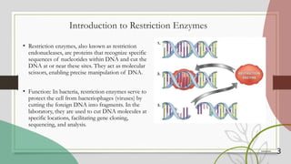
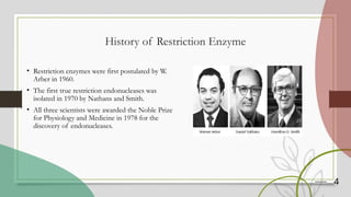
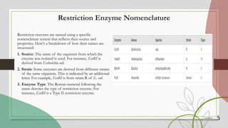
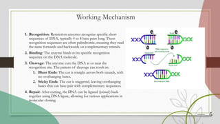
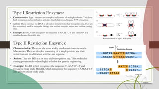
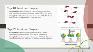
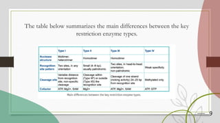
Restriction Enzymes and Their Types
Name : Anjani Kumari
University Roll No. :
24202891010003
Class Roll No. : 06
M.Sc 3rd sem
Botany Department
Table of Content
1. Introduction of Restriction Enzymes
2. History
3. Working Mechanism
4. Types
5. Application
6. Conclusion
3
Introduction to Restriction Enzymes
• Restriction enzymes, also known as restriction
endonucleases, are proteins that recognize specific
sequences of nucleotides within DNA and cut the
DNA at or near these sites. They act as molecular
scissors, enabling precise manipulation of DNA.
• Function: In bacteria, restriction enzymes serve to
protect the cell from bacteriophages (viruses) by
cutting the foreign DNA into fragments. In the
laboratory, they are used to cut DNA molecules at
specific locations, facilitating gene cloning,
sequencing, and analysis.
4
History of Restriction Enzyme
• Restriction enzymes were first postulated by W.
Arber in 1960.
• The first true restriction endonucleases was
isolated in 1970 by Nathans and Smith.
• All three scientists were awarded the Noble Prize
for Physiology and Medicine in 1978 for the
discovery of endonucleases.
5
Restriction Enzyme Nomenclature
Restriction enzymes are named using a specific
nomenclature system that reflects their source and
properties. Here's a breakdown of how their names are
structured:
1. Source: The name of the organism from which the
enzyme was isolated is used. For instance, EcoRI is
derived from Escherichia coli.
2. Strain: Some enzymes are derived from different strains
of the same organism. This is indicated by an additional
letter. For example, EcoRI is from strain R of E. coli.
3. Enzyme Type: The Roman numeral following the
name denotes the type of restriction enzyme. For
instance, EcoRI is a Type II restriction enzyme.
6
Working Mechanism
1. Recognition: Restriction enzymes recognize specific short
sequences of DNA, typically 4 to 8 base pairs long. These
recognition sequences are often palindromic, meaning they read
the same forwards and backwards on complementary strands.
2. Binding: The enzyme binds to its specific recognition
sequence on the DNA molecule.
3. Cleavage: The enzyme cuts the DNA at or near the
recognition site. The pattern of cleavage can result in:
1. Blunt Ends: The cut is straight across both strands, with
no overhanging bases.
2. Sticky Ends: The cut is staggered, leaving overhanging
bases that can base-pair with complementary sequences.
4. Repair: After cutting, the DNA can be ligated (joined) back
together using DNA ligase, allowing for various applications in
molecular cloning.
Type I Restriction Enzymes:
• Characteristics: Type I enzymes are complex and consist of multiple subunits. They have
both restriction and modification activities (methylation) and require ATP to function.
• Action: These enzymes cut DNA at a location distant from their recognition site. They are
less commonly used in molecular biology due to their complex nature and variable cutting
sites.
• Example: EcoKI, which recognizes the sequence 5’-GAATTC-3’ and cuts DNA at a
variable distance from this site.
Type II Restriction Enzymes:
• Characteristics: These are the most widely used restriction enzymes in
laboratories. They are simpler, consisting of a single protein, and their
restriction and modification activities are separate.
• Action: They cut DNA at or near their recognition site. This predictable
cutting pattern makes them highly valuable for genetic engineering.
• Example: EcoRI, which recognizes the sequence 5’-GAATTC-3’ and
produces sticky ends; HindIII, which recognizes the sequence 5’-AAGCTT-3’
and also produces sticky ends.
Restriction mode of type 1 R-M system
8
Type III Restriction Enzymes:
• Characteristics: These enzymes cut DNA at a site a few base pairs
away from their recognition sequence and require ATP for their action.
• Example: EcoP15I, which recognizes the sequence 5’-CCAGC-3’ and
cuts DNA at a site approximately 25 base pairs away.
Type IV Restriction Enzymes:
• Characteristics: These enzymes target modified DNA, such as
methylated or hydroxymethylated DNA, and are less common.
• Example: MspI, which recognizes the sequence 5’-C^CGG-3’ (where ^
indicates the cutting site) and is used for analyzing methylation patterns.
Restriction mode of type III R-M system
9
The table below summarizes the main differences between the key
restriction enzyme types.
10
Applications of Restriction Enzymes
• Genetic Engineering:
Restriction enzymes allow scientists to cut and paste DNA segments, facilitating gene cloning,
synthesis of recombinant proteins, and the creation of genetically modified organisms (GMOs).
• DNA Fingerprinting:
By cutting DNA into fragments and analyzing the patterns of these fragments, restriction enzymes are
used in forensic science to create DNA profiles for identification purposes.
• Gene Mapping:
Restriction enzymes help in mapping the locations of genes on chromosomes by cutting DNA into
fragments of known sizes, which can be analyzed using gel electrophoresis.
• Mutation Analysis:
Enzymes can be used to identify and study genetic mutations by comparing the patterns of DNA
fragments from mutant and wild-type organisms.
11
Conclusion
• Restriction enzymes are crucial tools in molecular biology and biotechnology. They
enable the precise cutting and manipulation of DNA, which has broad applications in
research, medicine, and agriculture.
• The advent of restriction enzymes has transformed genetic engineering, allowing for
detailed genetic studies and the development of novel biotechnological applications.
Thank You

Restriction Enzymes and Their Types.pptx

  • 1.
    Restriction Enzymes andTheir Types Name : Anjani Kumari University Roll No. : 24202891010003 Class Roll No. : 06 M.Sc 3rd sem Botany Department
  • 2.
    Table of Content 1.Introduction of Restriction Enzymes 2. History 3. Working Mechanism 4. Types 5. Application 6. Conclusion
  • 3.
    3 Introduction to RestrictionEnzymes • Restriction enzymes, also known as restriction endonucleases, are proteins that recognize specific sequences of nucleotides within DNA and cut the DNA at or near these sites. They act as molecular scissors, enabling precise manipulation of DNA. • Function: In bacteria, restriction enzymes serve to protect the cell from bacteriophages (viruses) by cutting the foreign DNA into fragments. In the laboratory, they are used to cut DNA molecules at specific locations, facilitating gene cloning, sequencing, and analysis.
  • 4.
    4 History of RestrictionEnzyme • Restriction enzymes were first postulated by W. Arber in 1960. • The first true restriction endonucleases was isolated in 1970 by Nathans and Smith. • All three scientists were awarded the Noble Prize for Physiology and Medicine in 1978 for the discovery of endonucleases.
  • 5.
    5 Restriction Enzyme Nomenclature Restrictionenzymes are named using a specific nomenclature system that reflects their source and properties. Here's a breakdown of how their names are structured: 1. Source: The name of the organism from which the enzyme was isolated is used. For instance, EcoRI is derived from Escherichia coli. 2. Strain: Some enzymes are derived from different strains of the same organism. This is indicated by an additional letter. For example, EcoRI is from strain R of E. coli. 3. Enzyme Type: The Roman numeral following the name denotes the type of restriction enzyme. For instance, EcoRI is a Type II restriction enzyme.
  • 6.
    6 Working Mechanism 1. Recognition:Restriction enzymes recognize specific short sequences of DNA, typically 4 to 8 base pairs long. These recognition sequences are often palindromic, meaning they read the same forwards and backwards on complementary strands. 2. Binding: The enzyme binds to its specific recognition sequence on the DNA molecule. 3. Cleavage: The enzyme cuts the DNA at or near the recognition site. The pattern of cleavage can result in: 1. Blunt Ends: The cut is straight across both strands, with no overhanging bases. 2. Sticky Ends: The cut is staggered, leaving overhanging bases that can base-pair with complementary sequences. 4. Repair: After cutting, the DNA can be ligated (joined) back together using DNA ligase, allowing for various applications in molecular cloning.
  • 7.
    Type I RestrictionEnzymes: • Characteristics: Type I enzymes are complex and consist of multiple subunits. They have both restriction and modification activities (methylation) and require ATP to function. • Action: These enzymes cut DNA at a location distant from their recognition site. They are less commonly used in molecular biology due to their complex nature and variable cutting sites. • Example: EcoKI, which recognizes the sequence 5’-GAATTC-3’ and cuts DNA at a variable distance from this site. Type II Restriction Enzymes: • Characteristics: These are the most widely used restriction enzymes in laboratories. They are simpler, consisting of a single protein, and their restriction and modification activities are separate. • Action: They cut DNA at or near their recognition site. This predictable cutting pattern makes them highly valuable for genetic engineering. • Example: EcoRI, which recognizes the sequence 5’-GAATTC-3’ and produces sticky ends; HindIII, which recognizes the sequence 5’-AAGCTT-3’ and also produces sticky ends. Restriction mode of type 1 R-M system
  • 8.
    8 Type III RestrictionEnzymes: • Characteristics: These enzymes cut DNA at a site a few base pairs away from their recognition sequence and require ATP for their action. • Example: EcoP15I, which recognizes the sequence 5’-CCAGC-3’ and cuts DNA at a site approximately 25 base pairs away. Type IV Restriction Enzymes: • Characteristics: These enzymes target modified DNA, such as methylated or hydroxymethylated DNA, and are less common. • Example: MspI, which recognizes the sequence 5’-C^CGG-3’ (where ^ indicates the cutting site) and is used for analyzing methylation patterns. Restriction mode of type III R-M system
  • 9.
    9 The table belowsummarizes the main differences between the key restriction enzyme types.
  • 10.
    10 Applications of RestrictionEnzymes • Genetic Engineering: Restriction enzymes allow scientists to cut and paste DNA segments, facilitating gene cloning, synthesis of recombinant proteins, and the creation of genetically modified organisms (GMOs). • DNA Fingerprinting: By cutting DNA into fragments and analyzing the patterns of these fragments, restriction enzymes are used in forensic science to create DNA profiles for identification purposes. • Gene Mapping: Restriction enzymes help in mapping the locations of genes on chromosomes by cutting DNA into fragments of known sizes, which can be analyzed using gel electrophoresis. • Mutation Analysis: Enzymes can be used to identify and study genetic mutations by comparing the patterns of DNA fragments from mutant and wild-type organisms.
  • 11.
    11 Conclusion • Restriction enzymesare crucial tools in molecular biology and biotechnology. They enable the precise cutting and manipulation of DNA, which has broad applications in research, medicine, and agriculture. • The advent of restriction enzymes has transformed genetic engineering, allowing for detailed genetic studies and the development of novel biotechnological applications.
  • 12.