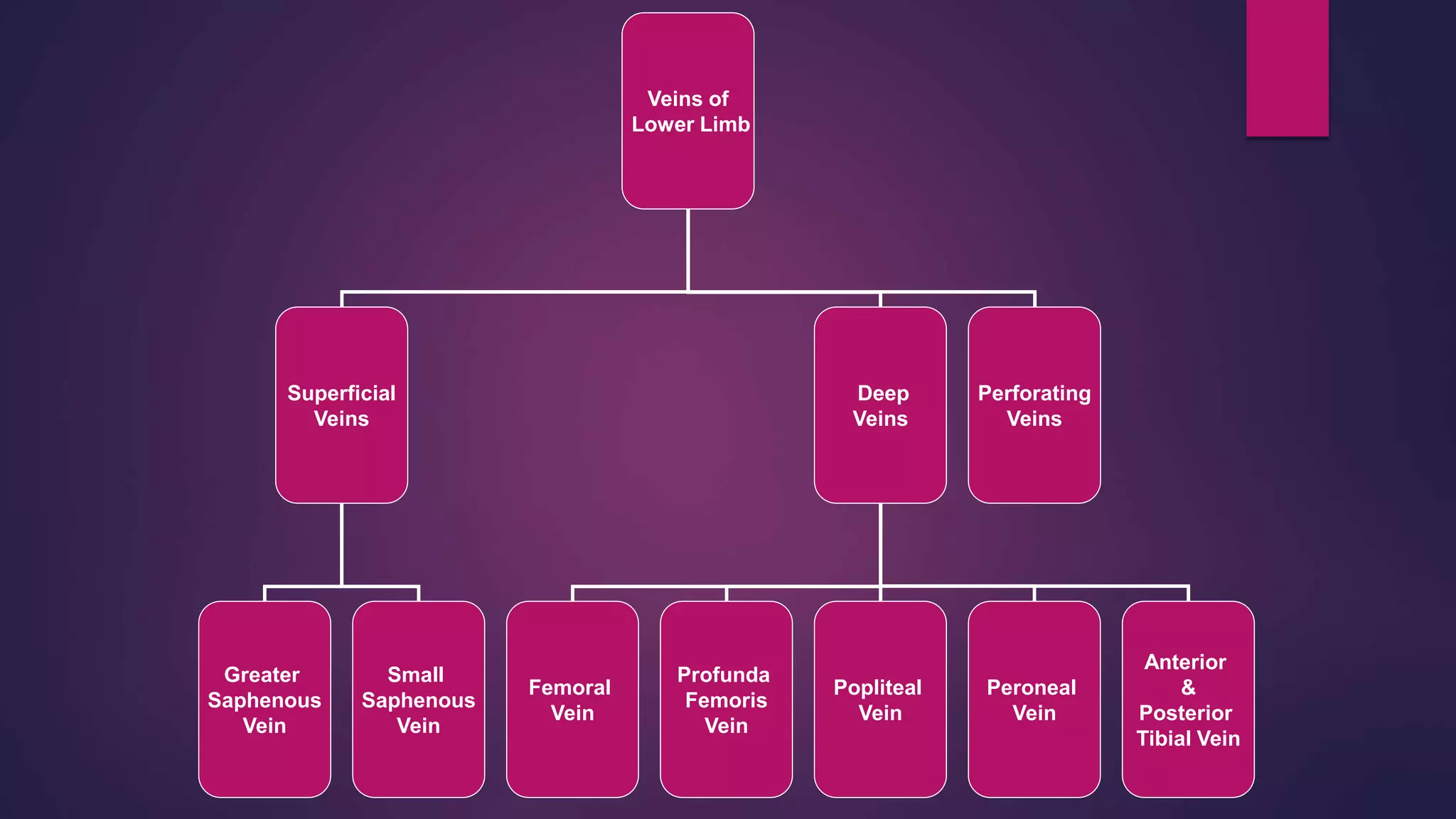
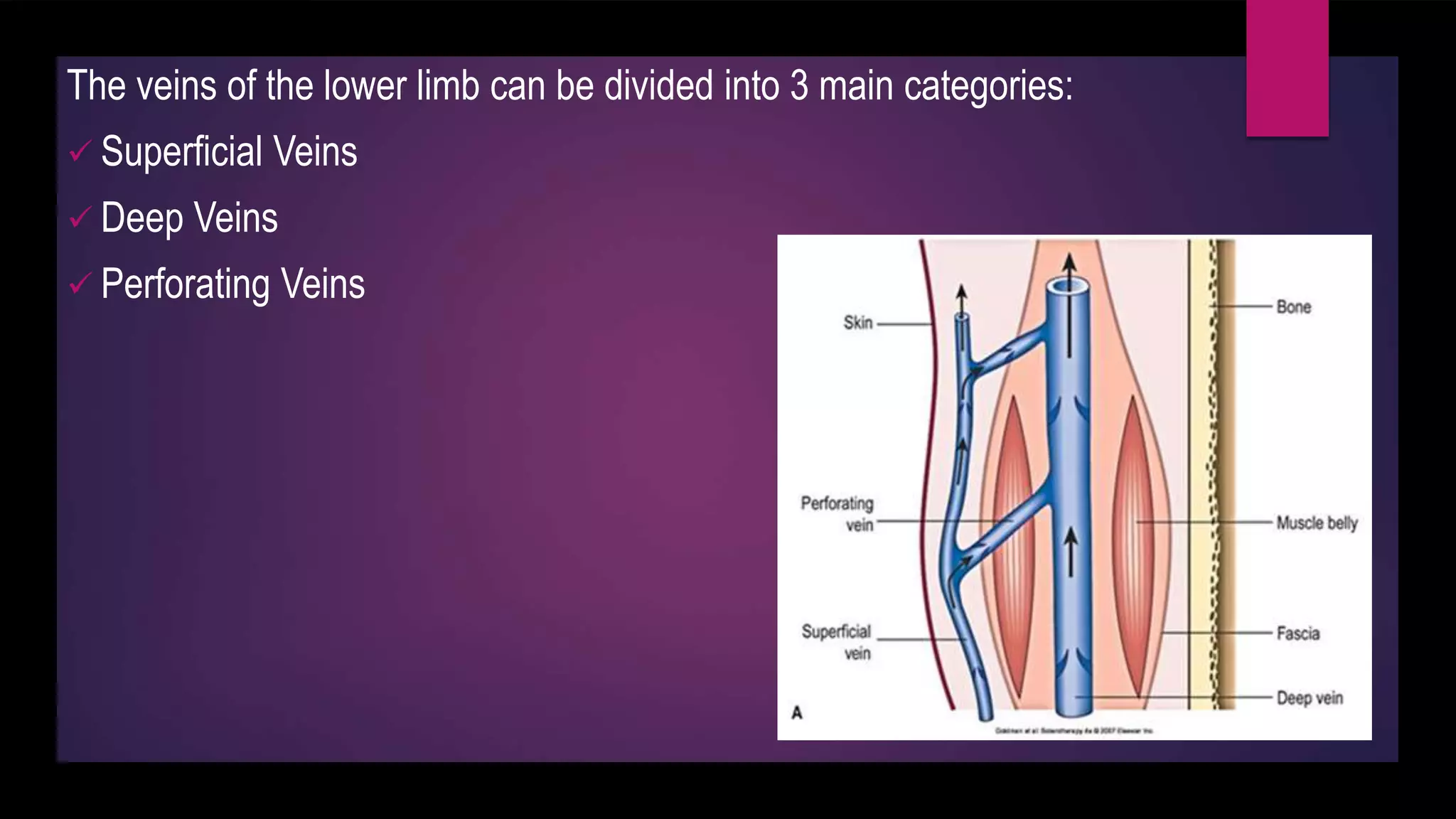
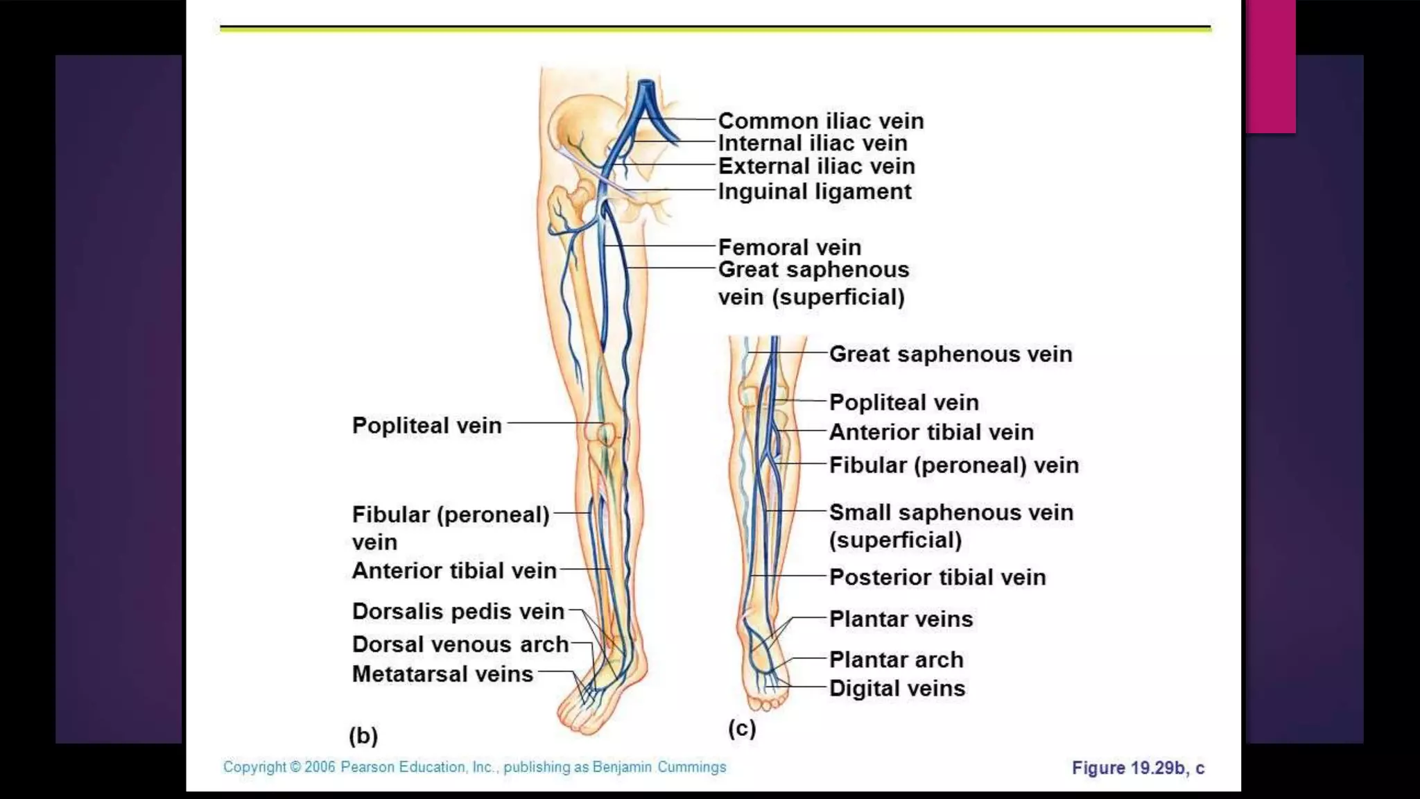
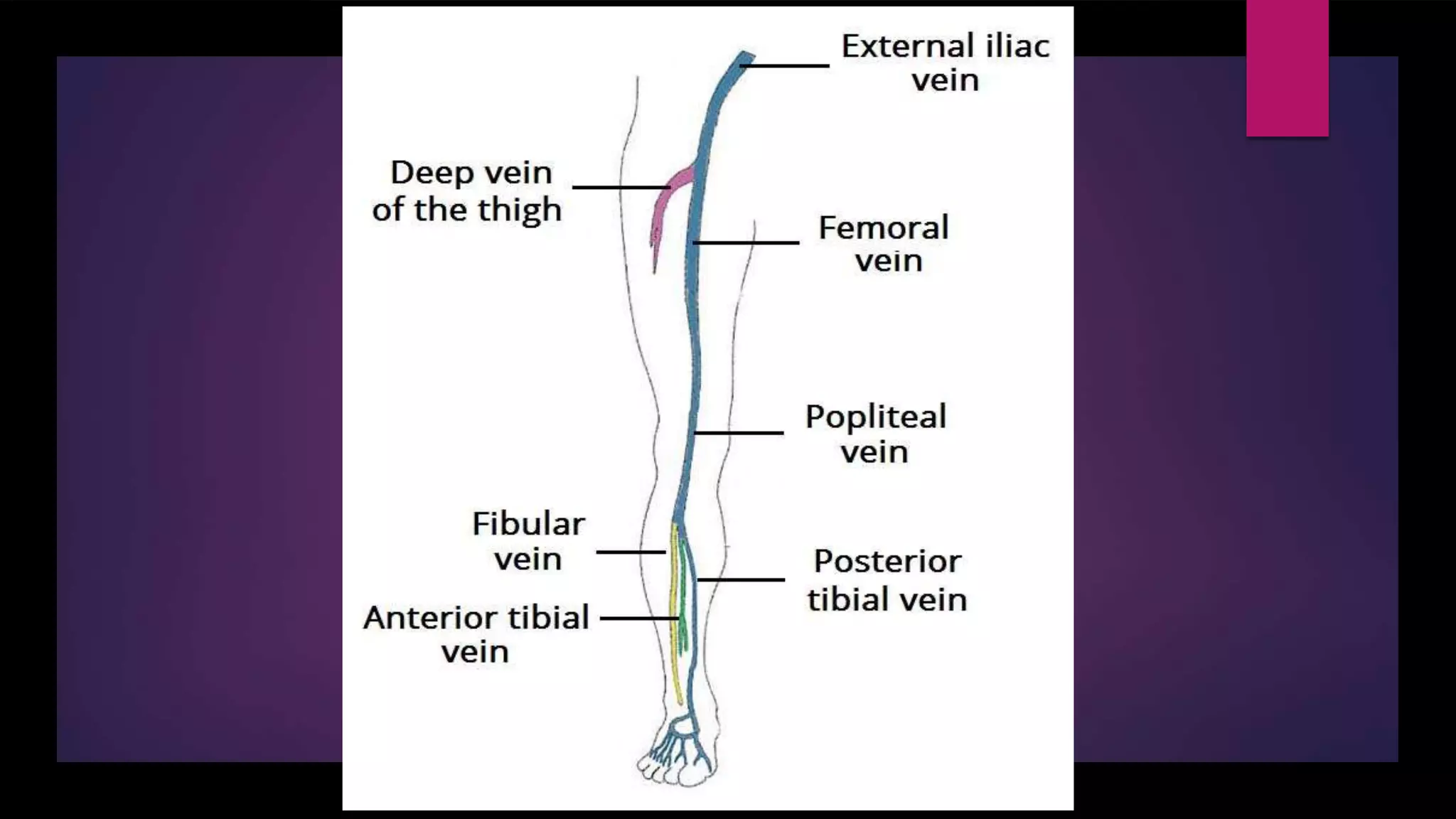
4. The veins of the lower limb can be divided into 3 main categories:
Superficial Veins
Deep Veins
Perforating Veins
5. SUPERFICIAL VEINS
These are beneath the skin in the superficial fascia, named as:
Great Saphenous Vein
Short Saphenous Vein
6. GREAT SAPHENOUS VEIN
Arises at medial aspect of dorsal venous arch when it merges with the dorsal vein of the big
toe
Passes anterior to medial malleolus
Courses superiorly in medial aspect of leg
Runs over the posterior border of the medial condyle of the femur
7.
8. It traverses the saphenous opening in the fascia lata 1.5 inches or 4cm below and lateral to the
pubic tubercle
It also anastomoses with the small saphenous vein
Empties into femoral vein
9. TRIBUTARIES
In the sole: medial marginal vein
In the lower leg : anastomoses freely with the small saphenous vein
In the knee: popliteal vein
In the thigh: posteromedial aspect gives accessory saphenous vein
Lateral and anterior r cutaneous veins of the thigh
At the saphenous opening, it receives: superficial epigastric vein
superficial circumflex iliac vein
superficial external pudendal vein
10. SHORT SAPHENOUS VEIN
Arises at lateral side of dorsal venous arch when it merges with
the dorsal vein of the little digit
Passes posterior to lateral malleolus
Passes superiorly at posterior aspect of calf
Ascends between the heads of gastrocnemius muscle
Pierces fascia of popliteal fossa
Drains into popliteal vein
11. PERFORATING VEINS
Allows the blood to flow only from superficial to deep veins.
To achieve compression, these veins have an oblique angle so that the back flow of blood is
prevented when the muscle contracts
Superficial to deep venous blood flow is important because it propels the blood to the heart
against gravity.
12. DEEP VEINS
ANTERIOR TIBIAL VEIN
The anterior tibial vein of the lower limb carries blood from the anterior compartment of the
leg to the popliteal vein which is formed when it joins with the posterior tibial vein.
The anterior tibial vein is accompanied by an anterior tibial artery.
13. POSTERIOR TIBIAL VEIN
The posterior tibial vein of the lower limb carries blood from the posterior
compartment and plantar surface of the foot to the popliteal
vein which it forms when it joins with the anterior tibial vein.
The posterior tibial vein is accompanied by posterior tibial artery.
14. PERONEAL VEIN
The fibular veins are deep veins that help carry blood from the lateral compartment of the leg.
They drain into the posterior tibial veins, which will in turn drain into the popliteal vein.
The fibular veins accompany the fibular artery.
15. POPLITEAL VEIN
Formed at the lower border of the popliteus muscle by the union of the anterior and posterior
tibial veins and continues as the femoral vein
16.
17. PROFUNDA FEMORIS VEIN
Profunda femoris vein (or deep femoral vein) is a large deep vein in the thigh.
It receives blood from the inner thigh and proceeds superiorly and medially running alongside
the profunda femoris artery to join the femoral vein.
18.
19. FEMORAL VEIN
The femoral vein is a blood vessel that accompanies the femoral artery in the femoral sheath.
It begins at the adductor canal (also known as hunter's canal) and is a continuation of
the popliteal vein.
It ends at the inferior margin of the inguinal ligament, where it becomes the external iliac vein.
22. SUPERFICIAL INGUINAL LYMPH
NODES
HORIZONTAL GROUP
Below the inguinal ligament
Medial members: These receive the lymph from:
Anterior abdominal wall below the umbilicus
Perineum
Urethra
Lower half of anal canal
23. Lateral members:
Receive the lymph from the back below the iliac crests
VERTICAL GROUP: Along the terminal part of great saphenous vein, receive lymph from:
Lower limb
Skin
Fascia
24. DEEP INGUINAL LYMPH NODES
Lie beneath the deep fascia along the medial side of femoral vein, receive the lymph from:
Superficial inguinal lymph nodes
Popliteal nodes