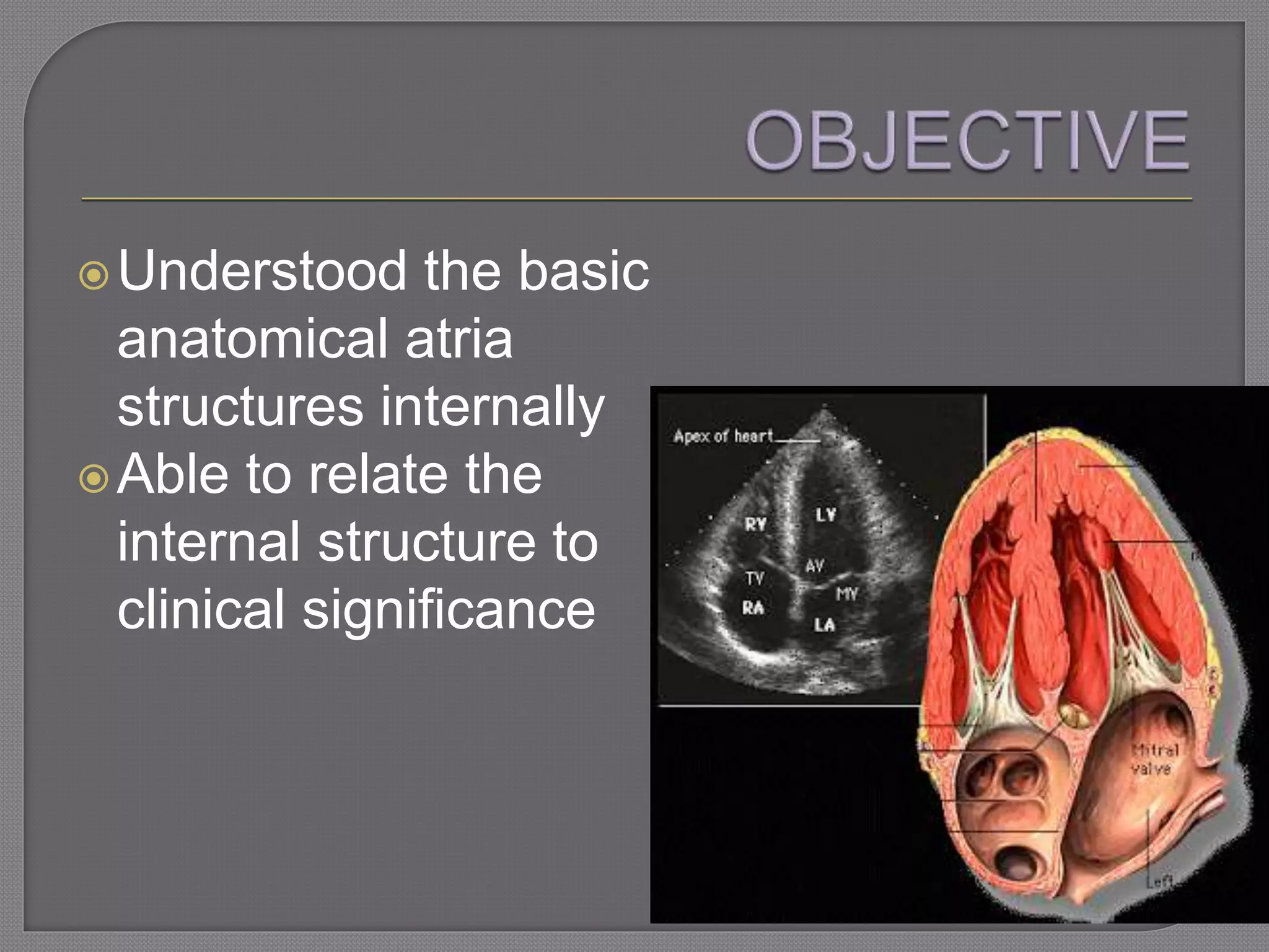
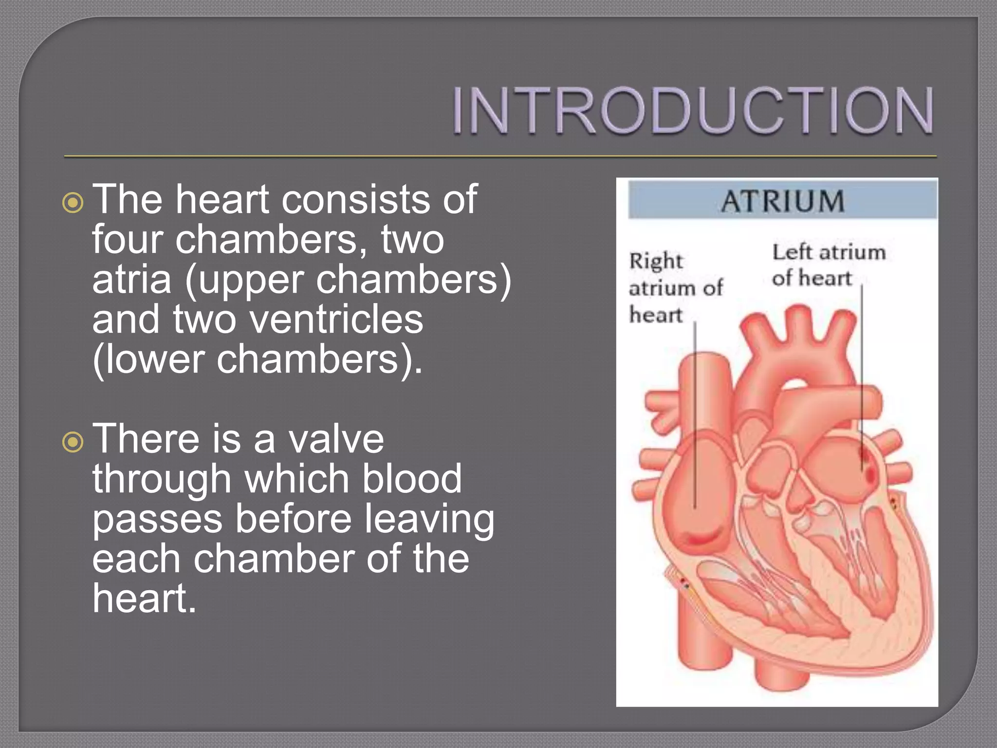
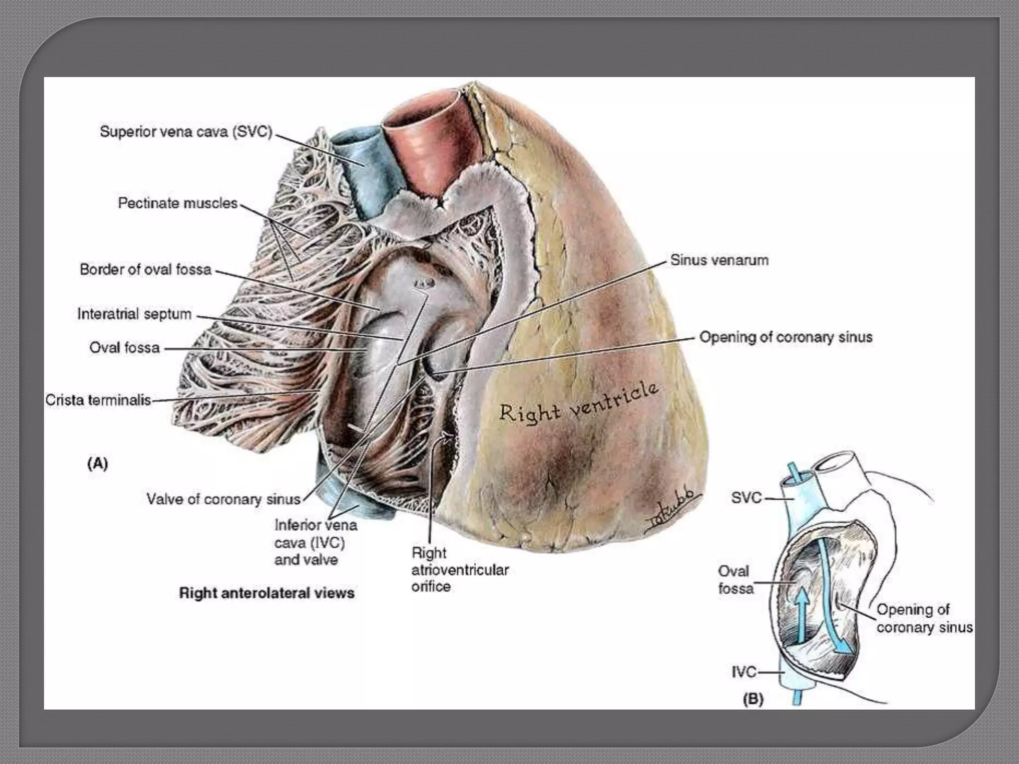
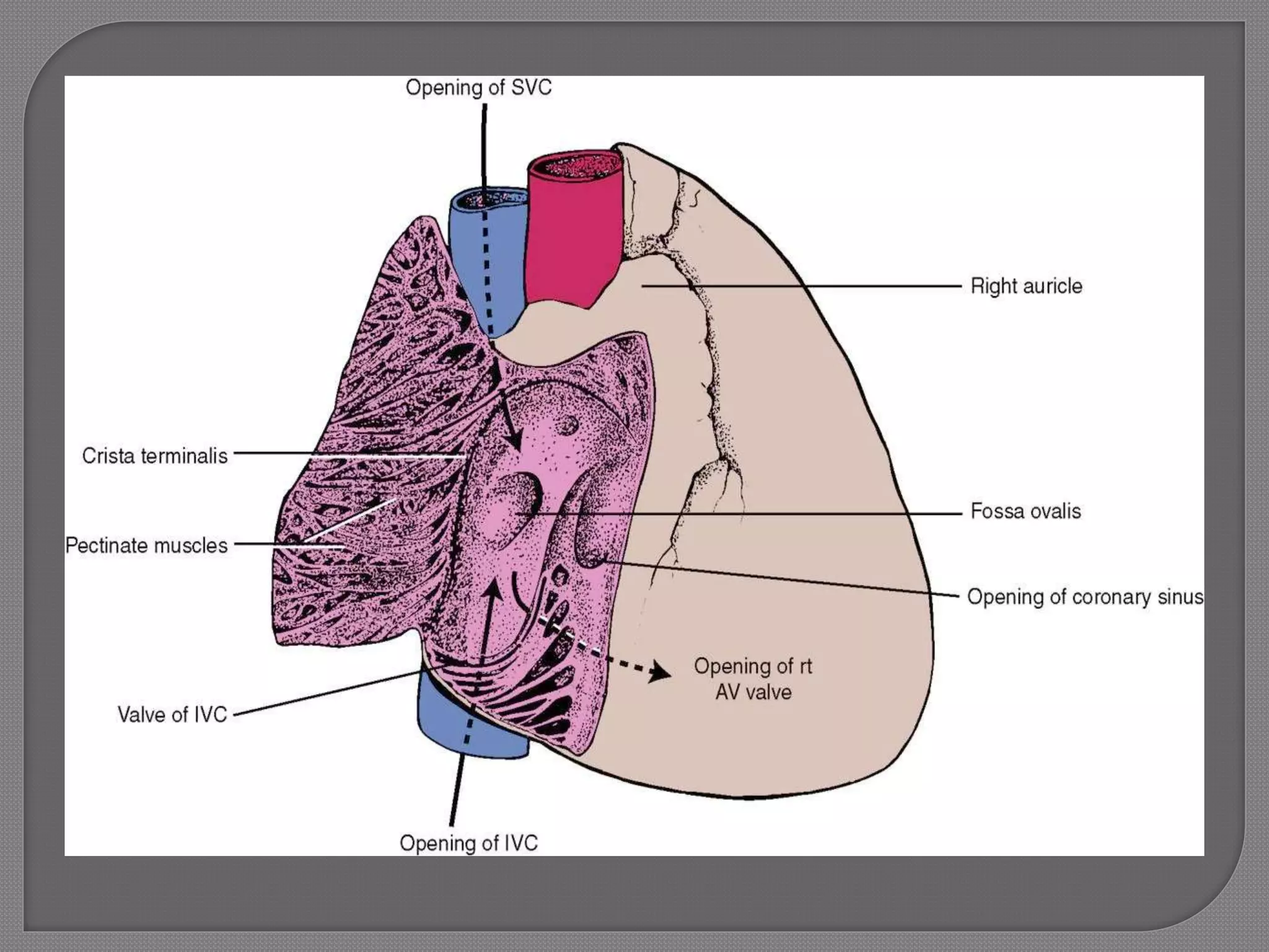
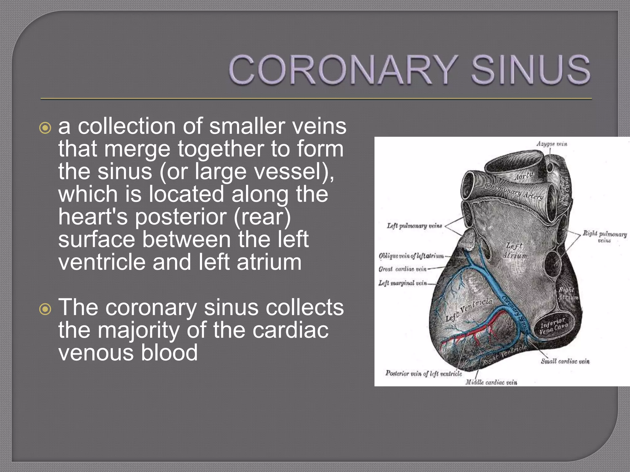
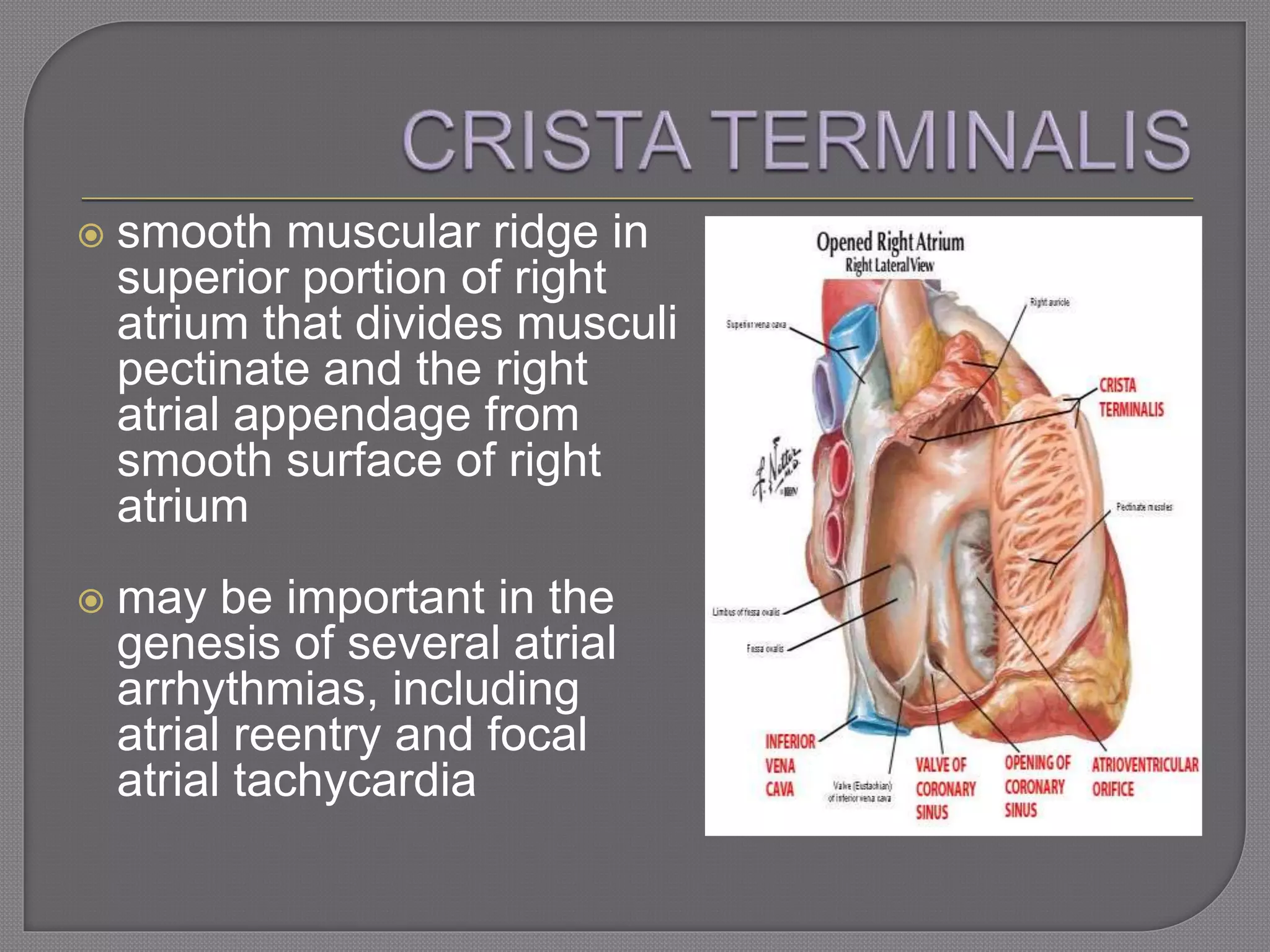
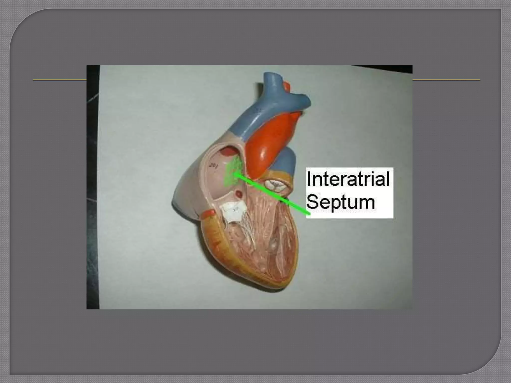
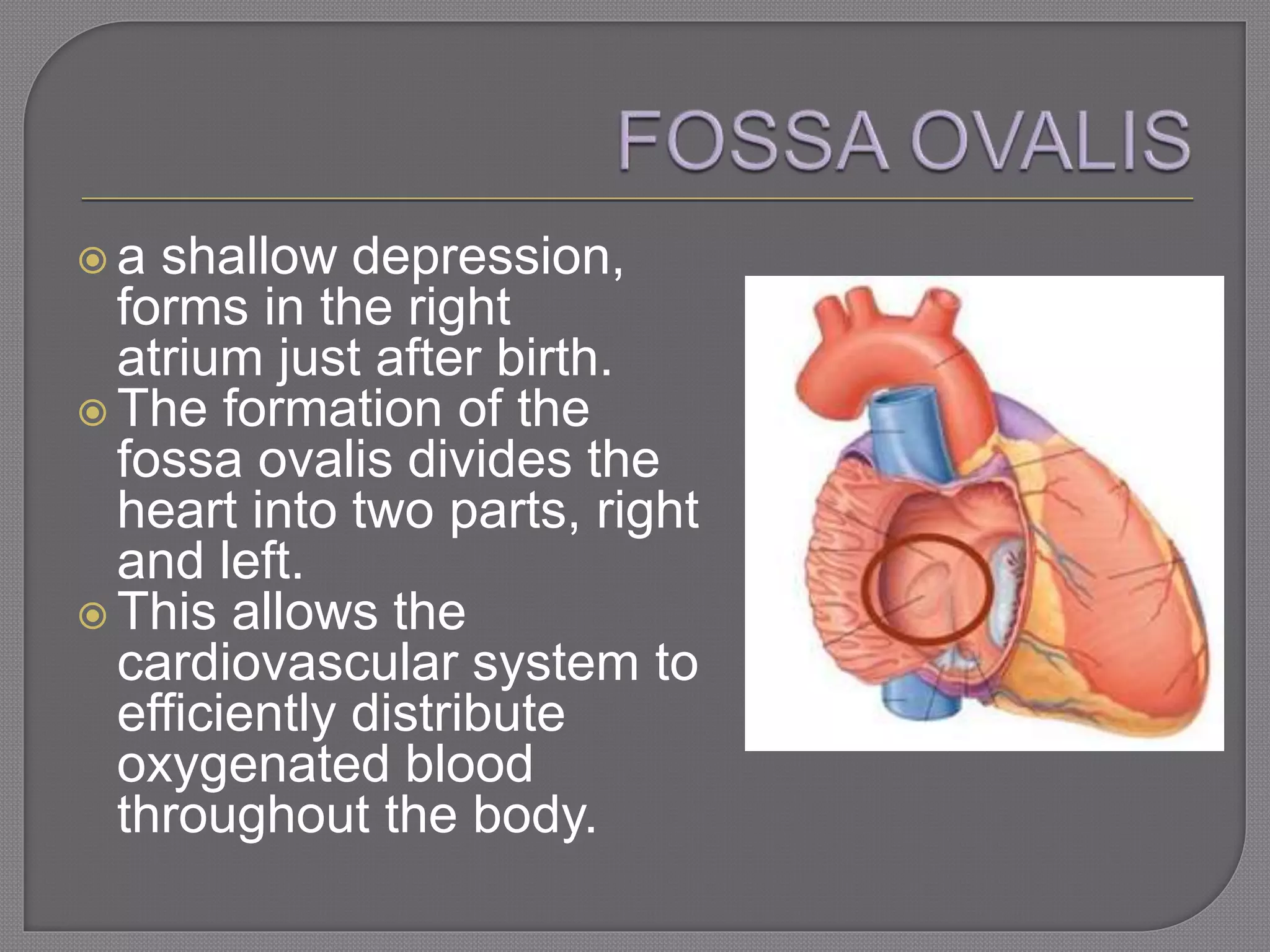
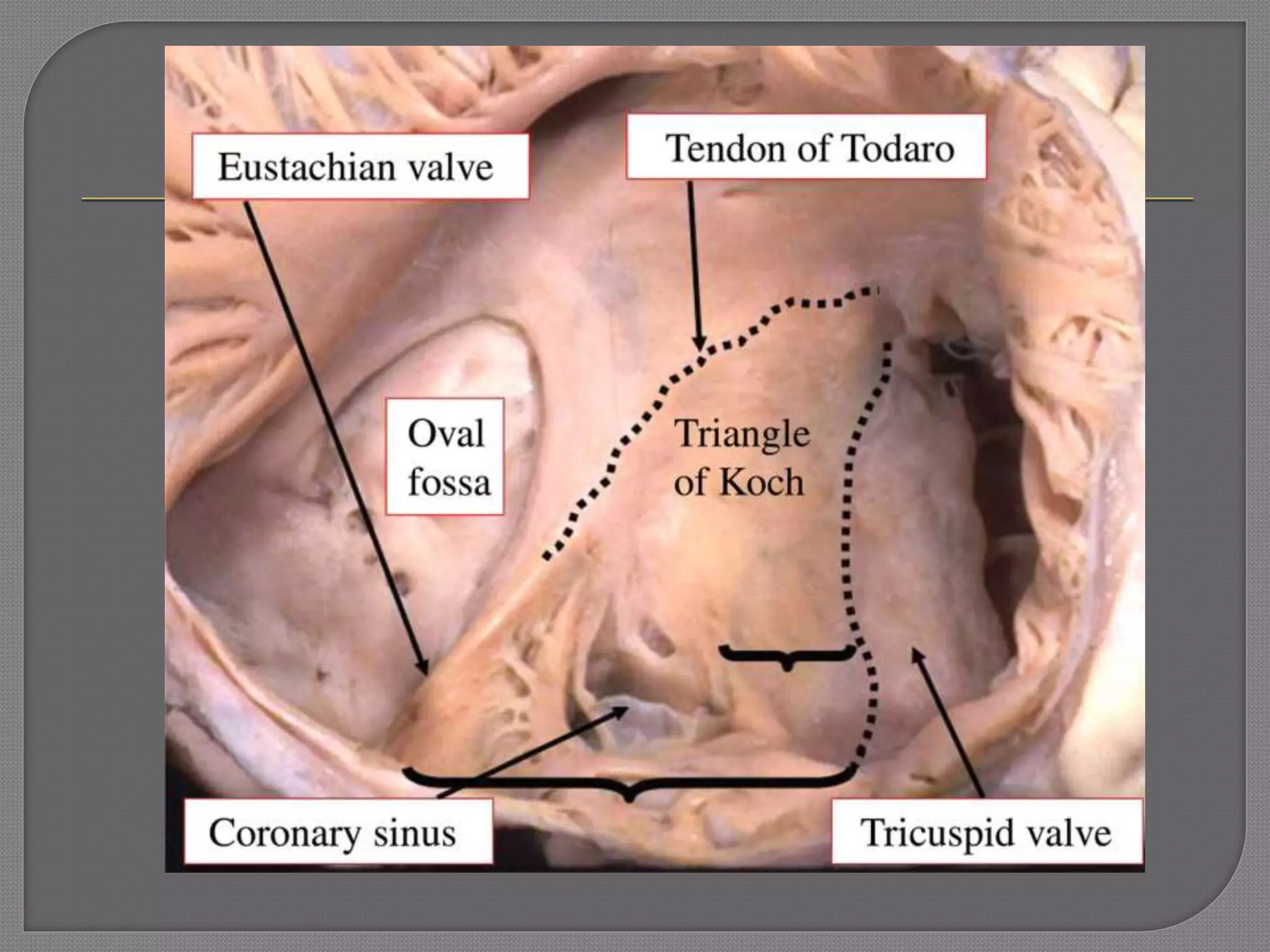
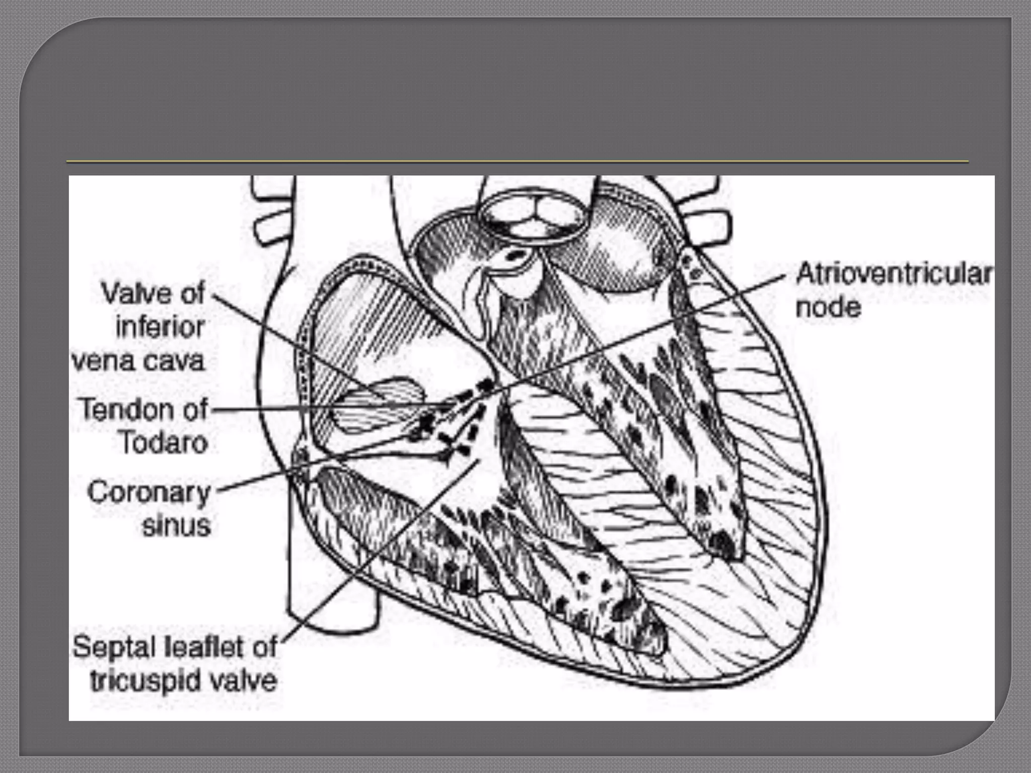
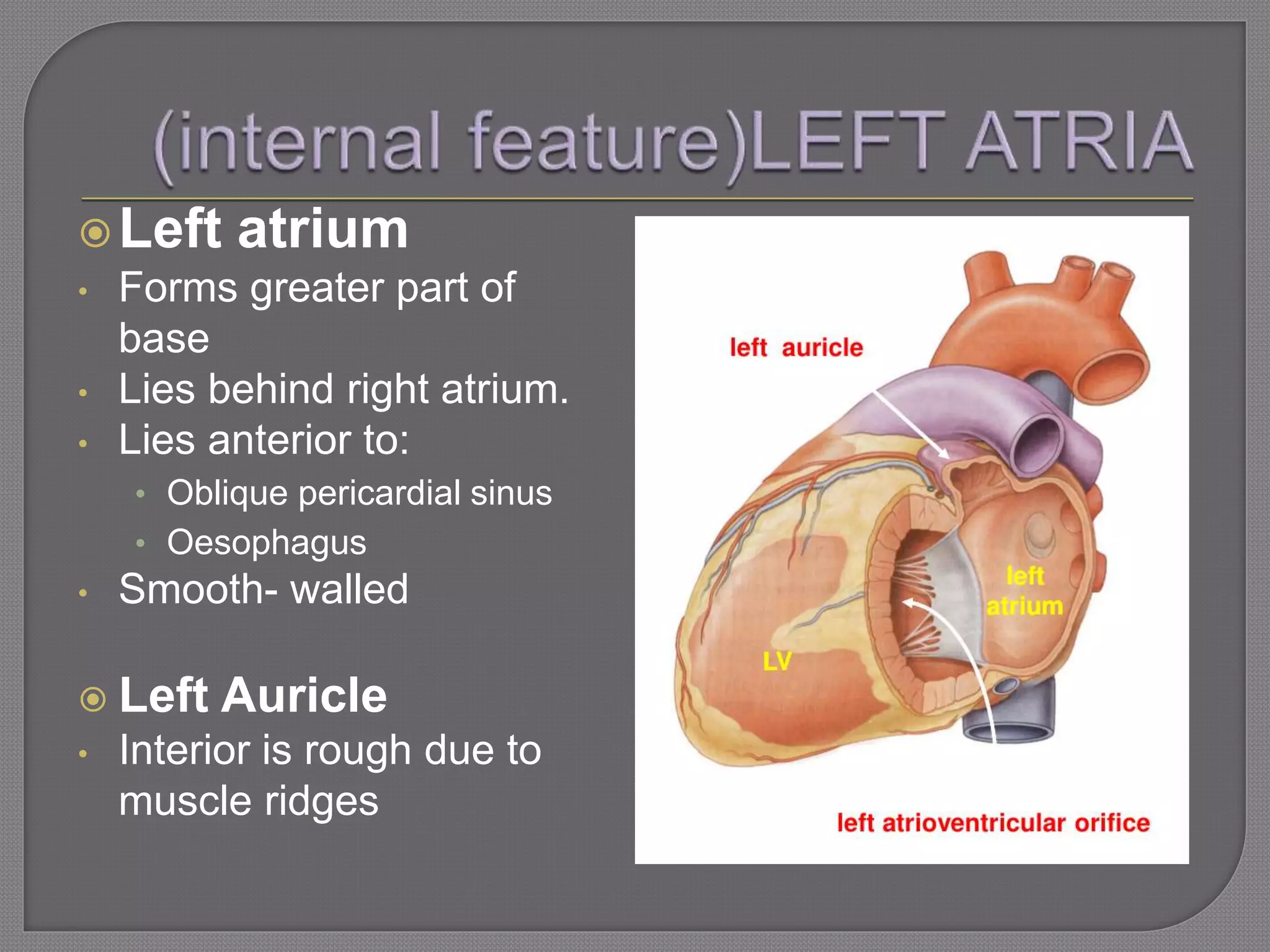
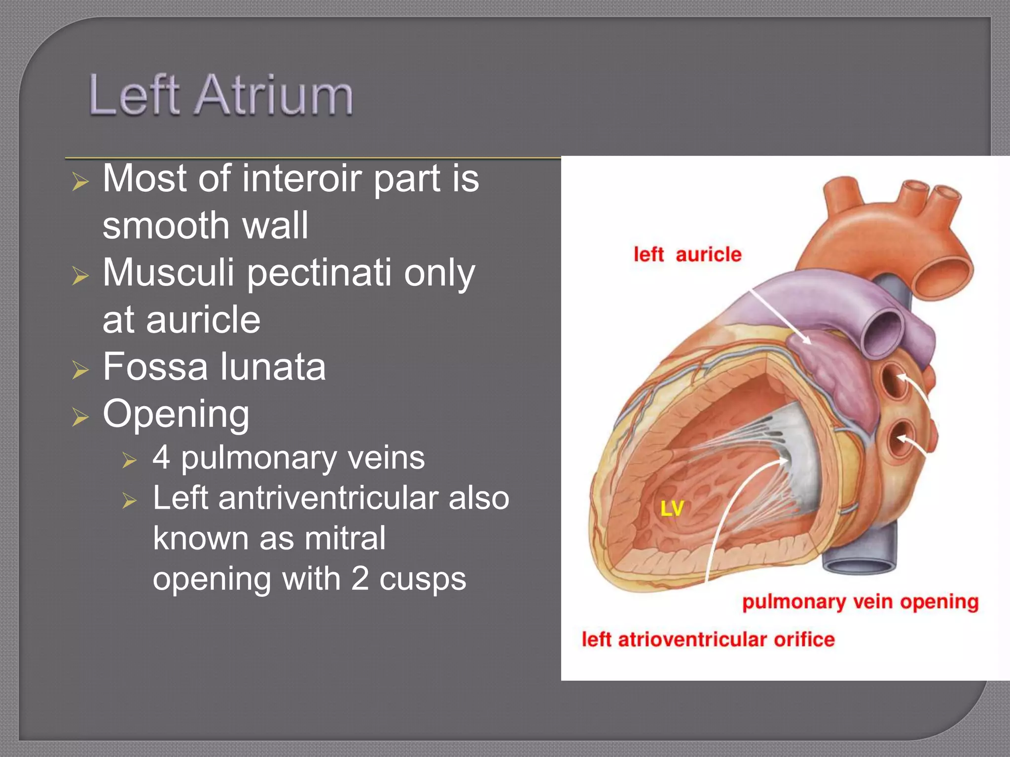
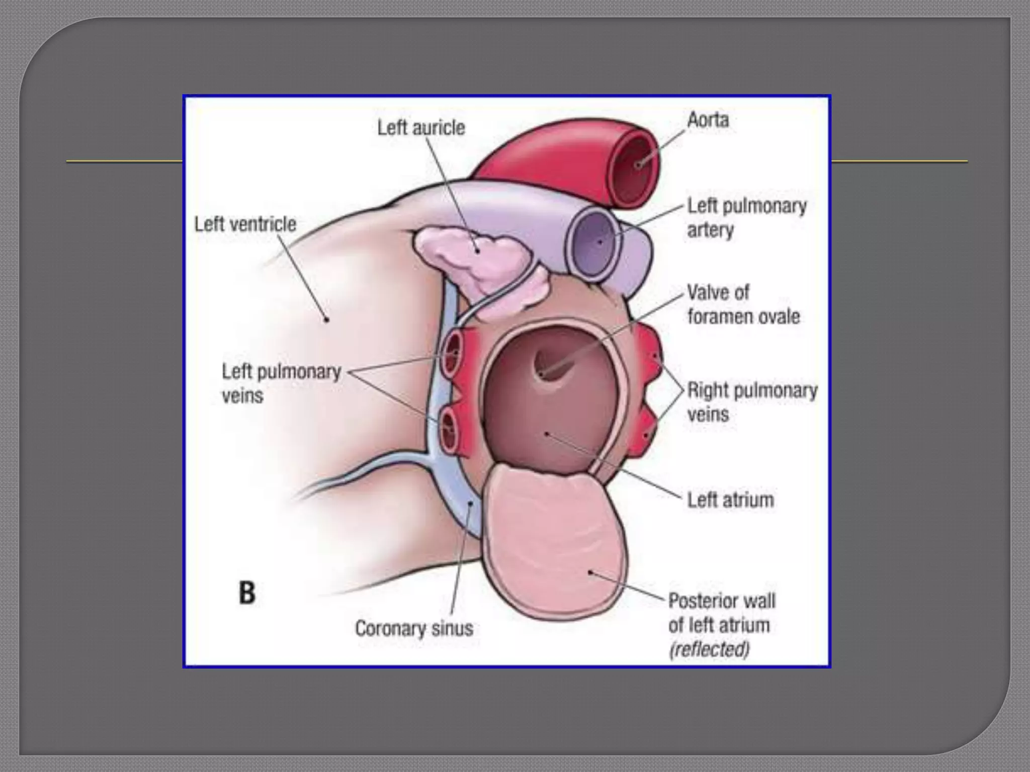
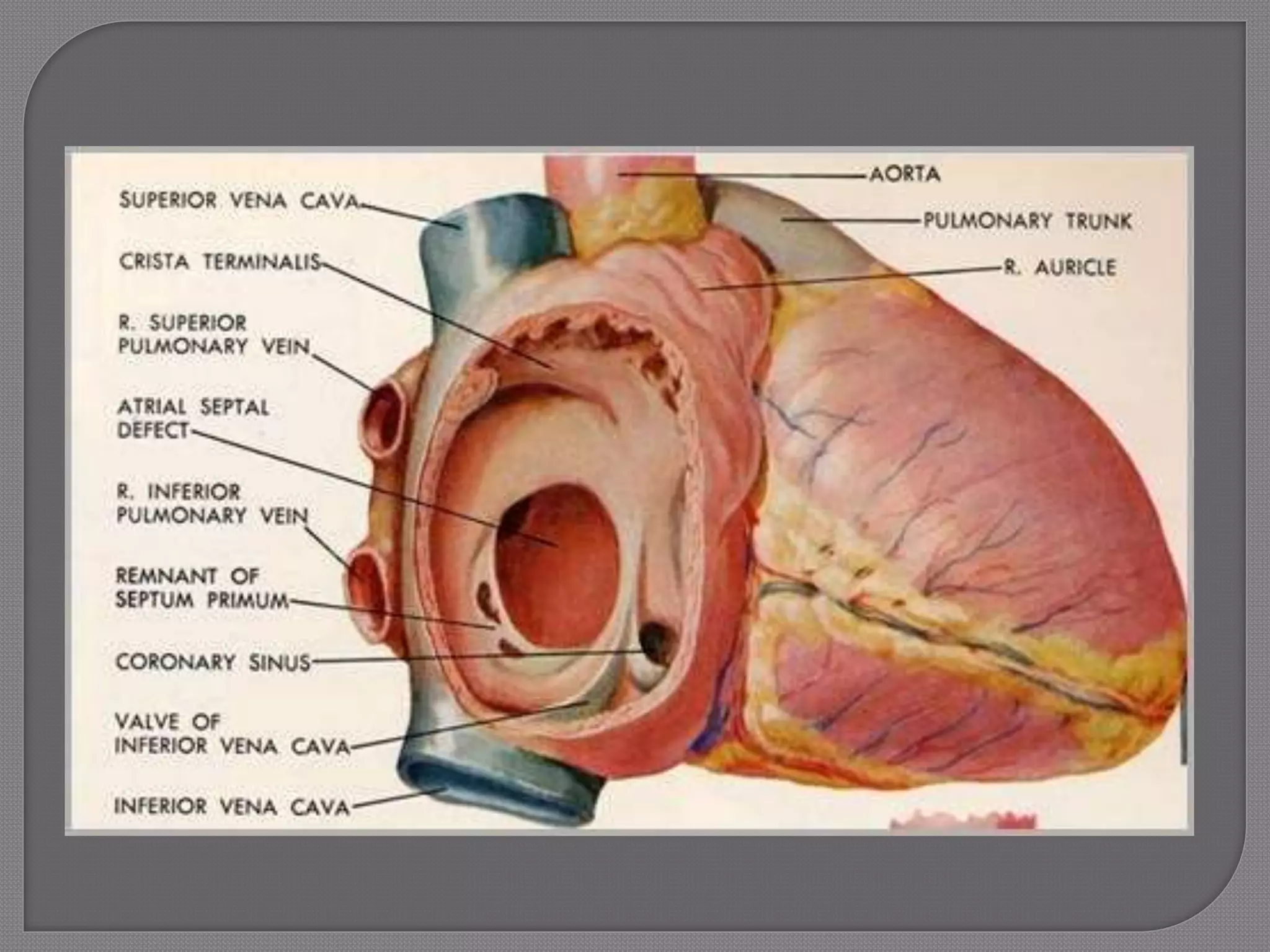
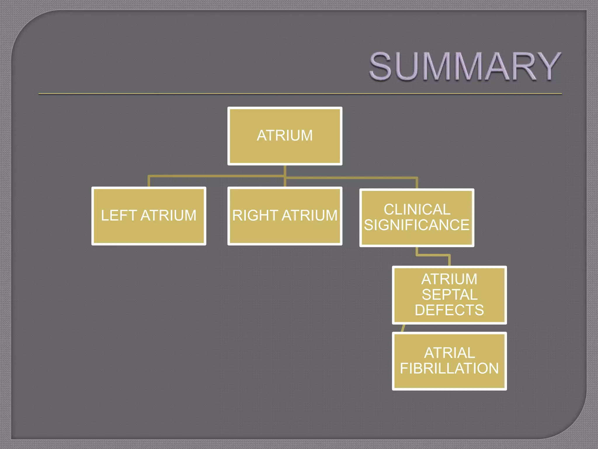
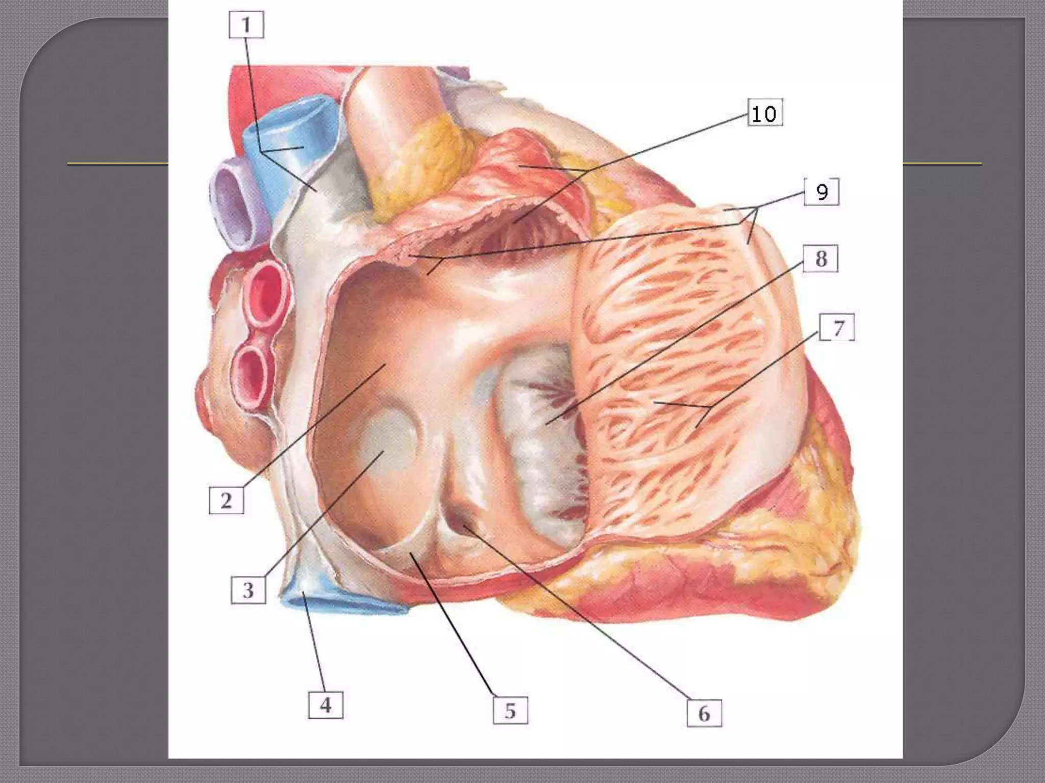
Name : NUR FARRA NAJWA BINTI ABDUL AZIM
Matric number : 082015100005
Understood the basic
anatomical atria
structures internally
Able to relate the
internal structure to
clinical significance
 The heart consists of
four chambers, two
atria (upper chambers)
and two ventricles
(lower chambers).
 There is a valve
through which blood
passes before leaving
each chamber of the
heart.
 Right Atrium: Structure
- Sinus Venarum (smooth-walled)
- Auricle (rough-walled)
- Pectinati muscles
 Orifices:
- SVC, IVC, Coronary sinus
- Crista Terminalis ("terminal crest")
- Interatrial septum
- Fossa Ovalis
- Annulus ovalis
- Right AV orifice
 a collection of smaller veins
that merge together to form
the sinus (or large vessel),
which is located along the
heart's posterior (rear)
surface between the left
ventricle and left atrium
 The coronary sinus collects
the majority of the cardiac
venous blood
 smooth muscular ridge in
superior portion of right
atrium that divides musculi
pectinate and the right
atrial appendage from
smooth surface of right
atrium
 may be important in the
genesis of several atrial
arrhythmias, including
atrial reentry and focal
atrial tachycardia
Sinuatrial node
• Lies in upper part of sulcus terminalis
• To the right to the opening of superior vena cava.
Atrioventricular node
• In lower part of atrial septum.
• Above the opening of coronary sinus
• Close to the attachment of tricuspid valve
 a shallow depression,
forms in the right
atrium just after birth.
 The formation of the
fossa ovalis divides the
heart into two parts, right
and left.
 This allows the
cardiovascular system to
efficiently distribute
oxygenated blood
throughout the body.
It is the prominent oval margin of the fossa
ovalis in the right atrium.
Form the upper margin of the fossa ovalis
It is most distinct above and at the sides of
the fossa ovalis and at the below, it is
deficient.
 Form by
• Tendon of Todaro (continuation of Eustachian valve of the
inferior vena cava and the Thebesian valve of the coronary
sinus)
• opening of the coronary sinus
• septal cusp of the tricuspid valve
 Angle of Triangel of Koch
• Coronary sinus orifice
• Tendon of Todaro
• Atrioventricular node
 The apex of the triangle of Koch is the location of
the atrioventricular node.
Left atrium
• Forms greater part of
base
• Lies behind right atrium.
• Lies anterior to:
• Oblique pericardial sinus
• Oesophagus
• Smooth- walled
 Left Auricle
• Interior is rough due to
muscle ridges
 Most of interoir part is
smooth wall
 Musculi pectinati only
at auricle
 Fossa lunata
 Opening
 4 pulmonary veins
 Left antriventricular also
known as mitral
opening with 2 cusps
Atrial septal defects
- Blood flows in reverse direction
- Reduce cardiac output
ATRIUM
LEFT ATRIUM RIGHT ATRIUM CLINICAL
SIGNIFICANCE
ATRIUM
SEPTAL
DEFECTS
ATRIAL
FIBRILLATION
ANY QUESTION ??

Internal feature of right and left atria

  • 1.
    Name : NURFARRA NAJWA BINTI ABDUL AZIM Matric number : 082015100005
  • 2.
    Understood the basic anatomicalatria structures internally Able to relate the internal structure to clinical significance
  • 3.
     The heartconsists of four chambers, two atria (upper chambers) and two ventricles (lower chambers).  There is a valve through which blood passes before leaving each chamber of the heart.
  • 4.
     Right Atrium:Structure - Sinus Venarum (smooth-walled) - Auricle (rough-walled) - Pectinati muscles  Orifices: - SVC, IVC, Coronary sinus - Crista Terminalis ("terminal crest") - Interatrial septum - Fossa Ovalis - Annulus ovalis - Right AV orifice
  • 8.
     a collectionof smaller veins that merge together to form the sinus (or large vessel), which is located along the heart's posterior (rear) surface between the left ventricle and left atrium  The coronary sinus collects the majority of the cardiac venous blood
  • 9.
     smooth muscularridge in superior portion of right atrium that divides musculi pectinate and the right atrial appendage from smooth surface of right atrium  may be important in the genesis of several atrial arrhythmias, including atrial reentry and focal atrial tachycardia
  • 11.
    Sinuatrial node • Liesin upper part of sulcus terminalis • To the right to the opening of superior vena cava. Atrioventricular node • In lower part of atrial septum. • Above the opening of coronary sinus • Close to the attachment of tricuspid valve
  • 13.
     a shallowdepression, forms in the right atrium just after birth.  The formation of the fossa ovalis divides the heart into two parts, right and left.  This allows the cardiovascular system to efficiently distribute oxygenated blood throughout the body.
  • 14.
    It is theprominent oval margin of the fossa ovalis in the right atrium. Form the upper margin of the fossa ovalis It is most distinct above and at the sides of the fossa ovalis and at the below, it is deficient.
  • 15.
     Form by •Tendon of Todaro (continuation of Eustachian valve of the inferior vena cava and the Thebesian valve of the coronary sinus) • opening of the coronary sinus • septal cusp of the tricuspid valve  Angle of Triangel of Koch • Coronary sinus orifice • Tendon of Todaro • Atrioventricular node  The apex of the triangle of Koch is the location of the atrioventricular node.
  • 18.
    Left atrium • Formsgreater part of base • Lies behind right atrium. • Lies anterior to: • Oblique pericardial sinus • Oesophagus • Smooth- walled  Left Auricle • Interior is rough due to muscle ridges
  • 19.
     Most ofinteroir part is smooth wall  Musculi pectinati only at auricle  Fossa lunata  Opening  4 pulmonary veins  Left antriventricular also known as mitral opening with 2 cusps
  • 21.
    Atrial septal defects -Blood flows in reverse direction - Reduce cardiac output
  • 23.
    ATRIUM LEFT ATRIUM RIGHTATRIUM CLINICAL SIGNIFICANCE ATRIUM SEPTAL DEFECTS ATRIAL FIBRILLATION
  • 26.