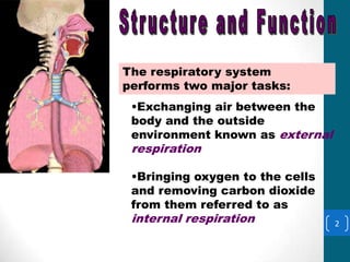
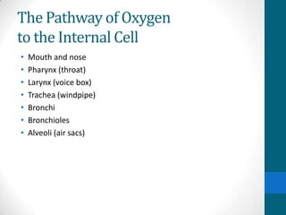
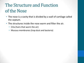
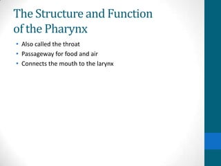
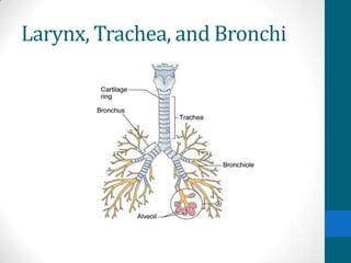
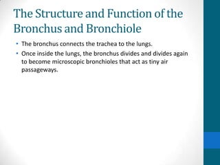
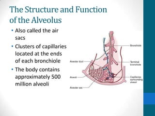
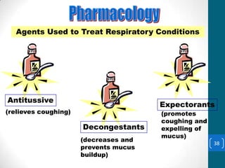
Respiratory System
Structure and Function
          The respiratory system
          performs two major tasks:
           •Exchanging air between the
           body and the outside
           environment known as external
           respiration

           •Bringing oxygen to the cells
           and removing carbon dioxide
           from them referred to as
           internal respiration            2
The Pathway of Oxygen
to the Internal Cell
•   Mouth and nose
•   Pharynx (throat)
•   Larynx (voice box)
•   Trachea (windpipe)
•   Bronchi
•   Bronchioles
•   Alveoli (air sacs)
The Structure and Function
of the Nose
• The nose is a cavity that is divided by a wall of cartilage called
  the septum.
• The structures inside the nose warm and filter the air.
  • Cilia (hairs that warm the air)
  • Mucous membranes (trap dust and bacteria)
The Structure and Function
of the Pharynx
• Also called the throat
• Passageway for food and air
• Connects the mouth to the larynx
The Structure and Function
of the Epiglottis
• Lid on the top of the larynx
• When food is swallowed, the lid closes so that food is directed
  down the esophagus and not into the lungs.
• Air passes over the open epiglottis and enters the larynx.
Larynx, Trachea, and Bronchi
The Structure and Function
of the Larynx
• Also called the voice
  box
• A tube made up of
  nine separate
  cartilages to maintain
  openness
• Lined with mucous
  membranes that
  form two folds called
  the vocal cords
The Structure and Function
of the Trachea
• Also called the windpipe
• Held open by C-shaped rings of cartilage
• The wall between the rings is elastic to adjust for body
  positions.
• Above the middle of the sternum, the trachea divides into two
  sections called bronchi.
The Structure and Function of the
Bronchus and Bronchiole
• The bronchus connects the trachea to the lungs.
• Once inside the lungs, the bronchus divides and divides again
  to become microscopic bronchioles that act as tiny air
  passageways.
The Structure and Function
of the Alveolus
• Also called the air
  sacs
• Clusters of capillaries
  located at the ends
  of each bronchiole
• The body contains
  approximately 500
  million alveoli
The Diaphragm and the Brain and
How They Relate to Breathing
• The main muscle of respiration is called the diaphragm.
• When the diaphragm contracts, it produces a vacuum that
  causes air to be drawn in.
• When the diaphragm relaxes, air is forced out of the lungs.
Occurrences That Alter
Breathing
• Coughing
  • Deep breaths followed by forceful exhalation that can clear
    mucus from the lower respiratory tract
• Hiccoughs
  • Caused by spasm of the diaphragm, possibly the result of an
    irritation to the diaphragm
Occurrences That Alter
Breathing
• Sneezing
  – Air is forced through the nose to clear the upper respiratory
    structures
• Yawning
  – Deep, prolonged breath that can be caused by a drop in oxygen
    levels
• Crying
  – A change in the breathing pattern that is in response to emotions
Diagnostic, Procedural &
 Laboratory Tests
Methods Used to
Diagnose Respiratory
Disorders:
•Auscultation
(stethoscope)

•Assessing respiratory
rate
                         Normal Adult
•Percussion              respiratory rate is 15
                         to 20 respirations per15
•Sputum analysis         minute.
Pulmonary Function Tests
 Pulmonary function tests measure the
 mechanics of breathing.
Peak flow meter
•measures the capacity for breathing

 Spirometer
•a pulmonary function testing machine that measures the
lungs volume and capacity

Forced Vital Capacity       Forced Expiratory Volume
Highest breathing            Shows breathing capacity at
capacity following the       different parts of the      16
deepest breath               respiratory cycle
Abnormalities/Masses
Abnormalities such as
masses and
restricted blood flow
within the lungs can
be detected via:
 •Chest x-rays
 •MRI
 •Lung scans            Structures of the
                        respiratory system can
                        be observed via:
                                                 17
                        •Endoscopy
                        •Bronchoscopy
Bronchoscopy and Chest X-Ray
• Bronchoscopy
  • A tube is inserted into the trachea to view the airways or to
    remove a foreign body
• Chest x-rays
  • Studies that tell the general health of the lungs and surrounding
    tissue
Laboratory Tests
Laboratory Tests
•Throat Cultures

•Sputum Sample


                   •Arterial Blood Gases

                   •Sweat Test

                                           19
Pathology
adenoiditis                                epiglottitis
                         bronchitis


                        Inflammatory
laryngitis               Conditions         pharyngitis


             rhinitis                 pneumonitis


 tonsillitis                                  sinusitis
                                                          20
                  laryngotracheobronchitis
Atelectasis and Bronchitis
• Atelectasis
  • Lack of air in the lungs resulting from collapse of the alveolus
• Bronchitis
  • Acute or chronic disease that results in inflammation of the
    bronchial walls and narrowing of the airways
Chronic Obstructive Pulmonary
Disease (COPD)
• A chronic condition that is usually the result of a combination
  of respiratory disorders
• A progressive disease that causes dyspnea, respiratory
  failure, and death
Emphysema and Epistaxis
• Emphysema
  • Irreversible enlargement of the air spaces in the lungs caused by
    destruction of the alveolar walls
  • Results in the inability to exchange oxygen and carbon dioxide
• Epistaxis
  • Nosebleeds
Laryngitis and Pleurisy
• Laryngitis
  • Acute or chronic inflammation of the vocal cords
• Pleurisy
  • Inflammation of the pleura that results as a complication of
    infections, pneumonia, tuberculosis, or injury
Paroxysmal Nocturnal Dyspnea
• Associated with chronic lung disease or left ventricular heart
  failure
• Individuals awaken at night with a sensation of suffocation
  that is probably caused by an accumulation of fluid in the
  lungs
Pneumonia and Pneumothorax
• Pneumonia
  • Acute infection of the lung tissues
  • The leading cause of death among patients already in a weakened
    state
• Pneumothorax
  • Air or gas that has accumulated between the two pleural
    layers, causing collapse of the lung tissue
Tuberculosis
• Acute or chronic bacterial lung infection that is highly
  contagious
• The body reacts to the bacteria by converting destroyed tissue
  into a cheeselike material that can develop into fiber optic
  obstruction of the lung cavities.
Upper Respiratory Infection
 Upper respiratory infection is a term that
 covers an infection of some or all of the
 respiratory tract.


Other Conditions:
                                                28
  •Croup            •Epistaxis     •Pertussis
  •Diptheria        •Rhinorrhea
Chronic Obstructive
 Pulmonary Disease
Chronic Obstructive Pulmonary Disease (COPD) is
a term for any disease with chronic obstruction of
the bronchial tubes and lungs such as:
           •Emphysema
           •Chronic Bronchitis


Asthma causes
narrowing of the bronchi
leading to                 Normal       Asthmatic
dyspnea, wheezing and      bronchiole   bronchiole, s
coughing.                               howing     29
                                        constriction
Allergic Rhinitis and Asthma
• Allergic rhinitis
  • A reaction of the eyes, nose, and sinuses to airborne allergens
• Asthma
  • A chronic disorder that causes swelling, inflammation, and
    constriction of the bronchi and bronchioles
  • Can be caused by exposure to allergens
Hemoptysis
Hemoptysis
Lung or bronchial hemorrhage that results in the spitting of
blood.

Cystic Fibrosis
Disease of the exocrine glands that causes secretion of
abnormally thick mucus which leads to chronic obstruction.

Atelectasis
Collapsed alveoli leading to collapse of a lung or part of a
lung.

Pneumonia                                                      31

 Acute infection of the alveoli.
Environmental Conditions
Conditions caused by environmental agents

                       Pneumoconiosis
                       •Caused by dust in the lungs
                       Anthracosis
                       •Caused by coal dust
                       Asbestosis
                       •Caused by asbestos particles
                       released during construction
                       of ships and buildings
                       Silicosis
                       •Caused by the silica dust     32
                       from grinding rocks or glass
Otorhinolaryngologists
Otorhinolaryngologists are physicians that
specialize in disorders of the upper respiratory
tract.


Surgical Removal Conditions
•Tonsillectomy                 •Laryngectomy

•Adenoidectomy                 •Pneumonectomy      33
                  •Lobectomy
Surgical Incisions
Surgical Incisions

•Laryngotracheotomy

•Sinusotomy

•Thoracotomy

•Tracheotomy                          Tracheostomy
                                      tube


 •Endotracheal intubation is the insertion of a
 tube through the nose or                            34
 mouth, pharynx, larynx and into the trachea
 to establish an airway.
Respiratory Distress Syndrome
•   Can kill infants between the ages of birth and 8 months of age
•   Normal breathing becomes rapid and shallow.
•   The nostrils flare and the sternum retracts.
•   The infant “grunts.”
Pharmacology

Antibiotics, antihistamines and anticoagulants are
used for respiratory disorders just as with other
system disorders.

 Medications specific to Respiratory Conditions:

Bronchodilators
•Dilate the bronchial walls

 Expectorants
 •Promote coughing and expulsion of mucus            36
Mechanical Devices
Mechanical Devices that aid in Respiration

 Ventilators
•Actually serve as a
breathing substitute for
patients who can not
breathe on their own.


 Nebulizers
•Deliver medication
through the mouth or
nose to ease breathing                       37
problems
Agents to Treat Respiratory
  Conditions Treat Respiratory Conditions
  Agents Used to


Antitussive
                                       Expectorants
(relieves coughing)                    (promotes
                                       coughing and
                      Decongestants    expelling of
                                       mucus)
                      (decreases and
                                                      38
                      prevents mucus
                      buildup)

Respiratory system2

  • 1.
  • 2.
    Structure and Function The respiratory system performs two major tasks: •Exchanging air between the body and the outside environment known as external respiration •Bringing oxygen to the cells and removing carbon dioxide from them referred to as internal respiration 2
  • 3.
    The Pathway ofOxygen to the Internal Cell • Mouth and nose • Pharynx (throat) • Larynx (voice box) • Trachea (windpipe) • Bronchi • Bronchioles • Alveoli (air sacs)
  • 4.
    The Structure andFunction of the Nose • The nose is a cavity that is divided by a wall of cartilage called the septum. • The structures inside the nose warm and filter the air. • Cilia (hairs that warm the air) • Mucous membranes (trap dust and bacteria)
  • 5.
    The Structure andFunction of the Pharynx • Also called the throat • Passageway for food and air • Connects the mouth to the larynx
  • 6.
    The Structure andFunction of the Epiglottis • Lid on the top of the larynx • When food is swallowed, the lid closes so that food is directed down the esophagus and not into the lungs. • Air passes over the open epiglottis and enters the larynx.
  • 7.
  • 8.
    The Structure andFunction of the Larynx • Also called the voice box • A tube made up of nine separate cartilages to maintain openness • Lined with mucous membranes that form two folds called the vocal cords
  • 9.
    The Structure andFunction of the Trachea • Also called the windpipe • Held open by C-shaped rings of cartilage • The wall between the rings is elastic to adjust for body positions. • Above the middle of the sternum, the trachea divides into two sections called bronchi.
  • 10.
    The Structure andFunction of the Bronchus and Bronchiole • The bronchus connects the trachea to the lungs. • Once inside the lungs, the bronchus divides and divides again to become microscopic bronchioles that act as tiny air passageways.
  • 11.
    The Structure andFunction of the Alveolus • Also called the air sacs • Clusters of capillaries located at the ends of each bronchiole • The body contains approximately 500 million alveoli
  • 12.
    The Diaphragm andthe Brain and How They Relate to Breathing • The main muscle of respiration is called the diaphragm. • When the diaphragm contracts, it produces a vacuum that causes air to be drawn in. • When the diaphragm relaxes, air is forced out of the lungs.
  • 13.
    Occurrences That Alter Breathing •Coughing • Deep breaths followed by forceful exhalation that can clear mucus from the lower respiratory tract • Hiccoughs • Caused by spasm of the diaphragm, possibly the result of an irritation to the diaphragm
  • 14.
    Occurrences That Alter Breathing •Sneezing – Air is forced through the nose to clear the upper respiratory structures • Yawning – Deep, prolonged breath that can be caused by a drop in oxygen levels • Crying – A change in the breathing pattern that is in response to emotions
  • 15.
    Diagnostic, Procedural & Laboratory Tests Methods Used to Diagnose Respiratory Disorders: •Auscultation (stethoscope) •Assessing respiratory rate Normal Adult •Percussion respiratory rate is 15 to 20 respirations per15 •Sputum analysis minute.
  • 16.
    Pulmonary Function Tests Pulmonary function tests measure the mechanics of breathing. Peak flow meter •measures the capacity for breathing Spirometer •a pulmonary function testing machine that measures the lungs volume and capacity Forced Vital Capacity Forced Expiratory Volume Highest breathing Shows breathing capacity at capacity following the different parts of the 16 deepest breath respiratory cycle
  • 17.
    Abnormalities/Masses Abnormalities such as massesand restricted blood flow within the lungs can be detected via: •Chest x-rays •MRI •Lung scans Structures of the respiratory system can be observed via: 17 •Endoscopy •Bronchoscopy
  • 18.
    Bronchoscopy and ChestX-Ray • Bronchoscopy • A tube is inserted into the trachea to view the airways or to remove a foreign body • Chest x-rays • Studies that tell the general health of the lungs and surrounding tissue
  • 19.
    Laboratory Tests Laboratory Tests •ThroatCultures •Sputum Sample •Arterial Blood Gases •Sweat Test 19
  • 20.
    Pathology adenoiditis epiglottitis bronchitis Inflammatory laryngitis Conditions pharyngitis rhinitis pneumonitis tonsillitis sinusitis 20 laryngotracheobronchitis
  • 21.
    Atelectasis and Bronchitis •Atelectasis • Lack of air in the lungs resulting from collapse of the alveolus • Bronchitis • Acute or chronic disease that results in inflammation of the bronchial walls and narrowing of the airways
  • 22.
    Chronic Obstructive Pulmonary Disease(COPD) • A chronic condition that is usually the result of a combination of respiratory disorders • A progressive disease that causes dyspnea, respiratory failure, and death
  • 23.
    Emphysema and Epistaxis •Emphysema • Irreversible enlargement of the air spaces in the lungs caused by destruction of the alveolar walls • Results in the inability to exchange oxygen and carbon dioxide • Epistaxis • Nosebleeds
  • 24.
    Laryngitis and Pleurisy •Laryngitis • Acute or chronic inflammation of the vocal cords • Pleurisy • Inflammation of the pleura that results as a complication of infections, pneumonia, tuberculosis, or injury
  • 25.
    Paroxysmal Nocturnal Dyspnea •Associated with chronic lung disease or left ventricular heart failure • Individuals awaken at night with a sensation of suffocation that is probably caused by an accumulation of fluid in the lungs
  • 26.
    Pneumonia and Pneumothorax •Pneumonia • Acute infection of the lung tissues • The leading cause of death among patients already in a weakened state • Pneumothorax • Air or gas that has accumulated between the two pleural layers, causing collapse of the lung tissue
  • 27.
    Tuberculosis • Acute orchronic bacterial lung infection that is highly contagious • The body reacts to the bacteria by converting destroyed tissue into a cheeselike material that can develop into fiber optic obstruction of the lung cavities.
  • 28.
    Upper Respiratory Infection Upper respiratory infection is a term that covers an infection of some or all of the respiratory tract. Other Conditions: 28 •Croup •Epistaxis •Pertussis •Diptheria •Rhinorrhea
  • 29.
    Chronic Obstructive PulmonaryDisease Chronic Obstructive Pulmonary Disease (COPD) is a term for any disease with chronic obstruction of the bronchial tubes and lungs such as: •Emphysema •Chronic Bronchitis Asthma causes narrowing of the bronchi leading to Normal Asthmatic dyspnea, wheezing and bronchiole bronchiole, s coughing. howing 29 constriction
  • 30.
    Allergic Rhinitis andAsthma • Allergic rhinitis • A reaction of the eyes, nose, and sinuses to airborne allergens • Asthma • A chronic disorder that causes swelling, inflammation, and constriction of the bronchi and bronchioles • Can be caused by exposure to allergens
  • 31.
    Hemoptysis Hemoptysis Lung or bronchialhemorrhage that results in the spitting of blood. Cystic Fibrosis Disease of the exocrine glands that causes secretion of abnormally thick mucus which leads to chronic obstruction. Atelectasis Collapsed alveoli leading to collapse of a lung or part of a lung. Pneumonia 31 Acute infection of the alveoli.
  • 32.
    Environmental Conditions Conditions causedby environmental agents Pneumoconiosis •Caused by dust in the lungs Anthracosis •Caused by coal dust Asbestosis •Caused by asbestos particles released during construction of ships and buildings Silicosis •Caused by the silica dust 32 from grinding rocks or glass
  • 33.
    Otorhinolaryngologists Otorhinolaryngologists are physiciansthat specialize in disorders of the upper respiratory tract. Surgical Removal Conditions •Tonsillectomy •Laryngectomy •Adenoidectomy •Pneumonectomy 33 •Lobectomy
  • 34.
    Surgical Incisions Surgical Incisions •Laryngotracheotomy •Sinusotomy •Thoracotomy •Tracheotomy Tracheostomy tube •Endotracheal intubation is the insertion of a tube through the nose or 34 mouth, pharynx, larynx and into the trachea to establish an airway.
  • 35.
    Respiratory Distress Syndrome • Can kill infants between the ages of birth and 8 months of age • Normal breathing becomes rapid and shallow. • The nostrils flare and the sternum retracts. • The infant “grunts.”
  • 36.
    Pharmacology Antibiotics, antihistamines andanticoagulants are used for respiratory disorders just as with other system disorders. Medications specific to Respiratory Conditions: Bronchodilators •Dilate the bronchial walls Expectorants •Promote coughing and expulsion of mucus 36
  • 37.
    Mechanical Devices Mechanical Devicesthat aid in Respiration Ventilators •Actually serve as a breathing substitute for patients who can not breathe on their own. Nebulizers •Deliver medication through the mouth or nose to ease breathing 37 problems
  • 38.
    Agents to TreatRespiratory Conditions Treat Respiratory Conditions Agents Used to Antitussive Expectorants (relieves coughing) (promotes coughing and Decongestants expelling of mucus) (decreases and 38 prevents mucus buildup)