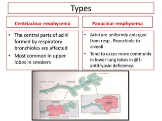
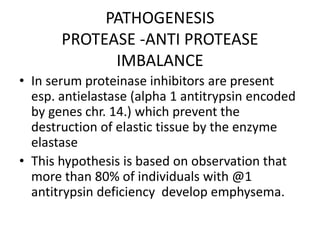
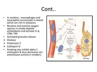
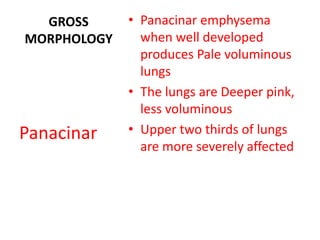
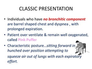
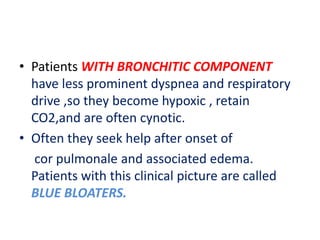
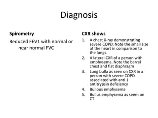
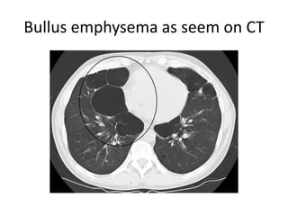
Emphysema
Presented By:
Aamir Sharif
M.Phil Human Pathology & Microbiology
SMC
Definition
• Emphysema is abnormal permanent
enlargement of air spaces distal to terminal
bronchioles accompanied by destruction of
their walls without obvious fibrosis
• Emphysema may be pure but in majority of
cases it is accompanied by chronic bronchitis
Types
Centriacinar emphysema
• The central parts of acini
formed by respiratory
bronchioles are affected
• Most common in upper
lobes in smokers
Panacinar emphysema
• Acini are uniformly enlarged
from resp . Bronchiole to
alveoli
• Tend to occur more commonly
in lower lung lobes in @1-
antitrypsin deficiency.
Types Cont..
Distal acinar emphysema
(paraseptal)
• The proximal portion of the
acinus is normal but distal part
is involoved.
• Localized along the pleura,
adjacent to perilobular septa
• More severe in upper half of
lung
• Bullae formation (multiple,
contigous, enlarged airspaces
<0.5 mm>2.0 cm cyst like
structure)
irregular emphysema
• Acinus is irregularly
involved.
• Associated with scarring
• Resulting from healed
inflammatory disaese.
• Often asymptomatic
PATHOGENESIS
PROTEASE -ANTI PROTEASE
IMBALANCE
• In serum proteinase inhibitors are present
esp. antielastase (alpha 1 antitrypsin encoded
by genes chr. 14.) which prevent the
destruction of elastic tissue by the enzyme
elastase
• This hypothesis is based on observation that
more than 80% of individuals with @1
antitrypsin deficiency develop emphysema.
Cont..
• In smokers , macrophages and
neutrophils accumulate in alveoli
which are rich in elastases.
• Nicotine and reactive oxygen
species in smoke deplete
antioxidants and activate IL-8,
LTB4, TNF
• Activated granules release
 Elastase
 Proteinase 3
 Cathepsin G
• Smoking also inhibit alpha 1
antitrypsin & thus decreases net
antielastase activity in smokers.
GROSS
MORPHOLOGY
• Panacinar emphysema
when well developed
produces Pale voluminous
lungs
• The lungs are Deeper pink,
less voluminous
• Upper two thirds of lungs
are more severely affected
Panacinar
MORPHOLOGY
• Histologically, there is thinning and destruction of
alveolar walls.
• With advanced disease adjacent alveoli become
confluent, creating large airspaces.
• Terminal and respiratory bronchioles may be
deformed bc of loss of septa.
• Small airways tend to collapse during expiration
due to loss of elastic tissue in septa which reduces
radial traction on the small airways.(an important
cause of chronic airflow obstruction in severe
emphysema)
CLASSIC PRESENTATION
• Individuals who have no bronchitic component
are barrel shaped chest and dyspnea , with
prolonged expiration.
• Patient over ventilate & remain well oxygenated,
called Pink Puffer
• Characteristic posture…sitting forward in
hunched over position attempting to
squeeze air out of lungs with each expiratory
effort.
• Patients WITH BRONCHITIC COMPONENT
have less prominent dyspnea and respiratory
drive ,so they become hypoxic , retain
CO2,and are often cynotic.
• Often they seek help after onset of
cor pulmonale and associated edema.
Patients with this clinical picture are called
BLUE BLOATERS.
Diagnosis
Spirometry
Reduced FEV1 with normal or
near normal FVC
CXR shows
1. A chest X-ray demonstrating
severe COPD. Note the small size
of the heart in comparison to
the lungs.
2. A lateral CXR of a person with
emphysema. Note the barrel
chest and flat diaphragm
3. Lung bulla as seen on CXR in a
person with severe COPD
associated with anti 1
antitrypsin deficiency
4. Bullous emphysema
5. Bullus emphysema as seem on
CT
A chest X-ray demonstrating severe COPD. Note the
small size of the heart in comparison to the lungs.
A lateral CXR of a person with emphysema. Note the
barrel chest and flat diaphragm
Lung bulla as seen on CXR in a person with severe
COPD associated with anti 1 antitrypsin deficiency
Bullous emphysema
Bullus emphysema as seem on CT

Emphysema

  • 1.
    Emphysema Presented By: Aamir Sharif M.PhilHuman Pathology & Microbiology SMC
  • 2.
    Definition • Emphysema isabnormal permanent enlargement of air spaces distal to terminal bronchioles accompanied by destruction of their walls without obvious fibrosis • Emphysema may be pure but in majority of cases it is accompanied by chronic bronchitis
  • 3.
    Types Centriacinar emphysema • Thecentral parts of acini formed by respiratory bronchioles are affected • Most common in upper lobes in smokers Panacinar emphysema • Acini are uniformly enlarged from resp . Bronchiole to alveoli • Tend to occur more commonly in lower lung lobes in @1- antitrypsin deficiency.
  • 4.
    Types Cont.. Distal acinaremphysema (paraseptal) • The proximal portion of the acinus is normal but distal part is involoved. • Localized along the pleura, adjacent to perilobular septa • More severe in upper half of lung • Bullae formation (multiple, contigous, enlarged airspaces <0.5 mm>2.0 cm cyst like structure) irregular emphysema • Acinus is irregularly involved. • Associated with scarring • Resulting from healed inflammatory disaese. • Often asymptomatic
  • 5.
    PATHOGENESIS PROTEASE -ANTI PROTEASE IMBALANCE •In serum proteinase inhibitors are present esp. antielastase (alpha 1 antitrypsin encoded by genes chr. 14.) which prevent the destruction of elastic tissue by the enzyme elastase • This hypothesis is based on observation that more than 80% of individuals with @1 antitrypsin deficiency develop emphysema.
  • 6.
    Cont.. • In smokers, macrophages and neutrophils accumulate in alveoli which are rich in elastases. • Nicotine and reactive oxygen species in smoke deplete antioxidants and activate IL-8, LTB4, TNF • Activated granules release  Elastase  Proteinase 3  Cathepsin G • Smoking also inhibit alpha 1 antitrypsin & thus decreases net antielastase activity in smokers.
  • 7.
    GROSS MORPHOLOGY • Panacinar emphysema whenwell developed produces Pale voluminous lungs • The lungs are Deeper pink, less voluminous • Upper two thirds of lungs are more severely affected Panacinar
  • 8.
    MORPHOLOGY • Histologically, thereis thinning and destruction of alveolar walls. • With advanced disease adjacent alveoli become confluent, creating large airspaces. • Terminal and respiratory bronchioles may be deformed bc of loss of septa. • Small airways tend to collapse during expiration due to loss of elastic tissue in septa which reduces radial traction on the small airways.(an important cause of chronic airflow obstruction in severe emphysema)
  • 9.
    CLASSIC PRESENTATION • Individualswho have no bronchitic component are barrel shaped chest and dyspnea , with prolonged expiration. • Patient over ventilate & remain well oxygenated, called Pink Puffer • Characteristic posture…sitting forward in hunched over position attempting to squeeze air out of lungs with each expiratory effort.
  • 10.
    • Patients WITHBRONCHITIC COMPONENT have less prominent dyspnea and respiratory drive ,so they become hypoxic , retain CO2,and are often cynotic. • Often they seek help after onset of cor pulmonale and associated edema. Patients with this clinical picture are called BLUE BLOATERS.
  • 11.
    Diagnosis Spirometry Reduced FEV1 withnormal or near normal FVC CXR shows 1. A chest X-ray demonstrating severe COPD. Note the small size of the heart in comparison to the lungs. 2. A lateral CXR of a person with emphysema. Note the barrel chest and flat diaphragm 3. Lung bulla as seen on CXR in a person with severe COPD associated with anti 1 antitrypsin deficiency 4. Bullous emphysema 5. Bullus emphysema as seem on CT
  • 12.
    A chest X-raydemonstrating severe COPD. Note the small size of the heart in comparison to the lungs.
  • 13.
    A lateral CXRof a person with emphysema. Note the barrel chest and flat diaphragm
  • 14.
    Lung bulla asseen on CXR in a person with severe COPD associated with anti 1 antitrypsin deficiency
  • 15.
  • 16.