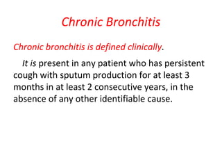
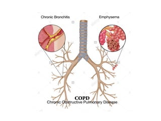
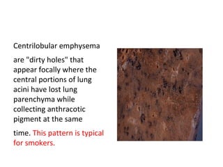
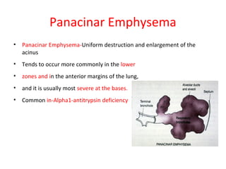
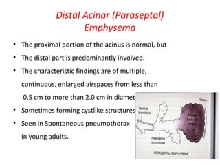
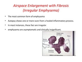
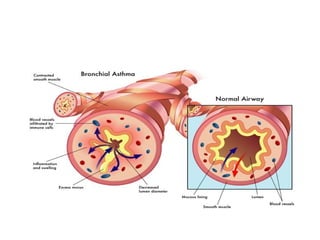
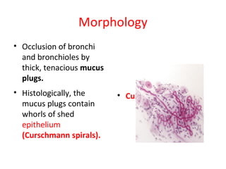
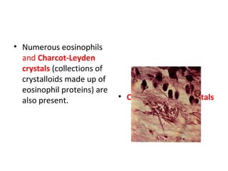
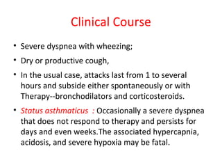
COPD
- BY SURAJ DHARA
(MMCH)
Anatomy of Tracheobronchial tree
OBSTRUCTIVE VS RESTRICTIVE
• Obstructive lung disease are characterized by
an increase in resistance to airflow due to
partial or complete obstruction at any level
from the trachea & larger bronchi to terminal
& respiratory bronchioles.
• These are contrasted with restrictive diseases,
which are characterized by reduced expansion
of lung parenchyma & decrease TLC.
Chronic Obstructive Pulmonary
Diseases (COPD)
• Emphysema
• Chronic Bronchitis
• Bronchial Asthma
• Bronchiectasis
• Bronchiolitis
Chronic Bronchitis
Chronic bronchitis is defined clinically.
It is present in any patient who has persistent
cough with sputum production for at least 3
months in at least 2 consecutive years, in the
absence of any other identifiable cause.
(1) Progress to chronic obstructive airway
disease
(2) Lead to cor pulmonale and heart failure
(3) Cause atypical metaplasia and dysplasia of
the respiratory epithelium and
(4) cancerous transformation.
Pathogenesis
• Tobacco smoke
• – 90% of patients are smokers.
• Grain, cotton, and silica dust
• Air pollution
• Infection
• – Bacterial and viral infections are important in
• triggering acute exacerbation of the disease.
• Others
• Simple chronic bronchitis: Most patients have
• The productive cough raises mucoid sputum, but
airflow is not obstructed.
• Chronic asthmatic bronchitis-chronic bronchitis may
demonstrate hyper-responsive airways with
intermittent bronchospasm and wheezing.
• Chronic obstructive bronchitis-Heavy smokers,
develops chronic outflow obstruction, usually with
evidence of associated emphysema.
• The earliest feature of chronic bronchitis is hyper secretion of mucus,
beginning in the large airways.
• Squamous metaplasia or dysplasia of bronchial epithelium.
• Histologically, in the trachea and larger bronchi there is enlargement
of the mucus-secreting glands.
• Bronchial or bronchiolar mucous plugging, inflammation and
fibrosis.
• Increased number of goblet cells and inflammatory cells in the
epithelium of peripheral airways.
• A normal Reid index is less than 0.4 and is increased in chronic
bronchitis.
• Chronic bronchitis. The lumen of
the bronchus is above. Note the
marked thickening of the mucous
gland layer (approximately twice
normal) and squamous metaplasia of
lung epithelium.
Clinical course
• Prominent cough
• Production of sputum
• (“Blue bloaters"). Hypercapnia, hypoxemia,
and cyanosis
• Pulmonary hypertension and cardiac failure
• Recurrent infections and respiratory failure
Complication
• Emphysema
• Cor pulmonale
• Bronchiectasis
• Bronchopneumonia
• Bronchogenic carcinoma of lung
Emphysema
• Emphysema is characterized by abnormal
permanent enlargement of the airspaces distal
to the terminal bronchioles, accompanied by
destruction of their walls without obvious
fibrosis.
CAUSES
• Smoking cigarettes is the most common
cause.
• Alpha 1 antitrypsin deficiency
Alpha 1 antitrypsin deficiency
• It is autosmal dominant disorder.
• It is mainly synthasized in liver.
• It is a serine protease inhibitor,
• It inhibits the proteases (particularly elastase)
secreted by neutrophils during inflammation.
• The defects is the result of alleles at the Pi
("protease-inhibitor") locus -- "M" is the
common allele,
• Homozygous Z type decreases the liver
synthesis of AAT.
• Emphysema develops at early age.
• Homozygous patients may also get cirrhosis
Pathogenesis of Emphysema
• Emphysema result from the destructive effect
of high protease activity in subjects with low
antiprotease activity.
• A consequence of two critical imbalances
• – The protease-antiprotease imbalance
• – Oxidant-antioxidant imbalance.
The chest cavity is opened at
autopsy to reveal numerous large
bullae apparent on the surface of
the lungs in a patient dying with
emphysema. Bullae are large
dilated airspaces that bulge out
from beneath the pleura.
Emphysema is characterized by
a loss of lung parenchyma by
destruction of alveoli so that
there is permanent dilation of
airspaces.
Pathogenesis of emphysema
• The protease-antiprotease imbalance and
oxidant-antioxidant imbalance are additive in
their effects and contribute to tissue damage.
α1-antitrypsin (α1-AT) deficiency can be either
congenital or
"functional" as a result of oxidative inactivation.
• Protease-Antiprotease Imbalance
Hypothesis
• 1.Genetic deficiency of the
antiprotease α1-antitrypsin
• 2.The effect of cigarette smoking in
the development of emphysema
 Increased elastase availability and
decreased antielastase.
Functional AAT deficiency
• Oxidant-Antioxidant Imbalance
• Tobacco smoke,
• Chemo tactic factor for
neutrophils and macrophages, in
turn they releases,Large number
of
Free radicals and elastases
 Inactivates AAT and Antioxidants.
Types of Emphysema
• According to its anatomic distribution within
• the lobule
• Four major types
• – Centriacinar
• – Panacinar
• – Paraseptal
• – Irregular
• Only the first two cause clinically significant
• airflow obstruction.
Centriacinar (Centrilobular)
Emphysema
• The central or proximal parts of the acini,
• Formed by respiratory bronchioles, are affected,
• Whereas distal alveoli are spared.
• The lesions are more common and
• Severe in the upper lobes,
• Particularly in the apical segments.
• Centriacinar emphysema occurs
• Predominantly in heavy smokers,
• Often in association with
chronic bronchitis.
Centrilobular emphysema
are "dirty holes" that
appear focally where the
central portions of lung
acini have lost lung
parenchyma while
collecting anthracotic
pigment at the same
time. This pattern is typical
for smokers.
Panacinar Emphysema
• Panacinar Emphysema-Uniform destruction and enlargement of the
acinus
• Tends to occur more commonly in the lower
• zones and in the anterior margins of the lung,
• and it is usually most severe at the bases.
• Common in-Alpha1-antitrypsin deficiency
Distal Acinar (Paraseptal)
Emphysema
• The proximal portion of the acinus is normal, but
• The distal part is predominantly involved.
• The characteristic findings are of multiple,
continuous, enlarged airspaces from less than
0.5 cm to more than 2.0 cm in diameter,
• Sometimes forming cystlike structures.
• Seen in Spontaneous pneumothorax
in young adults.
Airspace Enlargement with Fibrosis
(Irregular Emphysema)
• The most common form of emphysema
• Autopsy shows one or more scars from a healed inflammatory process.
• In most instances, these foci are irregular
• emphysema are asymptomatic and clinically insignificant.
Interstitial Emphysema
The entrance of air into the connective tissue stroma of
the lung, mediastinum, or subcutaneous tissue.
• Increase in intra-alveolar pressure (as with vomiting or
violent coughing) that causes a tear, with dissection of air
into the interstitium.
• A perforating injury (e.g., a fractured rib).
• Children with whooping cough and bronchitis,
• Patients on respirators who have partial obstruction to
the airways.
Compensatory Hyperinflation
(Emphysema)
• Surgical removal of a diseased lung or lobe.
• Designate dilation of alveoli but not
destruction of septal walls.
Bullous Emphysema
• Produces large
subpleural blebs or
bullae
• Spaces more than 1 cm
in diameter in the
distended state
• Most often subpleural,
and occur near the apex
• Pneumothorax
Bullous emphysema with large apical and
subpleural bullae.
Morphology
The diagnosis and classification of emphysema depend largely on
the macroscopic appearance of the lung.
 Panacinar emphysema
Produces pale, voluminous lungs that often obscure
the heart when the anterior chest wall is removed at
autopsy.
 Centriacinar emphysema
The upper two-thirds of the lungs is more severely
affected than the lower lungs and more pinkish and less
voluminous.
Microscopy
 Thinning and destruction of alveolar walls
 Adjacent alveoli become confluent, creating
large airspaces
 Loss of elastic tissue in the surrounding alveolar
septa
 The number of alveolar capillaries is diminished.
• Microscopically at high
magnification, the loss
of alveolar walls with
emphysema is
demonstrated.
• Remaining airspaces are
dilated.
• Pulmonary emphysema.
There is marked
enlargement of
airspaces, with thinning
and destruction of
alveolar septa.
Clinical features
• Inspection-barrel-chest,
• Dyspnea is usually the first symptom
• Prolonged expiration, sitting forward in a hunched-over position,
attempting to squeeze the air out of the lungs
• Because of prominent dyspnea and adequate oxygenation of
hemoglobin, these patients are sometimes called “Pink puffers.”
• Steadily progressive,
• Cough and wheezing,
• Weight loss,
• Pulmonary function tests, FVC is reduced and ratio of FEV1 to FVC is
reduced.
Complications
• Secondary pulmonary hypertension develops
gradually.
• Cor pulmonale
• Pneumothorax
• Respiratory failure. (respiratory acidosis,
hypoxia, and coma.)
• Anatomical distribution of pure chronic bronchitis and pure emphysema.
• In chronic bronchitis the small-airway disease (chronic bronchiolitis) results in airflow obstruction,
while the large-airway disease is primarily responsible for the mucus hyper secretion.
Bronchiectasis
• Bronchiectasis is the permanent dilation of
bronchi and bronchioles caused by
destruction of the muscle and elastic
supporting tissue, resulting from or associated
with chronic necrotizing infections.
• It is not a primary disease,
• It is secondary to persisting infection or
obstruction.
• The conditions that leads to bronchiactasis are,
• Bronchial obstruction causes
• Tumors
• Foreign bodies
• Occasionally impaction of mucus.
• In cystic fibrosis both obstruction and infection
accounts for bronchiectasis.
• In immunodeficiency states, increased susceptibility
to infections.
• Necrotizing, or suppurative, pneumonia, particularly
with virulent organisms such as Staphylococcus
aureus or Klebsiella species.
• Post-infective bronchiectasis was sometimes in
childhood pneumonias.
• Post-tubercular bronchiectasis continues to be a
significant cause of morbidity in endemic areas.
• Bronchiectasis is likely to develop because of an increased
susceptibility to repeated bacterial infections,
• Kartagener syndrome,
• It is an autosomal recessive disorder, is frequently
associated with
 Bronchiectasis,
 Sinusitis and
 Situs inversus
 Sterility in males.
Pathogenesis
• Two processes in the pathogenesis of
bronchiectasis:
1. Obstruction and
2. Chronic persistent infection.
Morphology
• Bronchiectatic involvement of the lungs
usually affects the lower lobes bilaterally,
particularly those air passages that are most
vertical,
• Foreign bodies, or tumors localized to a single
segment of the lung.
• The airways dilated as four times their usual
diameter.
• Intense acute and chronic inflammatory
exudate within the walls of the bronchi and
bronchioles - extensive areas of ulceration.
Culture - Staphylococci, Streptococci,
Pneumococci, enteric organisms, anaerobic
and microaerophilic bacteria,
• Children - Haemophilus influenzae and
Pseudomonas aeruginosa.
• Healing - the lining epithelium may regenerate
completely;
• Fibrosis of the bronchial and bronchiolar walls
and peribronchiolar fibrosis develop in
chronic cases.
• Might lead to lung abscess.
clinical manifestations
• Severe, persistent cough with expectoration of
mucopurulent, sometimes fetid, sputum.
• The sputum may contain flecks of blood;
• Frank hemoptysis can occur.
• Symptoms are often episodic and are precipitated by
upper respiratory tract infections.
• Clubbing of the fingers
• Pulmonary hypertension & (rarely) cor pulmonale.
• Metastatic brain abscesses.
BRONCHIAL ASTHMA
• Asthma is a chronic inflammatory disorder of the
airways that causes recurrent episodes of wheezing,
breathlessness, chest tightness, and cough,
particularly at night and/or early in the morning.
• It is a triad of intermittent and reversible airway
obstruction, chronic bronchial inflammation with
eosinophils, and bronchial smooth muscle cell
hypertrophy and hyperreactivity.
• Atopic ( extrinsic) asthma and
• 70% of cases are of atopic type.
• IgE and TH2-mediated immune responses to
environmental antigens.
• Non atopic (Intrinsic )asthma.
• Aspirin
• Pulmonary infections, especially those caused by
viruses;
• Cold
• Psychological stress
• Exercise
• Inhaled irritants
• Drug induced asthma …aspirin
• Occupational asthma :-
Exposure to fumes, organic chemical dust and
plastics.
Pathogenesis
• Type I hypersensitivity ("atopy"),
• Acute and chronic airway inflammation, and bronchial hyper-
responsiveness to a variety of stimuli.
• Inflammatory mediators: type 2 helper T (TH2)
• IL-4 : IgE production,
• IL-5 : Activates eosinophils,
• IL-13 : mucus production.
• Epithelial cells are activated to produce chemokines that
promote recruitment of more TH2 cells and eosinophils, other
leukocytes, and triggers the inflammation.
• Leukotrienes C4, D4, and E4: extremely potent
mediators that cause prolonged
bronchoconstriction, increase vascular
permeability, and increase mucin secretion.
• Platelet-activating factor: causes aggregation of
platelets and release of histamine from their
granules.
• Histamine: causes bronchospasm and increases
vascular permeability,
Type - 1 hypersensitivity
Morphology
• Occlusion of bronchi
and bronchioles by
thick, tenacious mucus
plugs.
• Histologically, the
mucus plugs contain
whorls of shed
epithelium
(Curschmann spirals).
• Curschmann spirals
• Numerous eosinophils
and Charcot-Leyden
crystals (collections of
crystalloids made up of
eosinophil proteins) are
also present. • Charcot-Leyden crystals
Morphology
• Airway remodeling -Thickening of the basement
membrane of the bronchial epithelium.
• Edema and an inflammatory infiltrate in the
bronchial walls, - eosinophils and mast cells.
• An increase in the size of the submucosal
glands.
• Hypertrophy of the bronchial muscle walls.
Clinical Course
• Severe dyspnea with wheezing;
• Dry or productive cough,
• In the usual case, attacks last from 1 to several
hours and subside either spontaneously or with
Therapy--bronchodilators and corticosteroids.
• Status asthmaticus : Occasionally a severe dyspnea
that does not respond to therapy and persists for
days and even weeks.The associated hypercapnia,
acidosis, and severe hypoxia may be fatal.

CHRONIC OBSTRUCTIVE PULMONARY DISEASES (COPD)

  • 1.
    COPD - BY SURAJDHARA (MMCH)
  • 2.
  • 3.
    OBSTRUCTIVE VS RESTRICTIVE •Obstructive lung disease are characterized by an increase in resistance to airflow due to partial or complete obstruction at any level from the trachea & larger bronchi to terminal & respiratory bronchioles. • These are contrasted with restrictive diseases, which are characterized by reduced expansion of lung parenchyma & decrease TLC.
  • 7.
    Chronic Obstructive Pulmonary Diseases(COPD) • Emphysema • Chronic Bronchitis • Bronchial Asthma • Bronchiectasis • Bronchiolitis
  • 9.
    Chronic Bronchitis Chronic bronchitisis defined clinically. It is present in any patient who has persistent cough with sputum production for at least 3 months in at least 2 consecutive years, in the absence of any other identifiable cause.
  • 11.
    (1) Progress tochronic obstructive airway disease (2) Lead to cor pulmonale and heart failure (3) Cause atypical metaplasia and dysplasia of the respiratory epithelium and (4) cancerous transformation.
  • 12.
    Pathogenesis • Tobacco smoke •– 90% of patients are smokers. • Grain, cotton, and silica dust • Air pollution • Infection • – Bacterial and viral infections are important in • triggering acute exacerbation of the disease. • Others
  • 13.
    • Simple chronicbronchitis: Most patients have • The productive cough raises mucoid sputum, but airflow is not obstructed. • Chronic asthmatic bronchitis-chronic bronchitis may demonstrate hyper-responsive airways with intermittent bronchospasm and wheezing. • Chronic obstructive bronchitis-Heavy smokers, develops chronic outflow obstruction, usually with evidence of associated emphysema.
  • 14.
    • The earliestfeature of chronic bronchitis is hyper secretion of mucus, beginning in the large airways. • Squamous metaplasia or dysplasia of bronchial epithelium. • Histologically, in the trachea and larger bronchi there is enlargement of the mucus-secreting glands. • Bronchial or bronchiolar mucous plugging, inflammation and fibrosis. • Increased number of goblet cells and inflammatory cells in the epithelium of peripheral airways. • A normal Reid index is less than 0.4 and is increased in chronic bronchitis.
  • 15.
    • Chronic bronchitis.The lumen of the bronchus is above. Note the marked thickening of the mucous gland layer (approximately twice normal) and squamous metaplasia of lung epithelium.
  • 16.
    Clinical course • Prominentcough • Production of sputum • (“Blue bloaters"). Hypercapnia, hypoxemia, and cyanosis • Pulmonary hypertension and cardiac failure • Recurrent infections and respiratory failure
  • 17.
    Complication • Emphysema • Corpulmonale • Bronchiectasis • Bronchopneumonia • Bronchogenic carcinoma of lung
  • 18.
    Emphysema • Emphysema ischaracterized by abnormal permanent enlargement of the airspaces distal to the terminal bronchioles, accompanied by destruction of their walls without obvious fibrosis.
  • 20.
    CAUSES • Smoking cigarettesis the most common cause. • Alpha 1 antitrypsin deficiency
  • 21.
    Alpha 1 antitrypsindeficiency • It is autosmal dominant disorder. • It is mainly synthasized in liver. • It is a serine protease inhibitor, • It inhibits the proteases (particularly elastase) secreted by neutrophils during inflammation.
  • 22.
    • The defectsis the result of alleles at the Pi ("protease-inhibitor") locus -- "M" is the common allele, • Homozygous Z type decreases the liver synthesis of AAT. • Emphysema develops at early age. • Homozygous patients may also get cirrhosis
  • 23.
    Pathogenesis of Emphysema •Emphysema result from the destructive effect of high protease activity in subjects with low antiprotease activity. • A consequence of two critical imbalances • – The protease-antiprotease imbalance • – Oxidant-antioxidant imbalance.
  • 24.
    The chest cavityis opened at autopsy to reveal numerous large bullae apparent on the surface of the lungs in a patient dying with emphysema. Bullae are large dilated airspaces that bulge out from beneath the pleura. Emphysema is characterized by a loss of lung parenchyma by destruction of alveoli so that there is permanent dilation of airspaces.
  • 26.
    Pathogenesis of emphysema •The protease-antiprotease imbalance and oxidant-antioxidant imbalance are additive in their effects and contribute to tissue damage. α1-antitrypsin (α1-AT) deficiency can be either congenital or "functional" as a result of oxidative inactivation.
  • 27.
    • Protease-Antiprotease Imbalance Hypothesis •1.Genetic deficiency of the antiprotease α1-antitrypsin • 2.The effect of cigarette smoking in the development of emphysema  Increased elastase availability and decreased antielastase. Functional AAT deficiency • Oxidant-Antioxidant Imbalance • Tobacco smoke, • Chemo tactic factor for neutrophils and macrophages, in turn they releases,Large number of Free radicals and elastases  Inactivates AAT and Antioxidants.
  • 28.
    Types of Emphysema •According to its anatomic distribution within • the lobule • Four major types • – Centriacinar • – Panacinar • – Paraseptal • – Irregular • Only the first two cause clinically significant • airflow obstruction.
  • 29.
    Centriacinar (Centrilobular) Emphysema • Thecentral or proximal parts of the acini, • Formed by respiratory bronchioles, are affected, • Whereas distal alveoli are spared. • The lesions are more common and • Severe in the upper lobes, • Particularly in the apical segments. • Centriacinar emphysema occurs • Predominantly in heavy smokers, • Often in association with chronic bronchitis.
  • 30.
    Centrilobular emphysema are "dirtyholes" that appear focally where the central portions of lung acini have lost lung parenchyma while collecting anthracotic pigment at the same time. This pattern is typical for smokers.
  • 31.
    Panacinar Emphysema • PanacinarEmphysema-Uniform destruction and enlargement of the acinus • Tends to occur more commonly in the lower • zones and in the anterior margins of the lung, • and it is usually most severe at the bases. • Common in-Alpha1-antitrypsin deficiency
  • 32.
    Distal Acinar (Paraseptal) Emphysema •The proximal portion of the acinus is normal, but • The distal part is predominantly involved. • The characteristic findings are of multiple, continuous, enlarged airspaces from less than 0.5 cm to more than 2.0 cm in diameter, • Sometimes forming cystlike structures. • Seen in Spontaneous pneumothorax in young adults.
  • 33.
    Airspace Enlargement withFibrosis (Irregular Emphysema) • The most common form of emphysema • Autopsy shows one or more scars from a healed inflammatory process. • In most instances, these foci are irregular • emphysema are asymptomatic and clinically insignificant.
  • 34.
    Interstitial Emphysema The entranceof air into the connective tissue stroma of the lung, mediastinum, or subcutaneous tissue. • Increase in intra-alveolar pressure (as with vomiting or violent coughing) that causes a tear, with dissection of air into the interstitium. • A perforating injury (e.g., a fractured rib). • Children with whooping cough and bronchitis, • Patients on respirators who have partial obstruction to the airways.
  • 35.
    Compensatory Hyperinflation (Emphysema) • Surgicalremoval of a diseased lung or lobe. • Designate dilation of alveoli but not destruction of septal walls.
  • 36.
    Bullous Emphysema • Produceslarge subpleural blebs or bullae • Spaces more than 1 cm in diameter in the distended state • Most often subpleural, and occur near the apex • Pneumothorax
  • 37.
    Bullous emphysema withlarge apical and subpleural bullae.
  • 38.
    Morphology The diagnosis andclassification of emphysema depend largely on the macroscopic appearance of the lung.  Panacinar emphysema Produces pale, voluminous lungs that often obscure the heart when the anterior chest wall is removed at autopsy.  Centriacinar emphysema The upper two-thirds of the lungs is more severely affected than the lower lungs and more pinkish and less voluminous.
  • 39.
    Microscopy  Thinning anddestruction of alveolar walls  Adjacent alveoli become confluent, creating large airspaces  Loss of elastic tissue in the surrounding alveolar septa  The number of alveolar capillaries is diminished.
  • 40.
    • Microscopically athigh magnification, the loss of alveolar walls with emphysema is demonstrated. • Remaining airspaces are dilated.
  • 41.
    • Pulmonary emphysema. Thereis marked enlargement of airspaces, with thinning and destruction of alveolar septa.
  • 42.
    Clinical features • Inspection-barrel-chest, •Dyspnea is usually the first symptom • Prolonged expiration, sitting forward in a hunched-over position, attempting to squeeze the air out of the lungs • Because of prominent dyspnea and adequate oxygenation of hemoglobin, these patients are sometimes called “Pink puffers.” • Steadily progressive, • Cough and wheezing, • Weight loss, • Pulmonary function tests, FVC is reduced and ratio of FEV1 to FVC is reduced.
  • 43.
    Complications • Secondary pulmonaryhypertension develops gradually. • Cor pulmonale • Pneumothorax • Respiratory failure. (respiratory acidosis, hypoxia, and coma.)
  • 44.
    • Anatomical distributionof pure chronic bronchitis and pure emphysema. • In chronic bronchitis the small-airway disease (chronic bronchiolitis) results in airflow obstruction, while the large-airway disease is primarily responsible for the mucus hyper secretion.
  • 45.
    Bronchiectasis • Bronchiectasis isthe permanent dilation of bronchi and bronchioles caused by destruction of the muscle and elastic supporting tissue, resulting from or associated with chronic necrotizing infections. • It is not a primary disease, • It is secondary to persisting infection or obstruction.
  • 46.
    • The conditionsthat leads to bronchiactasis are, • Bronchial obstruction causes • Tumors • Foreign bodies • Occasionally impaction of mucus. • In cystic fibrosis both obstruction and infection accounts for bronchiectasis.
  • 47.
    • In immunodeficiencystates, increased susceptibility to infections. • Necrotizing, or suppurative, pneumonia, particularly with virulent organisms such as Staphylococcus aureus or Klebsiella species. • Post-infective bronchiectasis was sometimes in childhood pneumonias. • Post-tubercular bronchiectasis continues to be a significant cause of morbidity in endemic areas.
  • 48.
    • Bronchiectasis islikely to develop because of an increased susceptibility to repeated bacterial infections, • Kartagener syndrome, • It is an autosomal recessive disorder, is frequently associated with  Bronchiectasis,  Sinusitis and  Situs inversus  Sterility in males.
  • 49.
    Pathogenesis • Two processesin the pathogenesis of bronchiectasis: 1. Obstruction and 2. Chronic persistent infection.
  • 50.
    Morphology • Bronchiectatic involvementof the lungs usually affects the lower lobes bilaterally, particularly those air passages that are most vertical, • Foreign bodies, or tumors localized to a single segment of the lung. • The airways dilated as four times their usual diameter.
  • 51.
    • Intense acuteand chronic inflammatory exudate within the walls of the bronchi and bronchioles - extensive areas of ulceration. Culture - Staphylococci, Streptococci, Pneumococci, enteric organisms, anaerobic and microaerophilic bacteria, • Children - Haemophilus influenzae and Pseudomonas aeruginosa.
  • 52.
    • Healing -the lining epithelium may regenerate completely; • Fibrosis of the bronchial and bronchiolar walls and peribronchiolar fibrosis develop in chronic cases. • Might lead to lung abscess.
  • 53.
    clinical manifestations • Severe,persistent cough with expectoration of mucopurulent, sometimes fetid, sputum. • The sputum may contain flecks of blood; • Frank hemoptysis can occur. • Symptoms are often episodic and are precipitated by upper respiratory tract infections. • Clubbing of the fingers • Pulmonary hypertension & (rarely) cor pulmonale. • Metastatic brain abscesses.
  • 54.
    BRONCHIAL ASTHMA • Asthmais a chronic inflammatory disorder of the airways that causes recurrent episodes of wheezing, breathlessness, chest tightness, and cough, particularly at night and/or early in the morning. • It is a triad of intermittent and reversible airway obstruction, chronic bronchial inflammation with eosinophils, and bronchial smooth muscle cell hypertrophy and hyperreactivity.
  • 56.
    • Atopic (extrinsic) asthma and • 70% of cases are of atopic type. • IgE and TH2-mediated immune responses to environmental antigens.
  • 57.
    • Non atopic(Intrinsic )asthma. • Aspirin • Pulmonary infections, especially those caused by viruses; • Cold • Psychological stress • Exercise • Inhaled irritants
  • 58.
    • Drug inducedasthma …aspirin • Occupational asthma :- Exposure to fumes, organic chemical dust and plastics.
  • 59.
    Pathogenesis • Type Ihypersensitivity ("atopy"), • Acute and chronic airway inflammation, and bronchial hyper- responsiveness to a variety of stimuli. • Inflammatory mediators: type 2 helper T (TH2) • IL-4 : IgE production, • IL-5 : Activates eosinophils, • IL-13 : mucus production. • Epithelial cells are activated to produce chemokines that promote recruitment of more TH2 cells and eosinophils, other leukocytes, and triggers the inflammation.
  • 60.
    • Leukotrienes C4,D4, and E4: extremely potent mediators that cause prolonged bronchoconstriction, increase vascular permeability, and increase mucin secretion. • Platelet-activating factor: causes aggregation of platelets and release of histamine from their granules. • Histamine: causes bronchospasm and increases vascular permeability,
  • 61.
    Type - 1hypersensitivity
  • 63.
    Morphology • Occlusion ofbronchi and bronchioles by thick, tenacious mucus plugs. • Histologically, the mucus plugs contain whorls of shed epithelium (Curschmann spirals). • Curschmann spirals
  • 64.
    • Numerous eosinophils andCharcot-Leyden crystals (collections of crystalloids made up of eosinophil proteins) are also present. • Charcot-Leyden crystals
  • 65.
    Morphology • Airway remodeling-Thickening of the basement membrane of the bronchial epithelium. • Edema and an inflammatory infiltrate in the bronchial walls, - eosinophils and mast cells. • An increase in the size of the submucosal glands. • Hypertrophy of the bronchial muscle walls.
  • 67.
    Clinical Course • Severedyspnea with wheezing; • Dry or productive cough, • In the usual case, attacks last from 1 to several hours and subside either spontaneously or with Therapy--bronchodilators and corticosteroids. • Status asthmaticus : Occasionally a severe dyspnea that does not respond to therapy and persists for days and even weeks.The associated hypercapnia, acidosis, and severe hypoxia may be fatal.