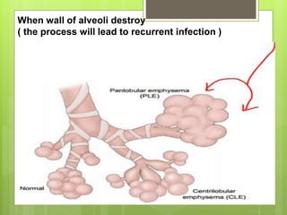
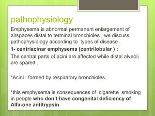
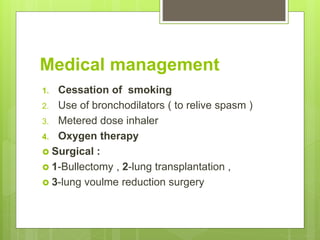
Emphysema
Prepared by :
akeel abd muslem
Content
Definition
causes
Types
Pathophysiology
Clinical manifestation (S&S)
Complication
Medical management
Nursing management
emphysema
 In emphysema: Damage and abnormal distention
of airspaces beyond the terminal bronchioles and
obstruction of walls of alveoli.
 there’s impaired carbon dioxide and oxygen
exchange , result from obstruction of walls of over
_ distended Alveoli ( enlargement of airspaces }
alveoli { .
 Destroyed by recurrent infection .
When wall of alveoli destroy
( the process will lead to recurrent infection )
Causes
1. Cigarrete smoking –or second hand smoking
2. Damaged bronchi tubes or lungs , or it maybe inherited .
3. Breathing in a-air pollution or b- toxic air
types
1- centrilobular emphysema
2- panlobular emphysema
3- distal acinar (paraseptal ) emphysema
4-Irregular emphysema
pathophysiology
Emphysema is abnormal permanent enlargement of
airspaces distal to terminal bronchioles , we discuss
pathophysiology according to types of disease .
1- centriacinar emphysema (centrilobular ) :
The central parts of acini are affected while distal alveoli
are spared .
*Acini : formed by respiratory bronchioles .
*this emphysema is consequences of cigarette smoking
in people who don’t have congenital deficiency of
Alfa-one antitrypsin
2- panlobular emphysema
Acini are uniformly enlarged from the level of
respiratory bronchioles to the terminal blind
alveoli .
*Occur commonly in lower lung zone .
* And occur in people with alfa- one
antitrypsin deficiency .
3- distal acinar (paraseptal )
emphysema
 Distal parts of acinus is involved ( while proximal
portion is normal )
 It’s more striking adjacent to pleura , along lobular
connective tissue septa , and at margins of lobules.
 The characteristic findings are the presence of multiple ,
continues enlargement of airspaces ( range in daimeter
from less than 0.5 mm to more than 2 cm , sometimes
form a cyst like structure called bullae .
4-Irregular emphysema
 Mixed and unclassified .
 The acini irregularly involved .
 Asymptomatic
 Usually as a complication from TB (
Tuberculosis ) or other various chronic
inflammatory disease .
 Healthy lung tissue produce substance called (alfa antitrypsin
)
 1-in response to infection : neutrophils and macrophages
release proteases enzymes (destructive enzymes )
 2- smoking reduce activity of alfa antitrypsin and increase
activity of proteases enzymes .
*Proteases destroy elastic fibers that act for elastic recoli ,
that needed in exhalation .
* As a result alveoli are over inflated as air becomes trapped
in lung , and increase air volume pushes diaphragm down
ward and disrupting chest shape and make breathing difficulty
.
* And finally , result in reduces lung tissue available to
external respiration , (and put patient at risk for hypoxemia
and hypoxia )
Pathogenesis of emphysema
Clinical manifestation
1-S.O.B (dyspnea)
2-cough
3-difficultybreathingwhile exertion
4-underweight
5-barrel chest
6-wheezing
7-morning headache
8-difficultysleeping
9-difficultyconcentration
10 sputum production
complication
1. Respiratory insufficiency or failure
2. Pneumonia
3. Pneumothorax
4. Chronic atelectasis
5. Pulmonary arterial hypertension (cor-
pulmonale) that lead to
RIGHT SIDED HEART FAIULURE
Medical management
1. Cessation of smoking
2. Use of bronchodilators ( to relive spasm )
3. Metered dose inhaler
4. Oxygen therapy
 Surgical :
 1-Bullectomy , 2-lung transplantation ,
 3-lung voulme reduction surgery
Nursing management
1. Adminster low flow oxygen as needed
2. Check vital signs
3. Ascultate lung sounds
4. Give medication as ordered
5. Provide fowler position
6. Assess skin color and temperature
7. Assess respiratory condition
8. And assess pulse oximetry
 Pulse oximetry : is a test used to measure the oxygen
level (oxygen saturation) of the blo
Reference
 Brunner book medical surgical nursing
 Slideshare.com
 www.google .com

Emphysema

  • 1.
  • 2.
  • 3.
    emphysema  In emphysema:Damage and abnormal distention of airspaces beyond the terminal bronchioles and obstruction of walls of alveoli.  there’s impaired carbon dioxide and oxygen exchange , result from obstruction of walls of over _ distended Alveoli ( enlargement of airspaces } alveoli { .  Destroyed by recurrent infection .
  • 4.
    When wall ofalveoli destroy ( the process will lead to recurrent infection )
  • 5.
    Causes 1. Cigarrete smoking–or second hand smoking 2. Damaged bronchi tubes or lungs , or it maybe inherited . 3. Breathing in a-air pollution or b- toxic air
  • 6.
    types 1- centrilobular emphysema 2-panlobular emphysema 3- distal acinar (paraseptal ) emphysema 4-Irregular emphysema
  • 7.
    pathophysiology Emphysema is abnormalpermanent enlargement of airspaces distal to terminal bronchioles , we discuss pathophysiology according to types of disease . 1- centriacinar emphysema (centrilobular ) : The central parts of acini are affected while distal alveoli are spared . *Acini : formed by respiratory bronchioles . *this emphysema is consequences of cigarette smoking in people who don’t have congenital deficiency of Alfa-one antitrypsin
  • 9.
    2- panlobular emphysema Aciniare uniformly enlarged from the level of respiratory bronchioles to the terminal blind alveoli . *Occur commonly in lower lung zone . * And occur in people with alfa- one antitrypsin deficiency .
  • 10.
    3- distal acinar(paraseptal ) emphysema  Distal parts of acinus is involved ( while proximal portion is normal )  It’s more striking adjacent to pleura , along lobular connective tissue septa , and at margins of lobules.  The characteristic findings are the presence of multiple , continues enlargement of airspaces ( range in daimeter from less than 0.5 mm to more than 2 cm , sometimes form a cyst like structure called bullae .
  • 11.
    4-Irregular emphysema  Mixedand unclassified .  The acini irregularly involved .  Asymptomatic  Usually as a complication from TB ( Tuberculosis ) or other various chronic inflammatory disease .
  • 12.
     Healthy lungtissue produce substance called (alfa antitrypsin )  1-in response to infection : neutrophils and macrophages release proteases enzymes (destructive enzymes )  2- smoking reduce activity of alfa antitrypsin and increase activity of proteases enzymes . *Proteases destroy elastic fibers that act for elastic recoli , that needed in exhalation . * As a result alveoli are over inflated as air becomes trapped in lung , and increase air volume pushes diaphragm down ward and disrupting chest shape and make breathing difficulty . * And finally , result in reduces lung tissue available to external respiration , (and put patient at risk for hypoxemia and hypoxia )
  • 13.
  • 14.
    Clinical manifestation 1-S.O.B (dyspnea) 2-cough 3-difficultybreathingwhileexertion 4-underweight 5-barrel chest 6-wheezing 7-morning headache 8-difficultysleeping 9-difficultyconcentration 10 sputum production
  • 15.
    complication 1. Respiratory insufficiencyor failure 2. Pneumonia 3. Pneumothorax 4. Chronic atelectasis 5. Pulmonary arterial hypertension (cor- pulmonale) that lead to RIGHT SIDED HEART FAIULURE
  • 16.
    Medical management 1. Cessationof smoking 2. Use of bronchodilators ( to relive spasm ) 3. Metered dose inhaler 4. Oxygen therapy  Surgical :  1-Bullectomy , 2-lung transplantation ,  3-lung voulme reduction surgery
  • 18.
    Nursing management 1. Adminsterlow flow oxygen as needed 2. Check vital signs 3. Ascultate lung sounds 4. Give medication as ordered 5. Provide fowler position 6. Assess skin color and temperature 7. Assess respiratory condition 8. And assess pulse oximetry  Pulse oximetry : is a test used to measure the oxygen level (oxygen saturation) of the blo
  • 19.
    Reference  Brunner bookmedical surgical nursing  Slideshare.com  www.google .com