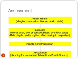
This document provides information on disorders of the respiratory system. It begins by describing the anatomy and functions of the respiratory system, including the conducting airways. It then discusses various upper and lower respiratory tract disorders like tonsillitis, pharyngitis, laryngitis, sinusitis, acute tracheo-bronchitis, and chronic bronchitis. For each disorder, it provides information on definition, causes, signs and symptoms, management, and nursing interventions. The document concludes with describing assessment techniques for respiratory disorders.
TUBERCULOSIS HAS BEEN EXCLUDED BECAUSE IN INDIA TUBERCULOSIS IS THE MOST COMMON CAUSE OF CHRONIC COUGH AND REST OTHER CAUSES OF CHRONIC COUGHS ARE IGNORED
Can AI do good? at 'offtheCanvas' India HCI preludeAlan Dix
Invited talk at 'offtheCanvas' IndiaHCI prelude, 29th June 2024.
https://www.alandix.com/academic/talks/offtheCanvas-IndiaHCI2024/
The world is being changed fundamentally by AI and we are constantly faced with newspaper headlines about its harmful effects. However, there is also the potential to both ameliorate theses harms and use the new abilities of AI to transform society for the good. Can you make the difference?
Hello everyone! I am thrilled to present my latest portfolio on LinkedIn, marking the culmination of my architectural journey thus far. Over the span of five years, I've been fortunate to acquire a wealth of knowledge under the guidance of esteemed professors and industry mentors. From rigorous academic pursuits to practical engagements, each experience has contributed to my growth and refinement as an architecture student. This portfolio not only showcases my projects but also underscores my attention to detail and to innovative architecture as a profession.
Dive into the innovative world of smart garages with our insightful presentation, "Exploring the Future of Smart Garages." This comprehensive guide covers the latest advancements in garage technology, including automated systems, smart security features, energy efficiency solutions, and seamless integration with smart home ecosystems. Learn how these technologies are transforming traditional garages into high-tech, efficient spaces that enhance convenience, safety, and sustainability.
Ideal for homeowners, tech enthusiasts, and industry professionals, this presentation provides valuable insights into the trends, benefits, and future developments in smart garage technology. Stay ahead of the curve with our expert analysis and practical tips on implementing smart garage solutions.
Expert Accessory Dwelling Unit (ADU) Drafting ServicesResDraft
Whether you’re looking to create a guest house, a rental unit, or a private retreat, our experienced team will design a space that complements your existing home and maximizes your investment. We provide personalized, comprehensive expert accessory dwelling unit (ADU)drafting solutions tailored to your needs, ensuring a seamless process from concept to completion.
Transforming Brand Perception and Boosting Profitabilityaaryangarg12
In today's digital era, the dynamics of brand perception, consumer behavior, and profitability have been profoundly reshaped by the synergy of branding, social media, and website design. This research paper investigates the transformative power of these elements in influencing how individuals perceive brands and products and how this transformation can be harnessed to drive sales and profitability for businesses.
Through an exploration of brand psychology and consumer behavior, this study sheds light on the intricate ways in which effective branding strategies, strategic social media engagement, and user-centric website design contribute to altering consumers' perceptions. We delve into the principles that underlie successful brand transformations, examining how visual identity, messaging, and storytelling can captivate and resonate with target audiences.
Methodologically, this research employs a comprehensive approach, combining qualitative and quantitative analyses. Real-world case studies illustrate the impact of branding, social media campaigns, and website redesigns on consumer perception, sales figures, and profitability. We assess the various metrics, including brand awareness, customer engagement, conversion rates, and revenue growth, to measure the effectiveness of these strategies.
The results underscore the pivotal role of cohesive branding, social media influence, and website usability in shaping positive brand perceptions, influencing consumer decisions, and ultimately bolstering sales and profitability. This paper provides actionable insights and strategic recommendations for businesses seeking to leverage branding, social media, and website design as potent tools to enhance their market position and financial success.
Between Filth and Fortune- Urban Cattle Foraging Realities by Devi S Nair, An...Mansi Shah
This study examines cattle rearing in urban and rural settings, focusing on milk production and consumption. By exploring a case in Ahmedabad, it highlights the challenges and processes in dairy farming across different environments, emphasising the need for sustainable practices and the essential role of milk in daily consumption.
Between Filth and Fortune- Urban Cattle Foraging Realities by Devi S Nair, An...
Unit II. Respiratory system disorders.pptx
1. 1
Unit II. Disorders of respiratory system
By Zebenay.W (MSc in Child health)
Contact Address:
zedo2015@gmail.com
2. Respiratory System
Its primary function is delivery of oxygen to the
lungs and removal of carbon dioxide from the
lungs.
2
3. Thoracic Cavity
The inside of the chest cage is called the thoracic
cavity.
Contained within the thoracic cavity are the lungs,
cone-shaped, porous organs encased in the
pleura, a thin, transparent double-layered serous
membrane lining the thoracic cavity.
3
4. The Anatomy of the Lungs
The right lung is larger than the left and is divided
into three sections or lobes: upper, middle, and
lower.
The left lung is divided into two lobes: upper and
lower.
The upper portion of the lungs is the apex; the
lower portion is the base.
4
5. Conducting Airways
The conducting airways are tubelike structures
that provide a passageway for air as it travels to
the lungs.
The conducting airways include the nasal
passages, mouth, pharynx, larynx, trachea,
bronchi, and bronchioles.
5
6. Pharynx Larynx Trachea
The conducting airways that connect nasal passages
and mouth to the lower parts of the respiratory tract.
The passageway for air entering and leaving the
trachea and containing the vocal cords.
Commonly known as the windpipe, this tube is
composed of connective tissue mucosa and smooth
muscle supported by C-shaped rings of cartilage.
6
7. Bronchi, Bronchioles
Two tubes, the right and left primary bronchi, that each
pass into its respective lung.
Within the lungs, the bronchi branch off into
increasingly smaller diameter tubes until they become
the terminal bronchioles.
7
8. Respiration
A process of gas exchange necessary to supply
cells with oxygen for carrying on metabolism, and
to remove carbon dioxide produced as a waste
by-product.
Two types of respiration: external and internal.
8
9. External & Internal Respiration
The exchange of gases between the inhaled air
and the blood in the pulmonary capillaries.
The exchange of gases at the cellular level
between tissue cells and blood in systemic
capillaries.
9
11. Assessment
Auscultation
(Listening for Normal and Adventitious Breath Sounds)
Palpation and Percussion
Inspection
(client's color, level of consciousness, emotional state)
(Rate, depth, quality, rhythm, effort relating to respiration)
Health History
(allergies, occupation, lifestyle, health habits)
11
12. The Normal Breath Sounds
1.Vesicular breath sound that is characterized by:
Inspiratory sounds lasting longer than expiratory
ones
Soft and low pitched
No pause between expiration and inspiration
Heard through inspiration and one–third of
expiration
Normally heard over most of both lungs
12
13. 2.Bronchial Breath
sound that is characterized by:
Loud and relatively high pitched
Expiratory sounds lasting longer than inspiratory
ones
Short silent period between inspiration and
expiration
The normal location is over the manubrium if heard at
all
3.Broncho-Vesicular Breath
sounds are characterized by:
Intermediate in intensity and pitch
Inspiratory and expiratory sounds are about equal in
duration
A silent gap between inspiration and expiration may or
may not be present
13
15. Terms
Normal breathing (Eupnea): - A respiratory rate of 10- 20
breaths per minute
Tachypnea: - Increased respiratory rate over 20 breaths per
minute
Bradypnea: -Decreased respiratory rate under 10 breaths per
minute
Apnea:- Total cessation of airflow to the lungs
Hyperpnea:-Increase in depth of respiration
Hyperventilation:-Increase in depth & rate of respiration
Hypoventilation: slow or irregular respiratory pattern with
shallow respirations
Hypoxia:- Situation in which there is lack of oxygen in the
body
Hypoxemia:-Oxygen deficiency in the blood.
15
16. Terms---
Hypercapnia:-is the retention of co2 in the blood i.e. the PaCO2
> 45mmHg at rest
Chyne-stokes respiration:- A cyclic pattern of progressively
deeper respirations , followed by progressively shallow
respirations, & a period of apnea
e.g. CHF, drug over dose , renal failure
Kussmal’s respiration: - Deep, regular, breaths, usually at a
rate greater than 20 per minute. E.g. DKA, renal failure,
metabolic acidosis states.
Respiratory system:- a system in which gas exchange occurs.
(The air flows through nasal and or oral passage to the pharynx,
larynx, trachea, bronchi, bronchioles, and to the alveoli)
16
17. Risk factors for respiratory disease
Smoking - is the single most important contribution to lung
d/se
Exposure to second hand smoke
Personal or family history of lung disease
Genetic make up
Allergens and environmental pollutants
Recreational and occupational exposure
17
18. Assessment of the respiratory system
1/ HX
Demographic data
Personal & family Hx
Occupational & socioeconomic status
Common manifestations ( cough, sputum, dyspnea, chest pain
)
2/Physical examination
V/S, HEENT, Digital clubbing
Chest examinations(inspection, palpation, percussion &
auscultation)
Breathing patterns
18
21. Upper respiratory tract disorders
Tonsillitis
Definition:- It is an inflammation & enlargement of the tonsil tissue
Causative agent:- Group A beta hemolytic streptococcus
- It can be viral in origin
- It is very common up to the age of 15 years
Predisposing factors:-
URTI, lowered immunity, pollution
Clinical features:- sore throat, dysphagia, headaches, earaches, voice
change & fever, snoring
Diagnostic evaluation
History
Physical examination / Enlarged hyperemic tonsils, tender and enlarged
cervical lymph nodes/
Culture & sensitivity
21
22. Nursing interventions:
-Analgesics such as paracetamol 500mg 2 tabs PRN
- Ampicillin/amoxacillin for 5-7 days
- Tepid sponge, high fluid intake,
- Bed rest and soft diet
- Warm saline gargles
Surgical mgt :- Tonsillectomy , Tonsiloadenectomy
Indications
- Recurrent tonsillitis not responding to antibiotic
- Obstruction to the airway
- Recurrent otitis media & hearing loss r/t tonsilitis
- After resolution of peritonsillar abscess
Complications
Quinsy (Peritonsillar abscess)
Laryngeal edema
Acute otitis media, sinustis, deep neck abscess, pneumonia
Septicemia, R-fever, RHD, acute nephritis
Chronic tonsillitis
22
23. Chronic tonsillitis
Def.;-It is due to recurrent acute tonsillitis attack.
Etiology: - similar to acute tonsillitis
C/M
- Recurrent pain - Cough - Enlarged cervical lymph node
R/X
- Nutritious diet & vitamins
- Antibiotic(Pencillins) & analgesics
- Tonsillectomy
23
24. Peritonsillar abscess/ Quinsy/
It is a collection of pus outside the tonsillar capsule
- It is usually unilateral
Etiology:- streptococcus group A
Risk factors: - Age between 20 – 50 years
- Male affected more than females
- Foreign body embeded
C/M: - Pain, - swelling, - redness, trismus - salivation
- Malaise, fever, -rupture - Cervical lymphadenopathy
DDX: - acute tonsillitis , Retro-pharyngeal abscess
Complications
-Para-pharyngeal abscess ,septicemia & hemorrhage
RX :-
Antibiotic, oral hygiene,incision and drainage
24
25. Pharyngitis
Defn :- It is a febrile inflammation of the throat
Causative agent:-
- Viral organism(adenoviruses,rhinoviruses)-70%
- Group A streptococcus is the most common bacterial organism
- Haemophilua influenza,staphylococus ,pneumococus
Clinical Features
Red pharyngeal membrane & tonsil
Enlarged & tender cervical lymph nodes
Fever, malaise, & sore throat
Odynophagia & dysphagia
hoarseness,& cough
Diagnosis: - clinically (hyperemia & swelling of pharynx)
Differential diagnosis: -
- Tonsillitis - Laryngitis - Diphtheria
25
26. Pharyngitis---
Complications
- Otitis media - mastoditis - meningitis
Medical Rx: - For bacterial causes
Ampicillin 500mg po Qid for10 days or
Erythromycin 500mg p.o. QID for 10days
Nursing interventions
Instruct bed rest during febrile stage of illness.
Liquid or soft diet is provided during acute stage of the disease.
Warm saline gargles or irrigation are used. Irrigating the throat
properly is an effective means of reducing spasm in the pharyngeal
muscle.
Acetaminophen 500mg 2 tabs at 6 hour interval.
Mouth care can be given.
26
27. Laryngitis
It is an inflammation of the larynx often occurs as a result
of voice abuse, exposure to dust, chemicals, smoke, &
other pollutant.
Etiology: - Almost always virus - Bacterial invasion may
be secondary.
Risk factors: - URTI
Vocal misuse and over use , Irritation / smoking, alcohol /
Seasonal changes
Iatrogenic / intubations and endo-laryngeal surgery /
Laryngitis is usually associated with acute rhinitis or
naso-pharyngitis. It is common in winter & is easily
transmitted.
27
28. Laryngitis---
C/F:- Hoarseness or complete loss of voice (aphonia)&
severe cough
Pain occurs in severe cases
Stridor may be present in children
Edema, exudates, congestion
Laryngitis can be the complication of chronic sinusitis & chronic
bronchitis.
Management
Resting the voice, avoid smoking, bed rest
Inhaling cool steam or aerosol
Treat secondary bacterial infection with antibiotics
28
30. Sinusitis
Is an inflammation of one or more sinuses. It occurs when infected
fluid/pus accumulates in the sinus
Causes
Allergy or virus
Bacteria (strep. Pneumoniae ,H.influenza,staphy,etc)
Maxillary & frontal sinuses are more affected
Predisposing factors
Nasal polyps & tumors
Deviated nasal septum
Facial trauma, dental infections
Chronically inhaled pollutants
30
31. Sinusitis---
S&sxs
Pain and sensation of pressure over the face
Tenderness
Nasal swelling and congestion
Ear pain and swelling & headache
Low grade fever
Purulent/bloody nasal discharge
Cough may be present
DX
HX & P/E-tenderness on percussion over the affected sinus
Paranasal sinus X-ray- Opacification & fluid
Coronal CT scanning/MRI- in chronic cases
31
32. Sinusitis---
Complications
Cellulitis
Osteomyelitis & bone destruction of the facial bones
Meningitis, brain abscess or epidural abscess
Ischemic infarction
Treatment & nursing interventions
Nasal decongestant (Oxymetazoline 0.05 %)
Anti allergic drugs and/or analgesics
Antibiotics amoxacillin 500 mg TID x 7ds
Hot application over the sinus
Nasal saline irrigation
Encourage high fluid intake
Surgery (Antral irrigation or endoscopic sinus surgery)
32
33. Lower respiratory tract disorders
Acute tracheo-bronchitis
Def: - Acute tracheobronchitis is an acute inflammation of the mucous mebranes
of the trachea & the bronchial tree that often follows infections of the upper
respiratory tract.
Causative agents
- It is often virus but the common bacterial causes are:- Streptococcus
pneumaniae, Haemephilus Influenza, Mycoplasma pneumonia
Predisposing factors
Pre- existing URTI
Inhalation of physical & chemical irritants gases & other air contaminants
Clinical manifestations
Initially, that patient has a dry, irritating cough & expectorates scanty
sputum
Sternal soreness from coughing
Fever, headache, & generalized malaise
As the infection progresses, inspiration may became noisy (inspiratory stridor)
& purulent sputum may be present.
33
34. Acute tracheo-bronchitis---
Medical management
Advice bed rest, steam inhalation
Advice fluid intake to thin the viscous & tenacious secretions
Dextromethorphan hydro-bromide 15-30mg P.O 3 to 4 times a day.
Codeine phosphate 10 – 20 mg P.O. 3-4 times a day
Antibiotic treatment
It is indicated, when bronchitis is complicated by bacterial infections
First line antibiotics
* Ampicillin 500mg P.O Qid, OR * Amoxicillin 500gm P.O tid, for 7 days
Alternative antibiotic
Erythromycin 500mg P.O Qid for 7 days OR
Tetracycline 500mg Qid, for 5-7 days OR
Co - trimoxazole 480 mg 2 tabs po bid for 7 days
Nursing interventions
- Encourage frequent coughing to remove secretions
- Advice the patient bed rest, steam inhalation & increase fluid intake
34
35. Chronic bronchitis
Def: - It is the presence of a productive cough that lasts 3 months a year for
two consecutive years, in the absence of major lung disease (WHO)
The accumulated secretions in the bronchioles interfere with effective
breathing
Causes: - The major causes are:-
Cigarette smoking
Exposure to pollution
Patients with chronic bronchitis are more susceptible to recurring infections
of the lower respiratory tract
Chronic bronchitis occurs mostly during the winter
Clinical manifestations
A chronic, productive cough in the winter months (recurrent coughing and
sputum production)
History of cigarette smoking & frequent respiratory infection
Production of thick, gelatinous sputum ( greater amount’s produced during
super imposed infection)
Wheezing and dyspnea as disease progresses
Recurrent acute respiratory infections followed by persistent cough
35
36. Chronic bronchitis---
Diagnostic evaluation
* A complete history, including family, environmental exposure to
irritating substance, & occupational history, and also history of
smoking (number of packs per day)
* Physical examination
* Chest x-ray
* Lung function studies
* Arterial blood gas analysis
Medical management
Remove bronchial secretions
Bronchodilators are prescribed to relieve bronchospasm & reduce air
way obstruction
Postural drainage
Increase fluid intake
The patient must stop smoking
36
37. Bronchiectasis
Def:- Bronchiectasis is a chronic, abnormal & permanent dilatation of
the bronchi and bronchioles
Pathology
The dilatation is due to destructive & inflammatory changes in the wall
of medium-sized air ways. The normal components of the wall,
including cartilage, muscle, & elastic tissue, are destroyed & may be
replaced by fibrous tissue. The dilated airways frequently contains
thick, purulent materials, while peripheral airways are often occluded
by secretion or obliterated& replaced by fibrous tissue.
Causes
- Pulmonary infections
- Obstruction of the bronchus
- Aspiration of foreign bodies & vomitus
- Pressure from tumors, dilated blood vessels & enlarged lymph nodes.
37
38. Bronchiectasis---
Predisposing factors
* Infection in early child hood (measles, influenza, tuberculosis, &
immune deficiency disorders )
* Surgery (when patient is unable to cough)
* Infection
Damage bronchia wall
Thick sputum production
Sputum obstruct bronchi
Wall of bronchi will be distended by cough
Distended air way
* S.aureus & Klebsiella & anaerobes remain important causes of
bronchiectasis when antibiotic treatment of pneumonia is not given
38
39. Bronchiectasis---
C/M: - Chronic cough (persistent or recurrent)
- Copious amount of purulent sputum
- Haemoptysis (50-70%)
- Clubbing of the fingers
P/E: Crackles, rhonchi,& wheezes may be heard
Diagnosis: - Bronchography & bronchoscope & CT Scan
Medical management
Antibiotic based on the results of culture & sensitivity
Postural drainage
Bronchodilators may be given
Surgical interventions
- Segmental resections - remove a segment of a lobe
- Lobectomy - remove lobe
39
40. Atelectasis
Defn It is collapse of an alveolus, a lobule, or larger lung units
Causes: - Obstruction of a branches by foreign body, a plug of
thick exudates, or hypoventilation
Risk factors
Splinting of the chest due to pain
Respiratory depression from opioids, sedatives, & muscle
relaxants & abdominal distention
Post operative patients, bedridden patients, pleural effusion,
pneumothorax,etc
40
41. Atelectasis---
C/M
- Marked dyspnea - Cyanosis( mainly central) - Pleural pain -
Tachycardia - Fever
- Signs of pulmonary infection may present
Medical mgt
Aspirate the plural effusion & pneumothorax
Mechanical ventilation may be necessary
Remove the causes & risk factors
Preventions
Encouraging the patient to cough, aspirate secretion, & postural
drainage
Turn the patient frequently
Coughing & deep breathing exercise postoperative
41
42. Pneumonia
Pneumonia is an inflammatory process of the lung parenchyma that
is commonly caused by infectious agent
Pneumonia is the most common cause of death
Classifications of pneumonia
There are three classification methods:-
* Etiological * Radiological * Clinical
Etiologic classifications
Infections - are the most common & most important
1/ Bacteria
a) Aerobic bacteria
Gram positive Gram negative
- Streptoccocus - E. coli
pneumoniae - Klebsiella
- Staphylococcus aureus - Pseudomonas
b) Anaerobic bacteria
2/ Virus 3/ Chlamydias 4/ Mycoplasmas 5/ Riketesiae
42
43. Pneumonia---
Radiologic /anatomic/ classification
1/ Lobar pneumonia
- If a substantial portion of one or more lobes is involved
2/ Broncho pneumonia
It is distributed in a fashion, having originated in one or more
localized areas with in the bronchi & extending to the adjacent
surrounding lung parenchyma
It is more common than lobar pneumonia
III) Clinical classifications
Community acquired pneumonia / CAP/
Hospital acquired pneumonia / Nosocomial/
Aspiration pneumonia
Pneumonia in immuno compromised individuals(PCP)
43
44. Community Acquired Pneumonia / CAP/
Occurs either in community setting or within the
first 48 hrs of hospitalization
Most common in people younger than 60 yrs
Most prevalent during winter & spring
Causative agents are: -
Typical or bacteria pneumonia
Streptococcus pneumonia / 50 – 60% /
Staphylococcus aureus
H. influenzae
Atypical pneumonia / 20%/
Ricketesia
Chlamydia
Mycoplasmas
Fungus
44
45. Hospital acquired pneumonia /Nosocomial/
Causative agents: -
Staph. auerus
Pseudomonas spp.
Gram negative enteric bacilli / E. col/, Klebsiella
If admitted patient developed pneumonia after 48 hour of
admission, it is most likely nosocomial
45
46. Clinical Manifestations
Sudden onset of shaking chills
Rapidly increase in body temperature 38-40 C
Chest pluratic pain increased by deep breathing
Patient looks severely ill with marked tachypnea
Shortness of breath
Orthopnea
Poor appetite
Diaphoresis &tires easily
Purulent sputum
46
47. Aspiration pneumonia
Predisposing factors for aspiration pneumonia are:
Coma / unconsciousness _ Seizure
Causative agents: - most of them are normal flora of oropharynx
They are anaerobic oral spp.
They can cause necrosis of lung / SUPPURATION/ as a result they
cause lung abscess
Clinical features: - Mimic pul. Tbc.
Fever, chillness
Weight loss
Pleuritic chest pain
Blood streaked sputum
47
48. Pneumonia in immuno compromised patients
Causative agents: -
Opportunistic / 50%/
Pneumocystic carinii pneumonia / PCP / = Pneoumocystosis
Pneumocystic Jirovecii pneumonia is a fungal infection of the lungs
Cytomegala virus / CMV /
Bacterial : - those mentioned
Clinical features:-
Cough : - first dry then rusty sputum / purulent/ after 24 hours
Fever
Pleurtic chest pain / aggravated by inspiration /
Tachycardia
Bronchial breath sound, dullness & some times crepitating
DX: - WBC & differential count - Sputum gram stain - Chest x – ray
Markers of severe pneumonia
Respiration rate greater than 30/ min / adult/
Cyanosis
Hypotension
Confusion age > 60 years
If more than or equal to two lobe is involved
Co – morbidity / asthma, DM. /
48
49. patient---
Complications
Para-pneumonic effusion - Empyema - Lung abscess
Lobar collapse - Sepsis
General management: -
Analgesics for pain
O2 administration
Specific management: -
For community acquired pneumonia / CAP/
RX: - Amoxicillin 500mg po. Tid 7 days OR
- Ampicillin 500mg po. Qid for 7 days
For severe CAP
Admit the patient
Crystalline penicillin 150,000 – 250.000 iu/ kg/ 24 hours, divided in to 6 doses.
For atypical pneumonia MX:-
Erythromycin 500mg p.o Qid for 7 days OR
Tetracycline 500mg po Qid for 7 days.
** Erythromycin covers all causes of CAP
49
50. Hospital acquired pneumonia
*For gram negative bacteria: - Gentamycin 80mg iv / im tid
for 7 – 10 days
* For gram positive bacteria (s.aureus): - cloxacillin 500mg
po / iv qid for 7 – 10 days
Aspiration pneumonia
Metronidazole 500mg po tid
Amoxicillin 500mg po / iv tid
Pneumonia in immuno - compromised
Pneumocystic Carnii Pneumonia / PCP/
*C/M of PCP includes:-
Shortness of breath / SOB/ - Dry cough - Fever
P/E: - Marked tachypnea & cyanosis
- Chest finding are minimal
RX:- Co – trimoxazole 480mg 4 tabs tid for 21 days
50
51. Lung abscess
Defn : It is a localized necrotic lesion of the lung parenchyma containing
purulent material, the lesion collapses & forms a cavity.
Causes :
Staphylococcus auerus: - is the most common aerobic organism.
Anaerobic organisms are much more prevalent
Risk factors:
Aspiration
Mechanical / functional obstruction of bronchi by tumor, foreign body, or
bronchial stenosis
Tuberculosis, chest trauma, necrotizing pneumonia, pulmonary embolism
C/M:-
Fever
Productive cough of moderate to copious amounts of foul – smelling sputum
Pleurisy, chest pain, dyspnea, weakness anorexia, weight loss
DX: - History
- Dullness on percussion & decreased or absent breath sounds
P/E :-
Crepitation , Chest x – ray, bronchoscope
Sputum culture
51
52. Lung abscess---
Medical MX: - Admit the patient
Cloxacillin 500mg IV Qid & change to po after 3 to 4 days
Postural drainage & chest physiotherapy
High protein & caloric diet
Duration of antibiotic is for 6 weeks
Surgical intervention is rare.
Preventions
Appropriate antibiotic therapy before any dental procedure
Oral hygiene
Appropriate treatment of pneumonia
Nursing interventions
Administer drugs as prescribed & monitor for any adverse effects.
Chest physiotherapy
Deep breathing & coughing exercise
Proper nutrition /high protein & calories diet/ intake encouragement.
Emotional support
Mouth care
52
53. Pulmonary emphysema
Defn: - It is an abnormal distention of the air spaces beyond the terminal
bronchioles with destruction of the wall of the alveoli & finally lung
losses its elasticity, or
- A complex and destructive lung disease wherein air
accumulates in the tissues of the lungs.
It is the end stage of a process that has progressed slowly for many
years
It is irreversible problem
Predisposing causes: -
Cigarette smoking ,air pollution, infection
C/M : - Onset is insidious, dyspnea, wheezing, tachypnea, anorexia,
weight loss, & weakness
Patient usually has a history of cigarette smoking and history of chronic cough
The symptoms are exacerbated with a respiratory infection
Barrel chest & hyper resonant on percussion, decreased breath sound with ronchi
53
54. Pulmonary emphysema---
DX: - History & physical examination - chest x - ray
Management
Bronchodilator:- given to dilate air way & these medications
include:
Aminophylline, 5mg/kg by slow i.v push over 5 minutes
Theophedrine (6mg/kg) 1 tab po tid
Salbutamol aerosol inhalation 2 puffs TID/QID
Treatment of infection
Patients with emphysema are susceptible to lung infections & must
be treated at the earliest signs of infection. The most common
organisms are S.pneumonia & H. influenza.
Ampicillin 500mg po Qid for 7 – 10 days OR
Amoxicillin 500mg po Tid for 7 – 10 days OR
CO – trimoxazole 960mg po bid for 7 days
Administer oxygen
54
55. Bronchial asthma
Defn: - It is an intermittent, reversible, obstructive air way disease in which the
trachea & bronchi respond in a hyperactive way to certain stimuli.
Airway hyper-responsiveness is defined as the exaggerated ability of the airways to
narrow in response to a variety of stimuli
Asthma is a chronic inflammatory disorder of the airways in which many cells and
cellular elements play a role in particular, mast cells, eosinophils, T lymphocytes,
macrophages, neutrophils, and epithelial cells
Asthma differ from COPD in that it is reversible process either
spontaneously or with treatment
Types of asthma
Allergic asthma
- It is caused by known allergen/ dust pollens, animals, dander, & food/
- Most of the allergens are airborne & seasonal
- Family history of allergies
- Post medical history of eczema
Idiopathic or non allergic asthma
It is not related to a specific allergens.
Aggravating factors include common cold respiratory tract infections, exercise, emotions, &
environmental pollutants.
Mixed asthma
It is the most common form of asthma
It has the characteristics of both allergic & idiopathic asthma
55
56. Bronchial asthma---
C/M - Cough - Dyspnea - Wheezing- most common C/Fs
Personal & or family history of allergic disease
cyanosis - diaphoresis - tachycardia - widened pulse
pressure
→ Hypoxemia
DX - Complete history & P/E
Chest x – ray :- over inflated lung
Sputum & blood study (IGE ) Tenacious, rubbery, & whitish
Treatment
I. Acute asthma attacks
- Administer concentrated oxygen by mask ( 6 liters/ min)
- Rehydrate the patient
Drugs Rx: –
I / Initial Mx:-
A/ First line
Salbutamol 2 puffs & repeat after 20 minute for the first hour OR
Aminophylline, 5mg/kg slow iv push over 5 minutes. The same dose could be repeated after 30
minutes
B/ Alternative Mx
- Adenaline, 1:1000, 0.5ml sc. Repeat after 30 minute to 1 hour if patient doesn’t respond.
- If response to initial therapy is poor, give the following
56
57. Bronchial asthma---
First line: -
Aminophylline drip load dose 3-5 mg/kg in dextrose & water
over 20 minutes. Then maintenance dose 0.6mg/kg hour in 5%
D/W
Plus Hydrocortisone, i.v 200mg stat AND/ OR Prednisolone,
40 – 60mg po in divide dose immediately after hydrocortisone
for 5 – 7 days.
II ) RX of chronic asthma
Intermittent Asthma
First Line: - Salbutamol, inhalation 2 puffs, 3 times a week
Alternative: - Thephedrine1 tab 3 times per day
Persistent Asthma
First Line: - Salbutamol inhalation 2 puffs x 3 / day OR
Theophedrine 100mg tid Plus Beclomethasone inhalation puff
daily for two weeks OR -Prednisalone, 0.5mg, po/ day.
57
58. Asthma
Prevention : allergic test to identify the substances
cause the symptoms and avoid it as possible
Complications
1. Asthmaticus
2. Rib fracture
3. Pneumonia
4. Atelectases
58
59. Asthma
Nursing Management
1. Immediate care based on severity of
symptoms
2. Assessment & Allergic History
3. Administer medication & observe patient
response
4. Antibiotics as prescribed for infection
5. Assist in intubations procedure if needed
6. Psychological support for patient & his family
59
60. Chronic Obstructive Pulmonary Disease /
COPD/
Disease state in which air flow is obstructed by
emphysema or bronchitis or both
The airway obstruction is usually progressive &
irreversible
Include:- chronic bronchitis, bronchiectasis, emphysema,
& asthma
Goal:-
Improvement in gas exchange
Achievement of airway clearance
Improvement in breathing pattern
Independence in self – care activities
Improvement in activity tolerance
Compliance with therapeutic program & home care
60
62. Chronic Obstructive Pulmonary Disease /
COPD/---
Nursing interventions
Administer bronchodilators as prescribed
Administer oxygen
Encourage fluid intake
Deep breathing exercise
Perform postural drainage
Encourage patient to begin to self bath, dress, & walk
Support patient in establishing a regular regimen of exercise
Discuss with patient about drugs
Potential complications of COPD
Atelectasis
Pneumothorax
Status asthmatics
Pulmonary hypertension
62
63. -
According to NICE-British Thoracic Society
Severe :- FEV1 < 30% predicted
Moderate:- FEV1 30 – 49 % predictetd
Mild air flow obstruction:-
FEV1 50 – 80% predicted
63
64. Pleurisy/Pleuritis
- It is an inflammation of both layers of the pleura or sac that encases
the lung.
C/M: - Severe, sharp, ‘’ knifelike’’ pain during inspiration
Friction rub
The pain may be localized or radiates to the shoulder / abdomen
Pleurisy may develop with: -
Pneumonia, URTI, Tuberculosis
Pulmonary embolism
Dx: - Chest X- rays
Sputum examination
Pleural fluid analysis/ pleural biopsy
Medical Mx: -
Treat the underlying disease /pneumonia. . .
Analgesics / Indometacin 25mg p.o tid for 1week
Nursing interventions
Turn frequently on the affected side to splint the chest wall / this lessen the
stretching of the pleura/
Emotional support & teaching
64
65. Empyema
It is a collection of purulent liquid / pus/ in the pleural cavity
It may occur if the lung abscess extends through pleural cavity
C/M: - Fever, anorexia, & weight loss
Night sweating ,chest pain , dyspnea
Dx: - 1) History & physical examination
On auscultation absence of breath sounds
On percussion – dullness
Chest x ray
Pleural fluid analysis (AFB, staining, protein, glucose, LDH)
Medical Mx: -
Thoracentesis: - if fluid is not too thick
Closed – chest drainage tube
Antibiotic
Nursing interventions
Resolution of empyema is prolonged process
Instruct breathing exercises
Provide care specific to drainage of pleural fluid
65
66. Pneumothorax
It is the accumulation of air in the pleural space occurring
spontaneously from injury or disease
In patients with chest trauma it is usually the result of a laceration
to the lung parenchyma, tracheo-bronchial tree or esophagus
Patient’s clinical status depends on the rate of air leakage and size
of wound and on previous respiratory condition
1. Tension pneumothorax: - occurs when air is drawn in to the pleural
space from a lacerated lung or through a small hole in the chest wall
Tension / pressure/ is built up with in the pleural space, which
caused the lung to collapse & the heart, great vessels, & trachea
shift toward unaffected parts
Impairs respiratory & circulatory function
Air enters but cannot leave
66
67. Pneumothorax---
Management
Temporary decompression with syringe and needle inserted in to
second intercostals space
Chest tube drainage of pleural space to evacuate air
2. Spontaneous pneumothorax
These occurs in healthy individuals; is usually due to rupture of a sub
pleural bleb of the lung
Treatment:- generally non-operative if pneumothorax is not too
extensive, needle aspiration or chest tube drainage may be necessary
Surgical intervention (thoracotomy) for patents with recurrent
spontaneous pneumothorax
C/M: - Air hunger, hypotension, tachycardia, profuse
diaphoresis, cyanosis
Medical Mx: -
Give o2
Withdraw air by inserting needle over 2nd intercostals space
Close the wound
67
68. Haemothorax
Defn: - Haemothorax is the accumulation of blood in the pleural
space.
Cause: - Trauma
C/M: - Dyspnea
Chest tightness
Haemoptysis
Signs of hypovolemic shock
Diagnostic tests
Chest x- ray
Thoracentesis
Bronchoscopy
Rx: - Chest tube is inserted to the fourth through sixth intercostals
space b/n posterior & anterior line of axilla to drain the blood
68
69. Pulmonary embolism
Refers to the obstruction of one or more pulmonary arteries by a thrombus that
originates somewhere in the venous system or in the right side of the heart
Risk factors
Venous stasis / slowing of blood flow in veins/ which may be due to:-
Prolonged immobilization / post operative/
Prolonged period of sitting
Varicose veins
Spinal cord injury
Hypercoagulability / due to release of tissue thromboplastin often injury /
surgery/ due to:-
- Injury -Tumor - Increased platelet count
Venous endothelial disease such as:- - Thrombophlebitis
Certain disease states such as:-
- Trauma - Postoperative / postpartum period
Other pre disposing factors include:-
- Pregnancy
- Oral contraceptive use
69
70. Pulmonary embolism---
C/M: - May be non specific
Chest pain is the commonest symptom
Dyspnea is the second most common symptom
Tachypnea & Haemoptysis
Tachycardia / rapid & weak pulse/
Cough & diaphoresis
Syncope & sudden death
** Multiple small emboli can lodge in the terminal pulmonary
arterioles, producing multiple small infarctions of the lungs
70
71. Pulmonary embolism---
Diagnostic evaluation :- Chest x – ray , Pulmonary angiography
Preventions:-
Prevent deep vein thrombosis / active leg exercise to avoid venous
stasis, early ambulation, & use of elastic stockings /.
Anticoagulant therapy
Avoid leaving IV catheter in veins for prolonged periods.
Elevate legs above level of heart
Advice against habits that increase venous stasis such as: -
Crossing leg
Sitting or lying down for prolonged period
Wearing constricting clothing
Emergency interventions
Oxygen administration to relieve hypoxemia, respiratory distress, &
cyanosis
Open IV infusion
Catheterize to monitor urinary output
IV diuretics
71
72. Pulmonary embolism---
Medical management
Anticogulation therapy
Heparin 5,000 units IV followed by continuous infusion of 1,000
unit per hour is used to prevent recurrence of emboli.
Surgical intervention
Pulmonary embolectomy
Nursing interventions
Prevention:- preventing thrombus formation / ambulation, active
& passive exercise to prevent venous stasis /
Pain management
Oxygen administration
Patient education
Coping with anxiety
72
73. Oxygen administration
Oxygen therapy- is the administration of oxygen as a medical
intervention, which can be for a variety of purposes in both
chronic and acute patient care.
Oxygen is essential for cell metabolism, and in turn, tissue
oxygenation is essential for all normal physiological functions
Room air only contains 21% oxygen, and increasing the fraction
of oxygen in the breathing gas increases the amount of oxygen
in the blood
High blood and tissue levels of oxygen can be helpful or
damaging, oxygen therapy should be used to benefit the patient
by increasing the supply of oxygen to the lungs and thereby
increasing the availability of oxygen to the body tissues,
especially when the patient is suffering from hypoxia and/or
hypoxaemia
73
75. Benefits of oxygen therapy---
Additional benefits of oxygen therapy
Increased clarity
Relieves nausea
Can prevent heart failure in people with severe lung disease
Allows the bodies organs to carry out normal functions
Long-term benefits of oxygen therapy
Prolongs life by reducing heart strain
Decreases shortness of breath
Makes exercise more tolerable
Results in fewer days of hospitalization
75
76. Indications of oxygen therapy
Oxygen is used as a medical treatment in both chronic and acute cases, and
can be used in hospital, pre-hospital or entirely out of hospital, dependant on
the needs of the patient and the views of the medical professional advising.
In COPD (Chronic obstructive pulmonary disease) patients with PaO2 ≤
65mmHg or SaO2 ≤ 90 %.
In emergency condition in resuscitation of major trauma, anaphylaxis, major
haemorrhage, shock, active convulsions ,Choking, cardiac arrest, foreign
inhalations ,carbon monoxide poisoning and hypothermia
It may also be indicated for any other patient where their illness causes
hypoxaemia (Severe pneumonia, cardiac illness & asthmatic cases e.t.c)
High concentration oxygen is used as home therapy to abort cluster headache
attacks, due to its vaso-constrictive effects
Oxygen can be administered also in general anesthesia technique
Indicated also as drug delivery route delivers nebulizable drugs such as
salbutamol or epinephrine into the airways by creating a vapor-mist from the
liquid form of the drug.
76
77. Indications for oxygen therapy---
Any individual with one or more of the following:
Peri and post cardiac or respiratory arrest
Hypoxia - diminished blood oxygen levels (oxygen
saturation levels of < 90%)
Acute and chronic hypoxemia (PaO2 < 65mmHg, SaO2 < 90%)
Signs and symptoms of shock
Low cardiac output and metabolic acidosis (HCO3 < 18mmol/l)
Chronic type two respiratory failure (hypoxia and hypercapnia)
77
78. Types of oxygen therapy
1. High concentration oxygen therapy – up to 60% results in the
reduced risk of hypoventilation and retention of carbon dioxide
High concentration oxygen therapy can have detrimental effects
on the respiratory system, particularly after prolonged usage and
can lead to respiratory distress due to absorption atelectasis
(collapse of alveolus due to blockage)
In the premature infant retrolental fibroplasias can be a side
effect due to vasoconstriction and could lead to permanent
blindness
78
79. Types of oxygen therapy---
2. Low concentration oxygen therapy (controlled oxygen
therapy) – used to correct hypoxaemia by using an accurate
amount of oxygen without depleting existing maintenance of
carbon dioxide and respiratory acidosis.
Blood gases should be used to measure the precise
concentration of oxygen
3. Long term oxygen therapy (LTOT) – the provision of
continuous oxygen therapy for patients with chronic
hypoxaemia, requirements vary between 24 hour
dependency and dependency during periods of sleep.
79
80. Types of oxygen therapy---
Long Term Oxygen Therapy (LTOT)
Principally aims to improve symptoms and prevent harm from
chronic hypoxaemia. Patient groups potentially affected by
chronic hypoxaemia include:
o Chronic lung disease
o Congenital heart disease with pulmonary hypertension
o Pulmonary hypertension secondary to respiratory disease
o Interstitial lung disease
o Obliterative bronchiolitis
o Cystic fibrosis and other causes of severe bronchiectasis
o Obstructive sleep apnea and other sleep related disorders
o Palliative care for symptom relief
80
81. Oxygen delivery systems
1. Nasal cannula
Also called nasal prongs
It is easy to apply and does not interfere with the client’s ability
to eat or talk
It delivers a relatively low concentration of oxygen which is
24% - 45% at flow rates of 2 to 6 liters per minute.
81
82. Administering oxygen by nasal cannula
1. Explain procedure to patient and review safety precautions necessary
when oxygen is in use. Place “No Smoking” sign in appropriate
areas
2. Perform hand hygiene
3. Connect nasal cannula to oxygen setup with humidification, if one is
in use. Adjust flow rate as ordered by physician. Check the oxygen is
flowing out of prongs.
4. Place the prongs in patient’s nostrils. Adjust according to type of
equipment:
a/ Over and behind each ear with adjuster comfortably under
chin or b/ Around patient’s head
5. Use gauze pads at ear beneath tubing as necessary
6. Encourage patient to breathe through nose with mouth closed.
7. Perform hand hygiene
8. Assess and chart patient’s response to therapy
9. Remove and clean cannula and assess nares at least every 8 hours
82
83. 2. Face mask
It cover the client’s nose and mouth
Exhalation ports on the sides of the mask allow exhaled carbon
dioxide to escape
Types of face masks
Simple Face Mask - Delivers oxygen concentrations from 40%
to 60% at liter flows of 5 to 8 liters per minute, respectively.
Partial Rebreather Mask – Delivers oxygen concentration of
60% to 90% at liter flows of 6 to 10 liters per minute,
respectively
Non Rebreather Mask – Delivers the highest oxygen
concentration possible 95% to 100% – by means other than
intubation or mechanical ventilation, at liter flows of 10 to 15
liters per minute
Venturi Mask – Delivers oxygen concentrations varying from
24% to 40% or 50% at liter flows of 4 to 10 liters per minute
83
85. Administering oxygen by mask
1. Explain procedure to patient and review safety precautions
necessary when oxygen is in use. Place No Smoking signs in
appropriate areas
2. Perform hand hygiene
3. Attach face mask to oxygen setup with humidification. Start
flow of oxygen to fill bag before placing mask over patient’s
nose and mouth
4. Position face mask over patient’s nose and mouth. Adjust it
with the elastic strap so mask fits snugly but comfortable on
face
5. Use gauze pads to reduce irrigation on patient’s ears and scalp
6. Perform hand hygiene
7. Remove mask and dry skin every 2 to 3 hours if oxygen is
running continuously. Do not powder around mask.
8. Assess and chart patient’s response to therapy
85
86. 3. Face Tent
It can replace oxygen masks when masks are poorly
tolerated by clients
It provides varying concentrations of oxygen such as 30% to
50% concentration of oxygen at 4 to 8 liters per minute
86
87. 4 . Transtracheal oxygen delivery
It may be used for oxygen-dependent clients
The client requires less oxygen (0.5 to 2 liters per minute)
because all of the low delivered enters the lungs
87
88. 5. Endotracheal tubes
Tubes inserted to the trachea
It provides oxygen up to 100 % at 2 to 6 liters per minute
Negative effects of oxygen therapy
Heart damage
Seizures
Loss of respiratory drive
Infant blindness
88
89. Oxygen therapy safety precautions
Teach family members and roommates to smoke only outside or in provided
smoking rooms away from the client
Place cautionary signs reading “No Smoking” Oxygen in use” on the clients
door, at the foot or head of the bed, and on the oxygen equipment
Instruct the client and visitors about the hazard of smoking with oxygen use
Make sure that electric devices (such as razors, hearing aids, radios,
televisions, and hearing pads) are in good working order to prevent the
occurrence of short-circuit sparks
Avoids materials that generate static electricity, such as woolen blankets and
synthetic fabrics. Cotton blankets should be used , and client and caregivers
should be advised to wear cotton fabrics
Avoid the use of volatile, flammable materials such as oils, greases, alcohol,
ether, and acetone(e.g. nail polish remover), near clients receiving oxygen
Ground electric monitoring equipment, suction machines and portable
diagnostic machines
Make known the location of the fire extinguishers, and make sure personnel
89
Quinsy throat inflammation: a severe inflammation of the throat near a tonsil that sometimes leads to the formation of an abscess that may require surgery.
Trismus: spasm of jaw: a sustained spasm of the jaw muscles, characteristic of the early stages of tetanus
Diphtheria : infectious disease of throat: a serious infectious disease, caused by a bacterium, Corynebacterium diphtheriae, that attacks the membranes of the throat and releases a toxin that damages the heart and the nervous system. The main symptoms are fever, weakness, and severe inflammation of the affected membranes.
Antra cavity in bone: a cavity within a bone, especially a sinus cavity
Emphysema: a disease of the airways characterized by destruction of the walls of over distended alveoli; is a category of COPD