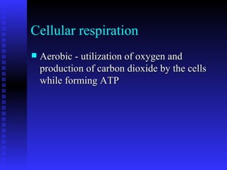
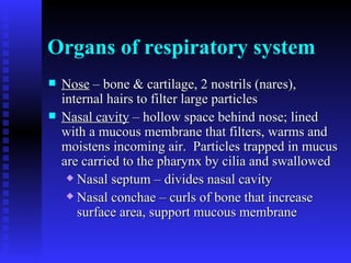
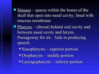
Respiratory System Passages filter air and transport it from outside body to lungs.  Microscopic air sacs are sites for gas exchange
Respiration 1.     Pulmonary ventilation  – movement of air in and out of lungs 2.    Exchange of gases between air in lungs and blood 3.    Transport of gases by blood between lungs and body cells 4.    Exchange of gases between blood and body cells
Cellular respiration Aerobic -  utilization of oxygen and production of carbon dioxide by the cells while forming ATP
Organs of respiratory system Nose  – bone & cartilage, 2 nostrils (nares), internal hairs to filter large particles   Nasal cavity  – hollow space behind nose; lined with a mucous membrane that filters, warms and moistens incoming air.  Particles trapped in mucus are carried to the pharynx by cilia and swallowed   Nasal septum – divides nasal cavity Nasal conchae – curls of bone that increase surface area, support mucous membrane
Sinuses  – spaces within the bones of the skull that open into nasal cavity; lined with mucous membrane   Pharynx  – (throat) behind oral cavity and between nasal cavity and larynx.  Passageway for air.  Aids in producing speech. Nasopharynx – superior portion Oropharynx – middle portion Laryngopharynx – inferior portion
Larynx  – contains vocal cords which produce sound by vibrating when air passes over them   Glottis – triangular opening in between vocal cords.  Can close to prevent food from entering Epiglottis – cartilage flap that covers glottis opening during swallowing
Trachea  – (windpipe) tube in the front of the neck; extends into thoracic cavity where it branches into left and right bronchi   Held open by cartilage rings.  Inner wall is lined with ciliated mucous membrane.  Filters air and moves trapped particles upward to pharynx to be swallowed
Bronchial Tree  – branched airways leading from trachea to alveoli in lungs.  Get progressively smaller   Primary bronchi – cartilage in walls similar to trachea; right and left divide into Secondary bronchi – continue to divide Bronchioles – no more cartilage, smooth muscle instead;  lead to Alveolar ducts – no muscle; lead to  Alveolar sacs – made of clusters of  Alveoli – microscopic air sacs surrounded by capillaries where gas exchange with blood takes place
Lungs soft, spongy, right lung has 3 lobes, left lung has 2 lobes   Occupy the pleural cavities Made of elastic tissue that stretches and recoils as we breathe Tissues must be moist to allow gas exchange Surfactants – keep alveolar surfaces from sticking together
Breathing mechanism - ventilation Inspiration – (inhalation) dome shaped diaphragm muscle contracts which decreases air pressure inside the lungs Air moves into the lungs because outside pressure is higher External intercostal muscles can also raise the ribs which further decreases internal pressure
Expiration (exhalation) Diaphragm relaxes returning to its dome shape Elastic recoil of lung tissue forces air out Internal intercostal muscles which pull ribs down and abdominal muscles which push up on the diaphragm can contract to exhale more forcefully
Air volume Tidal volume – the amount of air that enters and leaves during a respiration cycle Residual volume – air that remains in the lungs even after exhaling the maximum amount Vital capacity – the maximum amount a person can exhale after taking the deepest breath possible
Respiratory center Located in medulla oblongata Sensitive to the concentration of carbon dioxide, causes the urge to breathe Hyperventilation – breathing fast and deep, can let you hold your breath longer, but may make you pass out.  Don’t do it while swimming. Hypoxia – low oxygen levels reaching tissues

Respiratory system

  • 1.
    Respiratory System Passagesfilter air and transport it from outside body to lungs. Microscopic air sacs are sites for gas exchange
  • 2.
    Respiration 1.    Pulmonary ventilation – movement of air in and out of lungs 2.    Exchange of gases between air in lungs and blood 3.    Transport of gases by blood between lungs and body cells 4.    Exchange of gases between blood and body cells
  • 3.
    Cellular respiration Aerobic- utilization of oxygen and production of carbon dioxide by the cells while forming ATP
  • 4.
    Organs of respiratorysystem Nose – bone & cartilage, 2 nostrils (nares), internal hairs to filter large particles Nasal cavity – hollow space behind nose; lined with a mucous membrane that filters, warms and moistens incoming air. Particles trapped in mucus are carried to the pharynx by cilia and swallowed Nasal septum – divides nasal cavity Nasal conchae – curls of bone that increase surface area, support mucous membrane
  • 5.
    Sinuses –spaces within the bones of the skull that open into nasal cavity; lined with mucous membrane Pharynx – (throat) behind oral cavity and between nasal cavity and larynx. Passageway for air. Aids in producing speech. Nasopharynx – superior portion Oropharynx – middle portion Laryngopharynx – inferior portion
  • 6.
    Larynx –contains vocal cords which produce sound by vibrating when air passes over them Glottis – triangular opening in between vocal cords. Can close to prevent food from entering Epiglottis – cartilage flap that covers glottis opening during swallowing
  • 7.
    Trachea –(windpipe) tube in the front of the neck; extends into thoracic cavity where it branches into left and right bronchi Held open by cartilage rings. Inner wall is lined with ciliated mucous membrane. Filters air and moves trapped particles upward to pharynx to be swallowed
  • 8.
    Bronchial Tree – branched airways leading from trachea to alveoli in lungs. Get progressively smaller Primary bronchi – cartilage in walls similar to trachea; right and left divide into Secondary bronchi – continue to divide Bronchioles – no more cartilage, smooth muscle instead; lead to Alveolar ducts – no muscle; lead to Alveolar sacs – made of clusters of Alveoli – microscopic air sacs surrounded by capillaries where gas exchange with blood takes place
  • 9.
    Lungs soft, spongy,right lung has 3 lobes, left lung has 2 lobes Occupy the pleural cavities Made of elastic tissue that stretches and recoils as we breathe Tissues must be moist to allow gas exchange Surfactants – keep alveolar surfaces from sticking together
  • 10.
    Breathing mechanism -ventilation Inspiration – (inhalation) dome shaped diaphragm muscle contracts which decreases air pressure inside the lungs Air moves into the lungs because outside pressure is higher External intercostal muscles can also raise the ribs which further decreases internal pressure
  • 11.
    Expiration (exhalation) Diaphragmrelaxes returning to its dome shape Elastic recoil of lung tissue forces air out Internal intercostal muscles which pull ribs down and abdominal muscles which push up on the diaphragm can contract to exhale more forcefully
  • 12.
    Air volume Tidalvolume – the amount of air that enters and leaves during a respiration cycle Residual volume – air that remains in the lungs even after exhaling the maximum amount Vital capacity – the maximum amount a person can exhale after taking the deepest breath possible
  • 13.
    Respiratory center Locatedin medulla oblongata Sensitive to the concentration of carbon dioxide, causes the urge to breathe Hyperventilation – breathing fast and deep, can let you hold your breath longer, but may make you pass out. Don’t do it while swimming. Hypoxia – low oxygen levels reaching tissues