Download to read offline


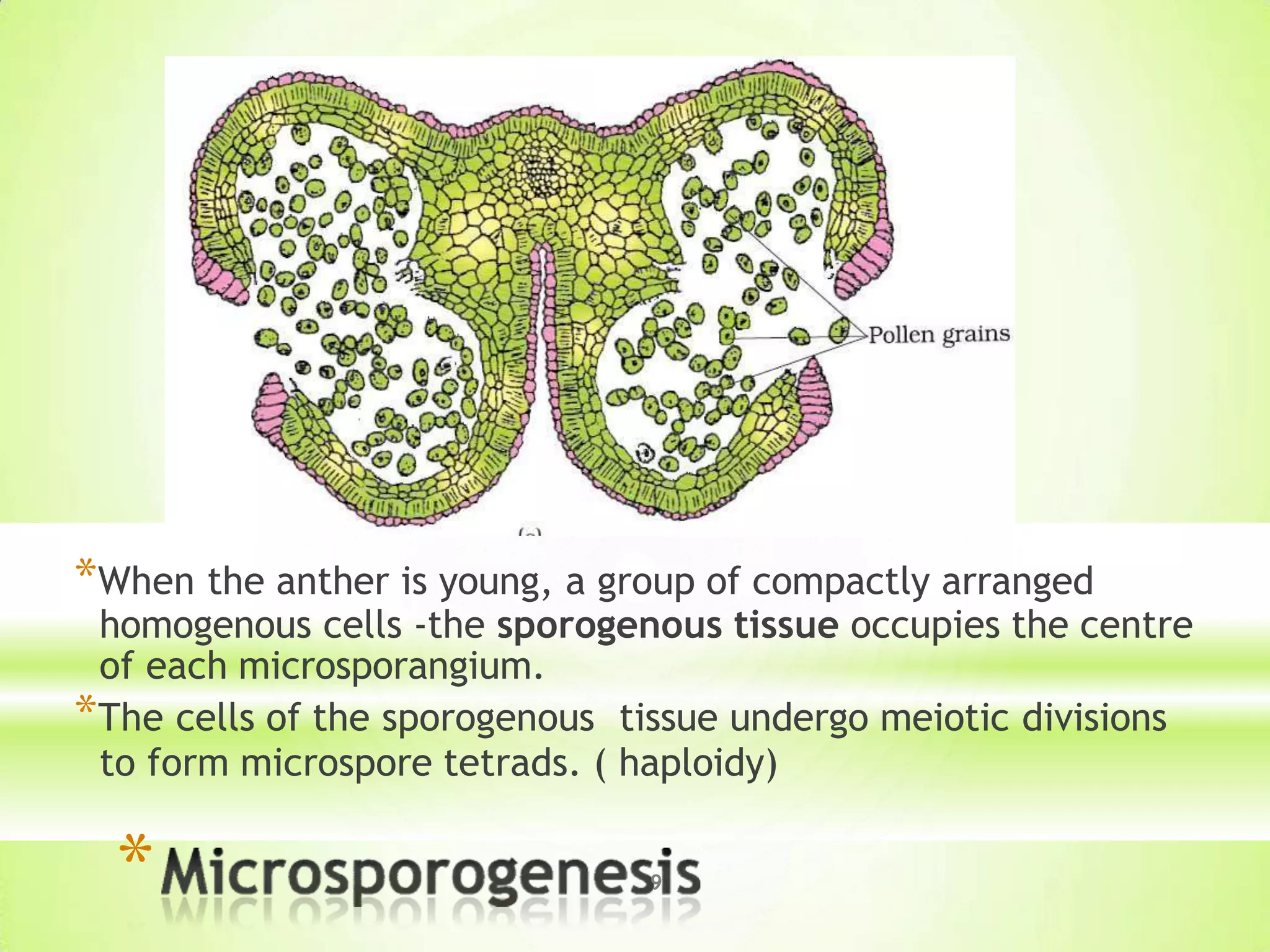


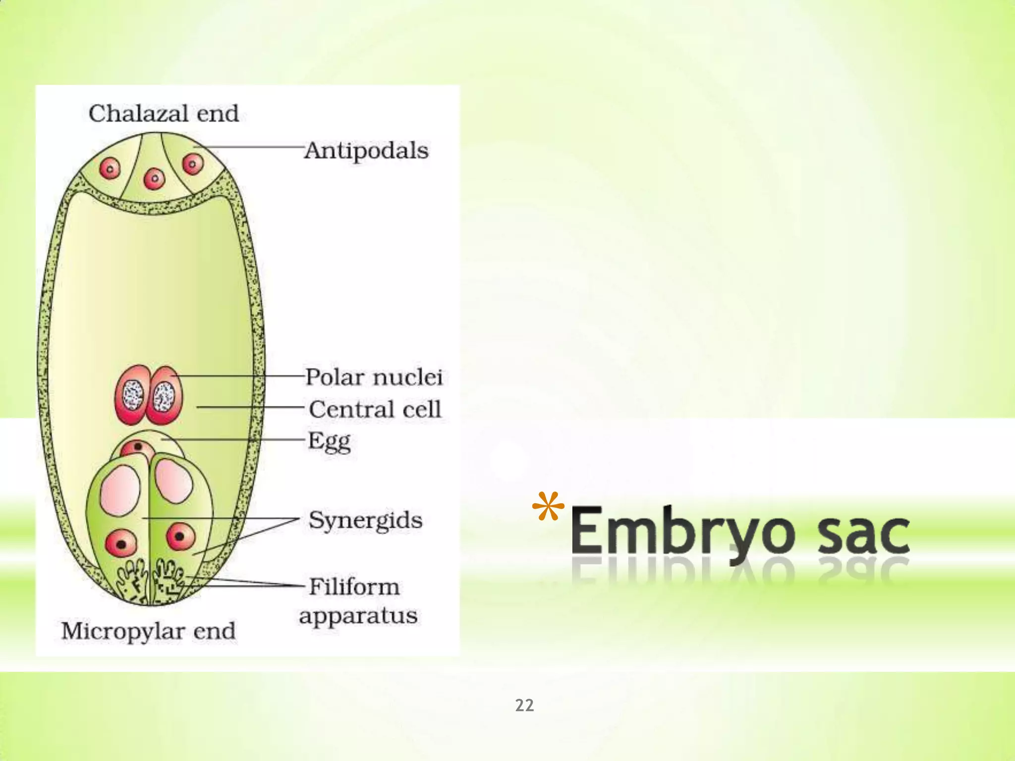


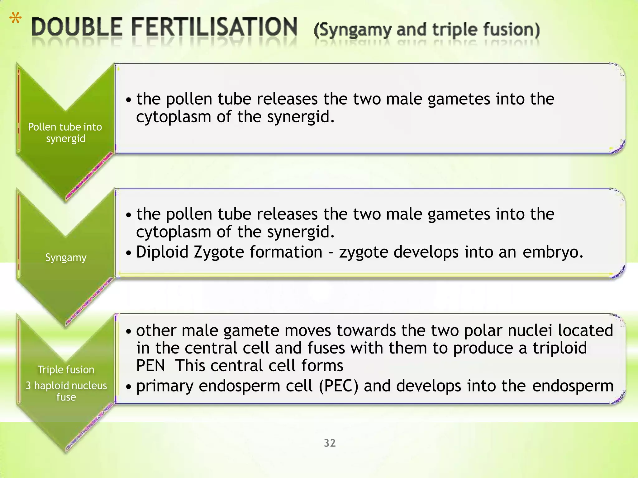
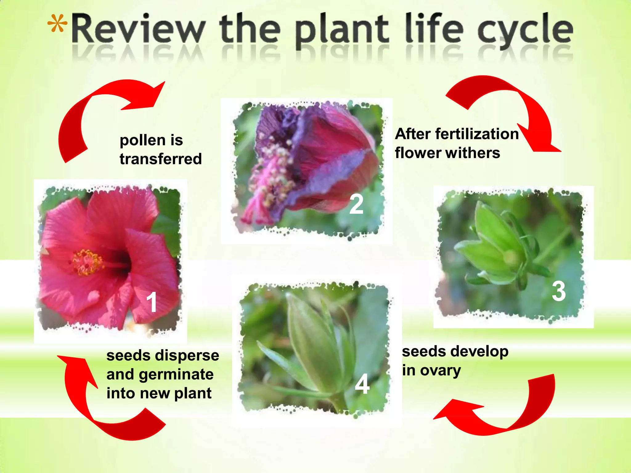


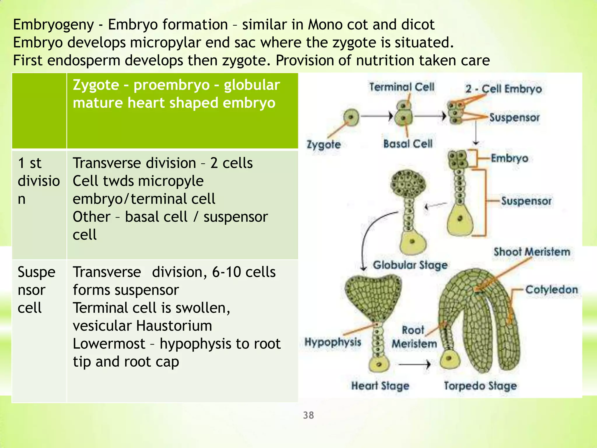

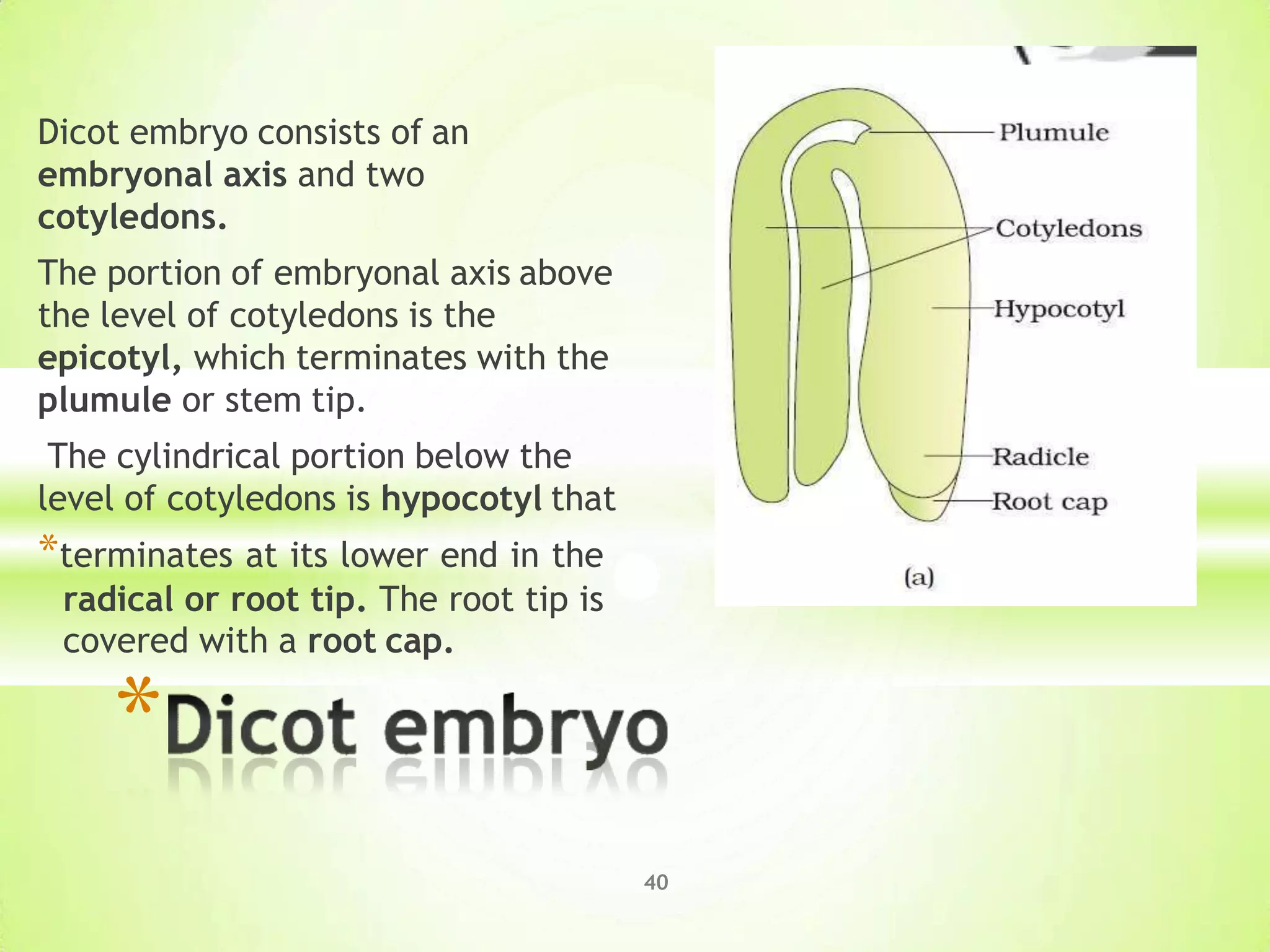

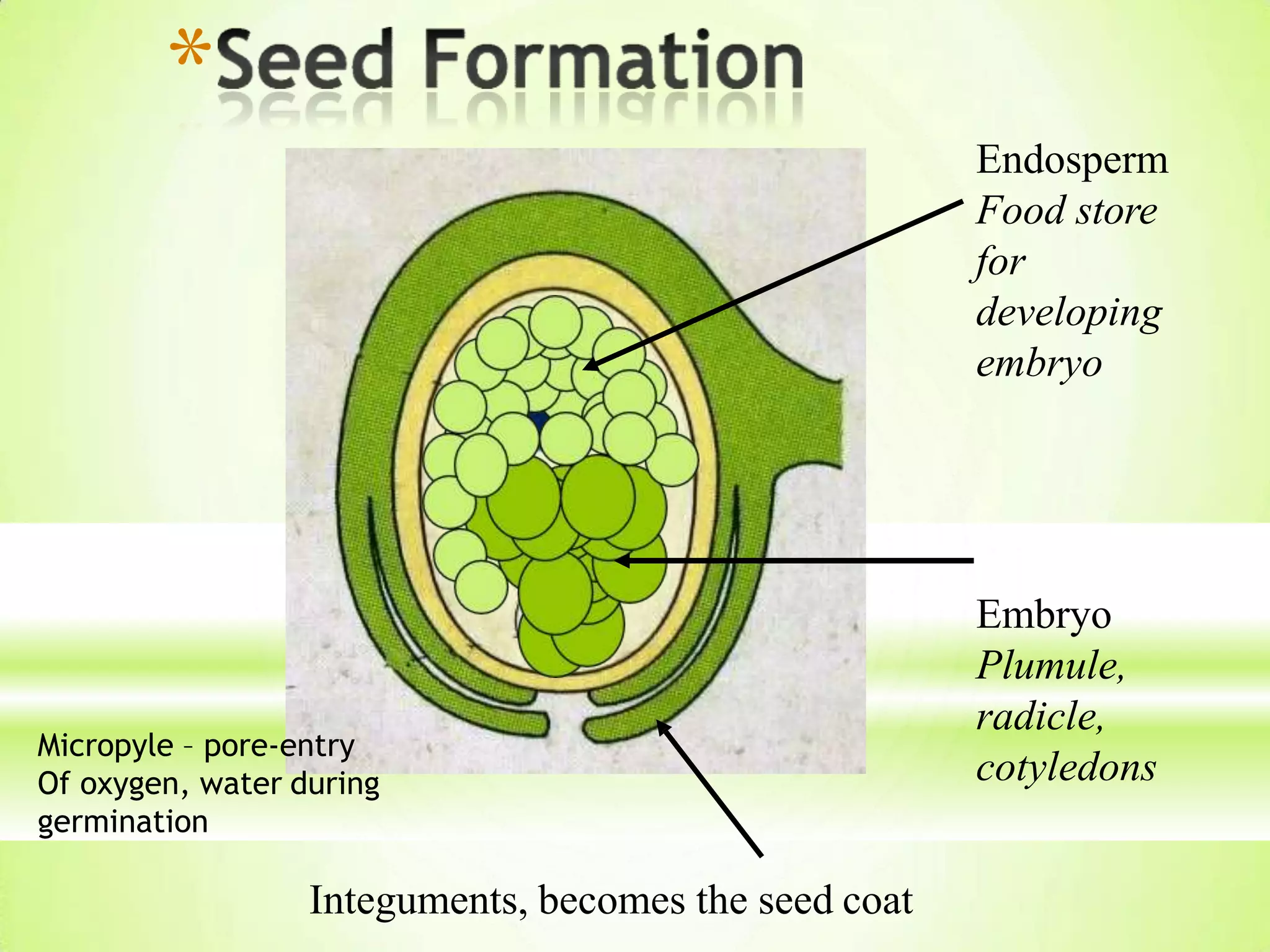

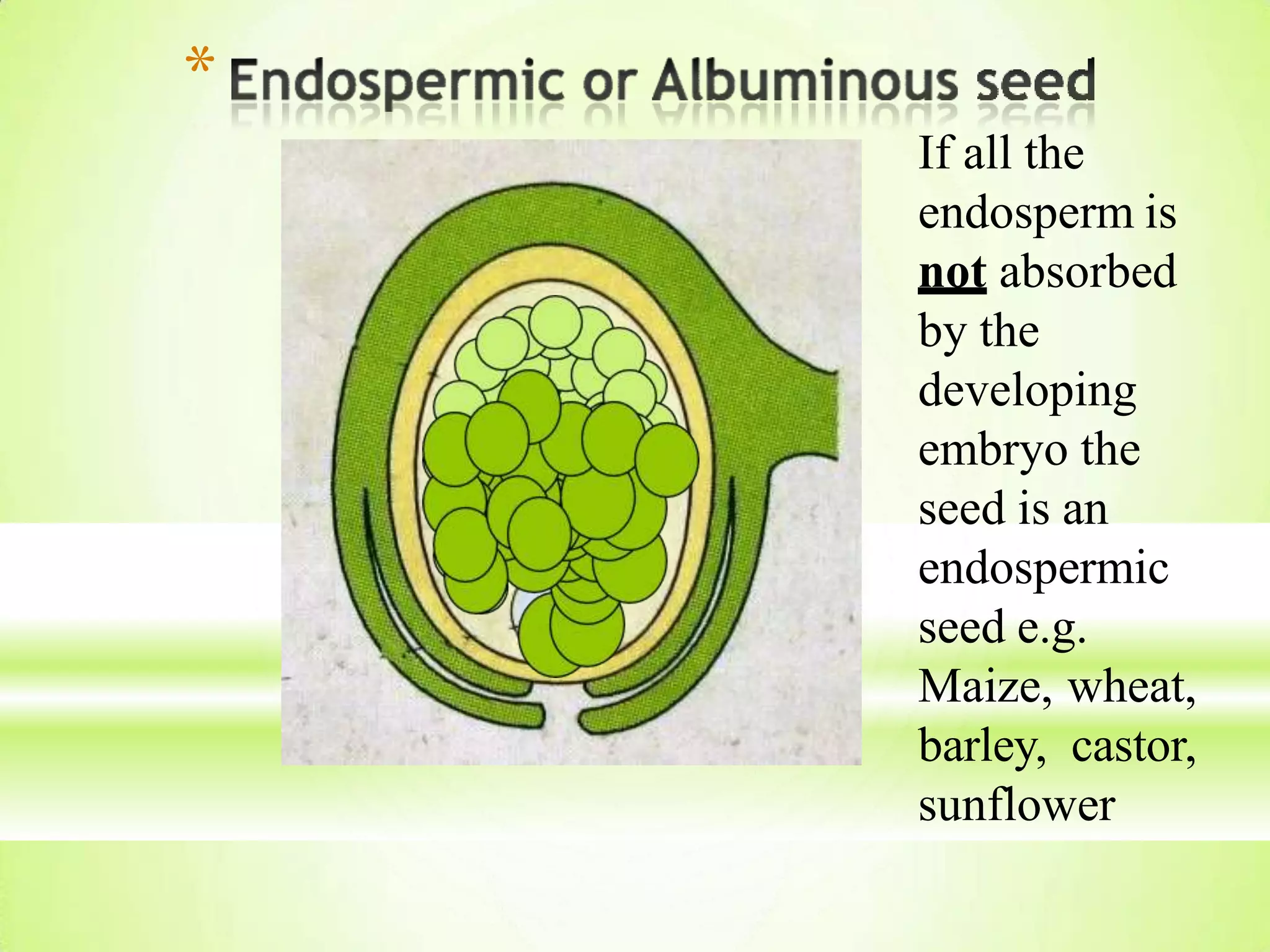
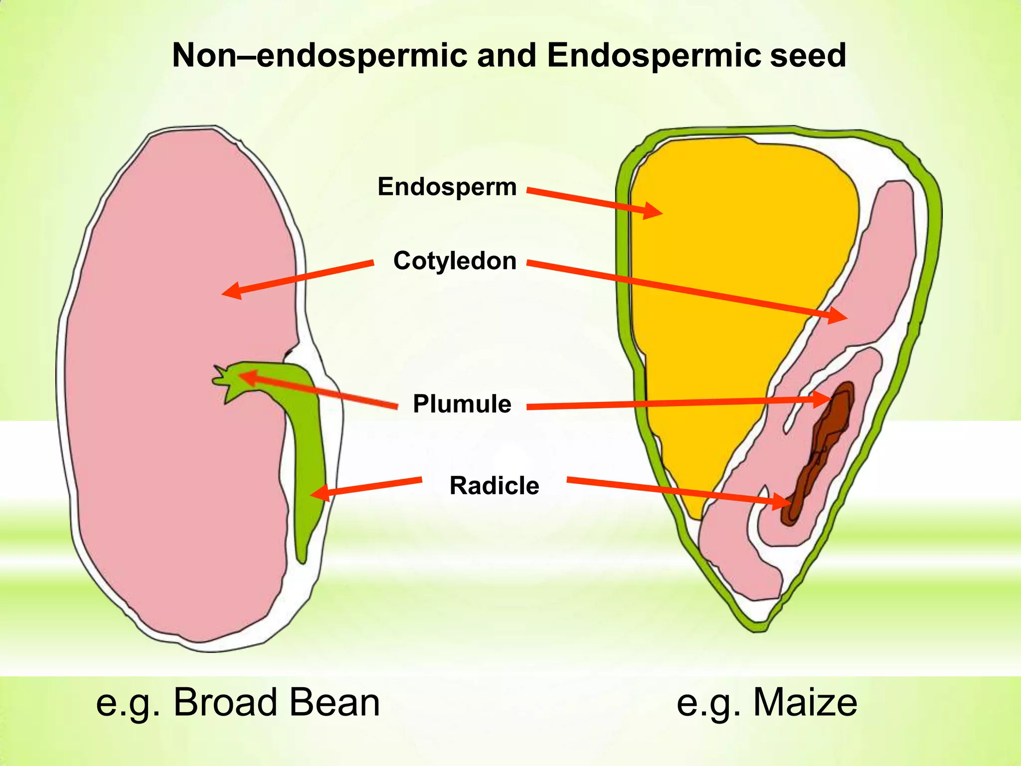
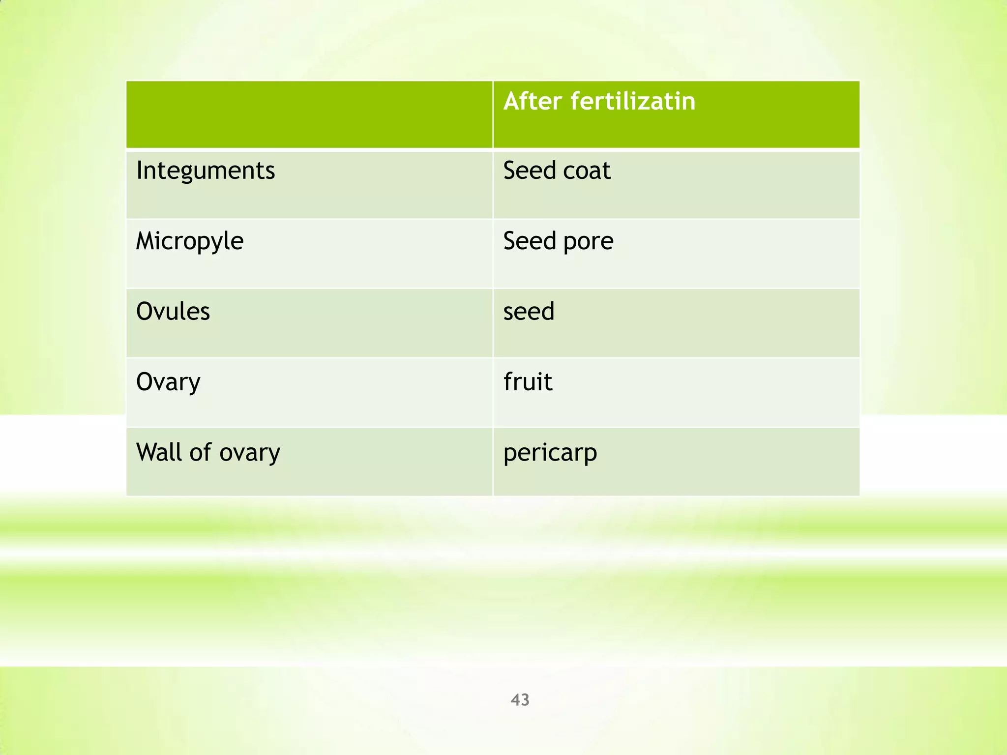


1. The document discusses the structure and development of flowers, stamens, anthers, pollen grains, pistils, ovules, and the process of fertilization. It describes the male and female reproductive organs in angiosperms as well as double fertilization. 2. Meiosis occurs in anthers and ovules to produce microspores that develop into pollen grains and megaspores that develop into embryo sacs, respectively. Fertilization involves the fusion of a pollen grain with an embryo sac to form a zygote and endosperm. 3. After fertilization, the zygote develops into an embryo while the endosperm provides nutrition. The ovary walls