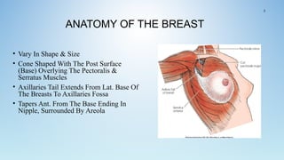
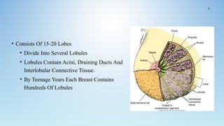
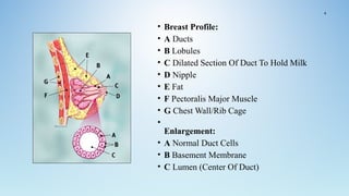
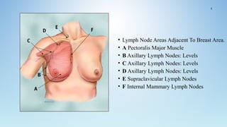
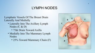
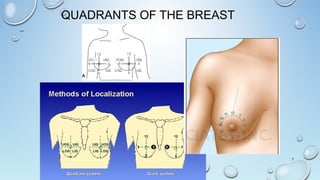
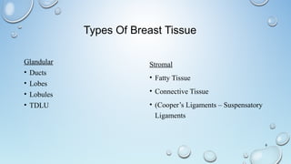
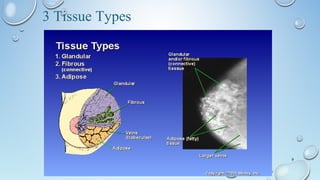
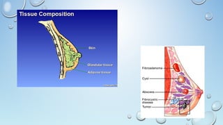
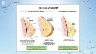
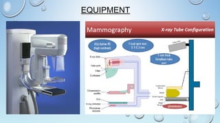
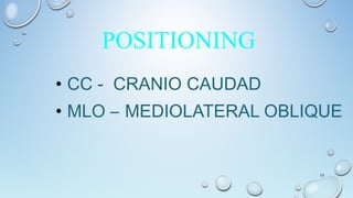
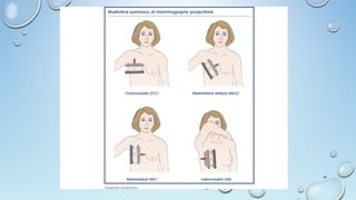
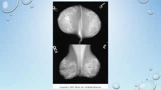
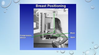
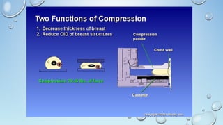
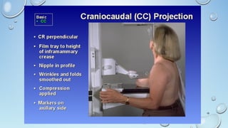
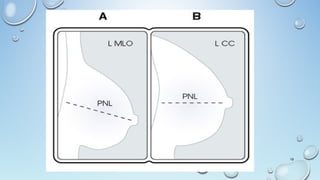
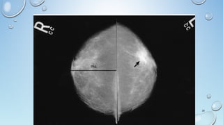
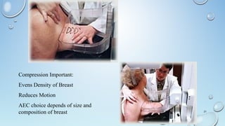
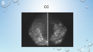
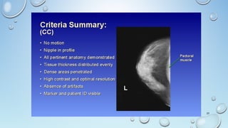
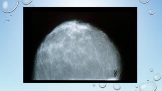
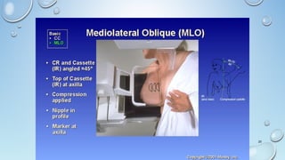
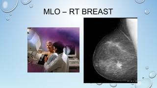
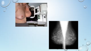
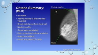
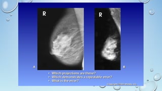
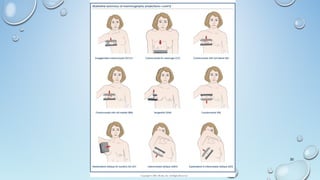
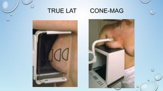
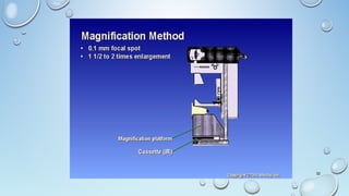
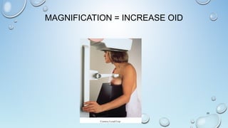
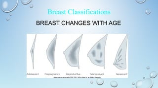
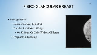
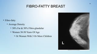
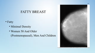
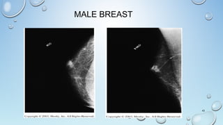
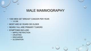
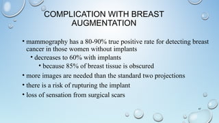
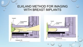
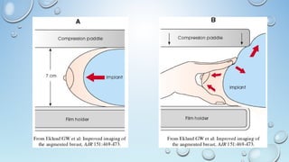
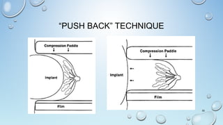
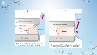
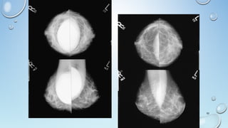
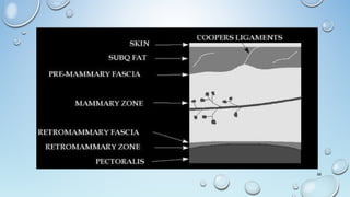
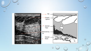
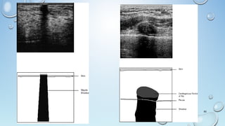
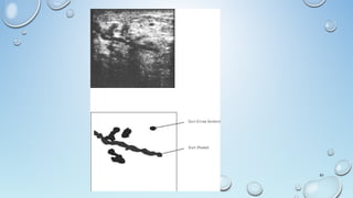
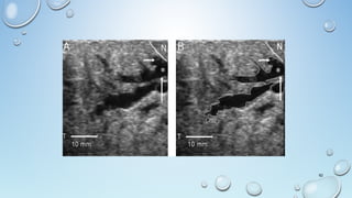
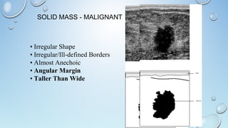
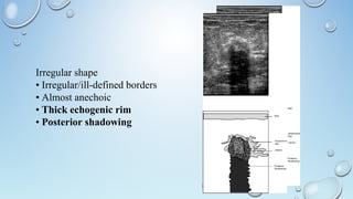
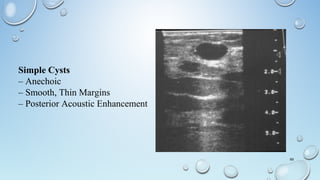
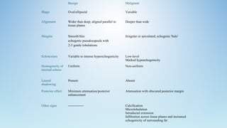
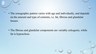
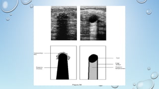
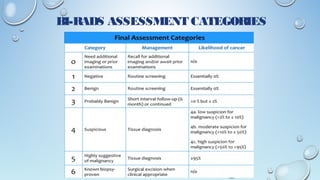
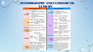
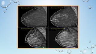
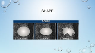
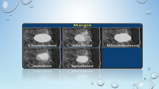
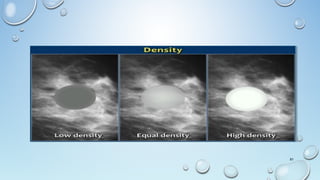
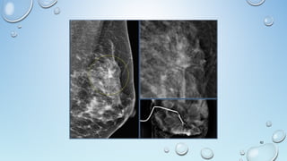
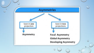
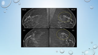
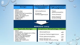
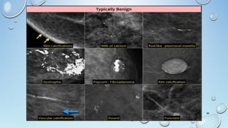
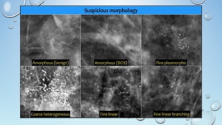
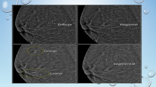
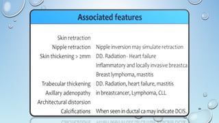
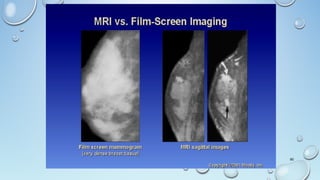
DR. VIBHAY PAREEK
RADIATION ONCOLOGY
JUPITER HOSPITAL
BREAST MAMMOGRAPHY
ANATOMY OF THE BREAST
• Vary In Shape & Size
• Cone Shaped With The Post Surface
(Base) Overlying The Pectoralis &
Serratus Muscles
• Axillaries Tail Extends From Lat. Base Of
The Breasts To Axillaries Fossa
• Tapers Ant. From The Base Ending In
Nipple, Surrounded By Areola
2
• Consists Of 15-20 Lobes
• Divide Into Several Lobules
• Lobules Contain Acini, Draining Ducts And
Interlobular Connective Tissue.
• By Teenage Years Each Breast Contains
Hundreds Of Lobules
3
• Breast Profile:
• A Ducts
• B Lobules
• C Dilated Section Of Duct To Hold Milk
• D Nipple
• E Fat
• F Pectoralis Major Muscle
• G Chest Wall/Rib Cage
•
Enlargement:
• A Normal Duct Cells
• B Basement Membrane
• C Lumen (Center Of Duct)
4
• Lymph Node Areas Adjacent To Breast Area.
• A Pectoralis Major Muscle
• B Axillary Lymph Nodes: Levels
• C Axillary Lymph Nodes: Levels
• D Axillary Lymph Nodes: Levels
• E Supraclavicular Lymph Nodes
• F Internal Mammary Lymph Nodes
5
LYMPH NODES
Lymphatic Vessels Of The Breast Drain
Laterally And Medially
• Laterally Into The Axillary Lymph
Nodes (C & D)
• 75& Drain Toward Axilla
• Medially Into The Mammary Lymph
Nodes
• 25% Toward Mammary Chain (F)
6
QUADRANTS OF THE BREAST
7
Types Of Breast Tissue
Glandular
• Ducts
• Lobes
• Lobules
• TDLU
Stromal
• Fatty Tissue
• Connective Tissue
• (Cooper’s Ligaments – Suspensatory
Ligaments
8
9
3 Tissue Types
10
11
EQUIPMENT
• CC - CRANIO CAUDAD
• MLO – MEDIOLATERAL OBLIQUE
13
POSITIONING
14
15
16
17
18
19
20
21
Compression Important:
Evens Density of Breast
Reduces Motion
AEC choice depends of size and
composition of breast
CC
22
23
24
25
26
MLO – RT BREAST
27
28
29
30
TRUE LAT CONE-MAG
31
32
MAGNIFICATION = INCREASE OID
33
BREAST CHANGES WITH AGE
34
Breast Classifications
FIBRO-GLANDULAR BREAST
• Fibro-glandular
• Dense With Very Little Fat
• Females 15-30 Years Of Age
• Or 30 Years Or Older Without Children
• Pregnant Or Lactating
35
FIBRO-FATTY BREAST
• Fibro-fatty
• Average Density
• 50% Fat & 50% Fibro-glandular
• Women 30-50 Years Of Age
• Or Women With 3 Or More Children
36
FATTY BREAST
• Fatty
• Minimal Density
• Women 50 And Older
(Postmenopausal), Men And Children
37
THE MALE BREAST
38
Male Mammography and Cancer
GYNECOMASTIA
• Benign Excessive Development Of Male Mammary Gland
• Occurs In 40% Of Male Cancer Pt’s
• Survival Rates With Treatment Are 97% For 5 Years
39
• Most Common Causes :
• Puberty (Hormonal Growth And Changes During
Adolescence)
• Estrogen Exposure (Female Hormone Present In The Body
And The Environment)
• Androgen Exposure (Body-building Hormones)
• Marijuana Use
• Medication Side Effects (Older Men)
• Klinefelter's Syndrome
40
GYNECOMASTIA
• Is A Benign Male Breast (Non-cancerous) Condition
• Some Men Who Have Prominent Breasts, Or Uneven Breasts, Often Feel
Some Embarrassment About Their Body Image.
• This Condition Can Also Cause Emotional Conflict Over Sexual Identity.
41
42
MALE BREAST
43
MALE MAMMOGRAPHY
• 1300 MEN GET BREAST CANCER PER YEAR
• 1/3 DIE
• MOST ARE 60 YEARS OR OLDER
• NEARLY ALL ARE PRIMARY TUMORS
• SYMPTOMS INCLUDE:
• NIPPLE RETRACTION
• CRUSTING
• DISCHARGE
• ULCERATION
44
MALE MASTECTOMY
45
BREAST IMPLANTS
IS IT WORTH THE RISK?
46
COMPLICATION WITH BREAST
AUGMENTATION
• mammography has a 80-90% true positive rate for detecting breast
cancer in those women without implants
• decreases to 60% with implants
• because 85% of breast tissue is obscured
• more images are needed than the standard two projections
• there is a risk of rupturing the implant
• loss of sensation from surgical scars
47
ELKLAND METHOD FOR IMAGING
WITH BREAST IMPLANTS
48
49
“PUSH BACK” TECHNIQUE
50
51
52
53
ULTRASOUND OF BREAST
APPLICATIONS
• Not Always Detect Cancers That Are Visualised Mammographically.
• Ultrasound Can Detect Clinically And Mammographically Occult Cancers
Particularly When There Is A Higher Possibility Of Cancer.
• New High-frequency Transducers
• Irregular Masses, Abnormal Dilated Ducts Or Clustered Foci Of Increased
Echogenicity With Increased Doppler Vascularity.
Patient PositionPatient Position
Medial LesionsMedial Lesions
• Patient Is Supine
• Ipsilateral Arm Is Placed Over The Patient’s Head.
Lateral LesionsLateral Lesions
• Patient Is Opposite.
Superior LesionsSuperior Lesions
• Patient Is SITTING
Apply Gentle Uniform Pressure With The UltrasoundApply Gentle Uniform Pressure With The Ultrasound
TransducerTransducer
Increase Transducer Pressure For:
– Greater Penetration
– Scanning The Subareolar Region.
Scanning Is Done In Three Directions.
1. Radial
2. Transverse
3. Longitudinal
• Localization Is By The Clock Face.
12
3
66
39
58
59
60
61
62
63
Lymph Node
• Solid nodule
• Ovoid
• Echogenic fatty hilum
• The Subcutaneous Fat LayerSubcutaneous Fat Layer Is Demonstrated Superficially As
Hypoechoic Tissue Compared To The Glandular Tissue From Which It Is
Separated By A Well-defined Scalloped Margin.
• Normal DuctsDucts Are Often Visible, Particularly In The Subareolar Region,
As Anechoic Tubular Structures.
• Deep To The Glandular Tissue, A Retromammary Fat LayerA Retromammary Fat Layer Is Usually
Visible And, Behind This, The Structures Of The Chest WallChest Wall.
64
• Symptomatic Breast Lumps In Women Aged Less Than 35 Years.
• Breast Lump Developing During Pregnancy Or Lactation.
• Assessment Of Mammographic Abnormality (± Further Mammographic
Views)
• Assessment Of MRI Or Scintimammography Detected Lesions.
• Clinical Breast Mass With Negative Mammograms.
• Breast Inflammation.
• The Augmented Breast (Together With MRI).
• Breast Lump In A Male (Together With Mammography).
• Guidance Of Needle Biopsy Or Localisation.
• Follow-up Of Breast Cancer Treated With Adjuvant Chemotherapy. 65
Benign CharacteristicsBenign Characteristics
• Ellipsoid Shape
• Thin Definable Capsule
• Two Or Three Lobulations
• Hyperechogenicity.
SOLID MASS - MALIGNANT
• Irregular Shape
• Irregular/Ill-defined Borders
• Almost Anechoic
• Angular Margin
• Taller Than Wide
Irregular shape
• Irregular/ill-defined borders
• Almost anechoic
• Thick echogenic rim
• Posterior shadowing
69
Simple Cysts
– Anechoic
– Smooth, Thin Margins
– Posterior Acoustic Enhancement
Benign Malignant
Shape Oval/ellipsoid Variable
Alignment Wider than deep; aligned parallel to
tissue planes
Deeper than wide
Margins Smooth/thin
echogenic pseudocapsule with
2-3 gentle lobulations
Irregular or spiculated; echogenic 'halo'
Echotexture Variable to intense hyperechogenicity Low-level
Marked hypoechogenicity
Homogeneity of
internal echoes
Uniform Non-uniform
Lateral
shadowing
Present Absent
Posterior effect Minimum attenuation/posterior
enhancement
Attenuation with obscured posterior margin
Other signs -------------- Calcification
Microlobulation
Intraductal extension
Infiltration across tissue planes and increased
echogenicity of surrounding fat
• The sonographic pattern varies with age and individually, and depends
on the amount and type of contents, i.e. fat, fibrous and glandular
tissues.
• The fibrous and glandular components are variably echogenic, while
fat is hypoechoic.
BI-RADS
• BIRADS Stands For Breast Imaging- Reporting And Data
System Which Is A Widely Accepted Risk Assessment And
Quality Assurance Tool In Mammography, Ultrasound And
MRI.
73
74
BI-RADS ASSESSMENT CATEGORIES
MAMMOGRAPHY ANDULTRASOUND
LEXICON
77
MASS
78
A 'Mass' is a space occupying 3D lesion seen in two different
projections.
If a potential mass is seen in only a single projection it should be called
a 'asymmetry' until its three-dimensionality is confirmed.
Shape: oval (may include 2 or 3 lobulations), round or irregular
Margins: circumscribed, obscured, microlobulated, indistinct,
spiculated
Density: high, equal, low or fat-containing.
SHAPE
79
80
81
82
83
84
85
86
87
88
89
90
91

Radiology day 1 mammography

  • 1.
    DR. VIBHAY PAREEK RADIATIONONCOLOGY JUPITER HOSPITAL BREAST MAMMOGRAPHY
  • 2.
    ANATOMY OF THEBREAST • Vary In Shape & Size • Cone Shaped With The Post Surface (Base) Overlying The Pectoralis & Serratus Muscles • Axillaries Tail Extends From Lat. Base Of The Breasts To Axillaries Fossa • Tapers Ant. From The Base Ending In Nipple, Surrounded By Areola 2
  • 3.
    • Consists Of15-20 Lobes • Divide Into Several Lobules • Lobules Contain Acini, Draining Ducts And Interlobular Connective Tissue. • By Teenage Years Each Breast Contains Hundreds Of Lobules 3
  • 4.
    • Breast Profile: •A Ducts • B Lobules • C Dilated Section Of Duct To Hold Milk • D Nipple • E Fat • F Pectoralis Major Muscle • G Chest Wall/Rib Cage • Enlargement: • A Normal Duct Cells • B Basement Membrane • C Lumen (Center Of Duct) 4
  • 5.
    • Lymph NodeAreas Adjacent To Breast Area. • A Pectoralis Major Muscle • B Axillary Lymph Nodes: Levels • C Axillary Lymph Nodes: Levels • D Axillary Lymph Nodes: Levels • E Supraclavicular Lymph Nodes • F Internal Mammary Lymph Nodes 5
  • 6.
    LYMPH NODES Lymphatic VesselsOf The Breast Drain Laterally And Medially • Laterally Into The Axillary Lymph Nodes (C & D) • 75& Drain Toward Axilla • Medially Into The Mammary Lymph Nodes • 25% Toward Mammary Chain (F) 6
  • 7.
  • 8.
    Types Of BreastTissue Glandular • Ducts • Lobes • Lobules • TDLU Stromal • Fatty Tissue • Connective Tissue • (Cooper’s Ligaments – Suspensatory Ligaments 8
  • 9.
  • 10.
  • 11.
  • 12.
  • 13.
    • CC -CRANIO CAUDAD • MLO – MEDIOLATERAL OBLIQUE 13 POSITIONING
  • 14.
  • 15.
  • 16.
  • 17.
  • 18.
  • 19.
  • 20.
  • 21.
    21 Compression Important: Evens Densityof Breast Reduces Motion AEC choice depends of size and composition of breast
  • 22.
  • 23.
  • 24.
  • 25.
  • 26.
  • 27.
  • 28.
  • 29.
  • 30.
  • 31.
  • 32.
  • 33.
  • 34.
    BREAST CHANGES WITHAGE 34 Breast Classifications
  • 35.
    FIBRO-GLANDULAR BREAST • Fibro-glandular •Dense With Very Little Fat • Females 15-30 Years Of Age • Or 30 Years Or Older Without Children • Pregnant Or Lactating 35
  • 36.
    FIBRO-FATTY BREAST • Fibro-fatty •Average Density • 50% Fat & 50% Fibro-glandular • Women 30-50 Years Of Age • Or Women With 3 Or More Children 36
  • 37.
    FATTY BREAST • Fatty •Minimal Density • Women 50 And Older (Postmenopausal), Men And Children 37
  • 38.
    THE MALE BREAST 38 MaleMammography and Cancer
  • 39.
    GYNECOMASTIA • Benign ExcessiveDevelopment Of Male Mammary Gland • Occurs In 40% Of Male Cancer Pt’s • Survival Rates With Treatment Are 97% For 5 Years 39
  • 40.
    • Most CommonCauses : • Puberty (Hormonal Growth And Changes During Adolescence) • Estrogen Exposure (Female Hormone Present In The Body And The Environment) • Androgen Exposure (Body-building Hormones) • Marijuana Use • Medication Side Effects (Older Men) • Klinefelter's Syndrome 40
  • 41.
    GYNECOMASTIA • Is ABenign Male Breast (Non-cancerous) Condition • Some Men Who Have Prominent Breasts, Or Uneven Breasts, Often Feel Some Embarrassment About Their Body Image. • This Condition Can Also Cause Emotional Conflict Over Sexual Identity. 41
  • 42.
  • 43.
  • 44.
    MALE MAMMOGRAPHY • 1300MEN GET BREAST CANCER PER YEAR • 1/3 DIE • MOST ARE 60 YEARS OR OLDER • NEARLY ALL ARE PRIMARY TUMORS • SYMPTOMS INCLUDE: • NIPPLE RETRACTION • CRUSTING • DISCHARGE • ULCERATION 44
  • 45.
  • 46.
    BREAST IMPLANTS IS ITWORTH THE RISK? 46
  • 47.
    COMPLICATION WITH BREAST AUGMENTATION •mammography has a 80-90% true positive rate for detecting breast cancer in those women without implants • decreases to 60% with implants • because 85% of breast tissue is obscured • more images are needed than the standard two projections • there is a risk of rupturing the implant • loss of sensation from surgical scars 47
  • 48.
    ELKLAND METHOD FORIMAGING WITH BREAST IMPLANTS 48
  • 49.
  • 50.
  • 51.
  • 52.
  • 53.
  • 54.
    APPLICATIONS • Not AlwaysDetect Cancers That Are Visualised Mammographically. • Ultrasound Can Detect Clinically And Mammographically Occult Cancers Particularly When There Is A Higher Possibility Of Cancer. • New High-frequency Transducers • Irregular Masses, Abnormal Dilated Ducts Or Clustered Foci Of Increased Echogenicity With Increased Doppler Vascularity.
  • 55.
    Patient PositionPatient Position MedialLesionsMedial Lesions • Patient Is Supine • Ipsilateral Arm Is Placed Over The Patient’s Head. Lateral LesionsLateral Lesions • Patient Is Opposite. Superior LesionsSuperior Lesions • Patient Is SITTING
  • 56.
    Apply Gentle UniformPressure With The UltrasoundApply Gentle Uniform Pressure With The Ultrasound TransducerTransducer Increase Transducer Pressure For: – Greater Penetration – Scanning The Subareolar Region. Scanning Is Done In Three Directions. 1. Radial 2. Transverse 3. Longitudinal
  • 57.
    • Localization IsBy The Clock Face. 12 3 66 39
  • 58.
  • 59.
  • 60.
  • 61.
  • 62.
  • 63.
    63 Lymph Node • Solidnodule • Ovoid • Echogenic fatty hilum
  • 64.
    • The SubcutaneousFat LayerSubcutaneous Fat Layer Is Demonstrated Superficially As Hypoechoic Tissue Compared To The Glandular Tissue From Which It Is Separated By A Well-defined Scalloped Margin. • Normal DuctsDucts Are Often Visible, Particularly In The Subareolar Region, As Anechoic Tubular Structures. • Deep To The Glandular Tissue, A Retromammary Fat LayerA Retromammary Fat Layer Is Usually Visible And, Behind This, The Structures Of The Chest WallChest Wall. 64
  • 65.
    • Symptomatic BreastLumps In Women Aged Less Than 35 Years. • Breast Lump Developing During Pregnancy Or Lactation. • Assessment Of Mammographic Abnormality (± Further Mammographic Views) • Assessment Of MRI Or Scintimammography Detected Lesions. • Clinical Breast Mass With Negative Mammograms. • Breast Inflammation. • The Augmented Breast (Together With MRI). • Breast Lump In A Male (Together With Mammography). • Guidance Of Needle Biopsy Or Localisation. • Follow-up Of Breast Cancer Treated With Adjuvant Chemotherapy. 65
  • 66.
    Benign CharacteristicsBenign Characteristics •Ellipsoid Shape • Thin Definable Capsule • Two Or Three Lobulations • Hyperechogenicity.
  • 67.
    SOLID MASS -MALIGNANT • Irregular Shape • Irregular/Ill-defined Borders • Almost Anechoic • Angular Margin • Taller Than Wide
  • 68.
    Irregular shape • Irregular/ill-definedborders • Almost anechoic • Thick echogenic rim • Posterior shadowing
  • 69.
    69 Simple Cysts – Anechoic –Smooth, Thin Margins – Posterior Acoustic Enhancement
  • 70.
    Benign Malignant Shape Oval/ellipsoidVariable Alignment Wider than deep; aligned parallel to tissue planes Deeper than wide Margins Smooth/thin echogenic pseudocapsule with 2-3 gentle lobulations Irregular or spiculated; echogenic 'halo' Echotexture Variable to intense hyperechogenicity Low-level Marked hypoechogenicity Homogeneity of internal echoes Uniform Non-uniform Lateral shadowing Present Absent Posterior effect Minimum attenuation/posterior enhancement Attenuation with obscured posterior margin Other signs -------------- Calcification Microlobulation Intraductal extension Infiltration across tissue planes and increased echogenicity of surrounding fat
  • 71.
    • The sonographicpattern varies with age and individually, and depends on the amount and type of contents, i.e. fat, fibrous and glandular tissues. • The fibrous and glandular components are variably echogenic, while fat is hypoechoic.
  • 73.
    BI-RADS • BIRADS StandsFor Breast Imaging- Reporting And Data System Which Is A Widely Accepted Risk Assessment And Quality Assurance Tool In Mammography, Ultrasound And MRI. 73
  • 74.
  • 75.
  • 76.
  • 77.
  • 78.
    MASS 78 A 'Mass' isa space occupying 3D lesion seen in two different projections. If a potential mass is seen in only a single projection it should be called a 'asymmetry' until its three-dimensionality is confirmed. Shape: oval (may include 2 or 3 lobulations), round or irregular Margins: circumscribed, obscured, microlobulated, indistinct, spiculated Density: high, equal, low or fat-containing.
  • 79.
  • 80.
  • 81.
  • 82.
  • 83.
  • 84.
  • 85.
  • 86.
  • 87.
  • 88.
  • 89.
  • 90.
  • 91.

Editor's Notes

  • #7 A pectoralis major muscle B axillary lymph nodes: levels C axillary lymph nodes: levels D axillary lymph nodes: levels E supraclavicular lymph nodes F internal mammary lymph nodes