Downloaded 135 times

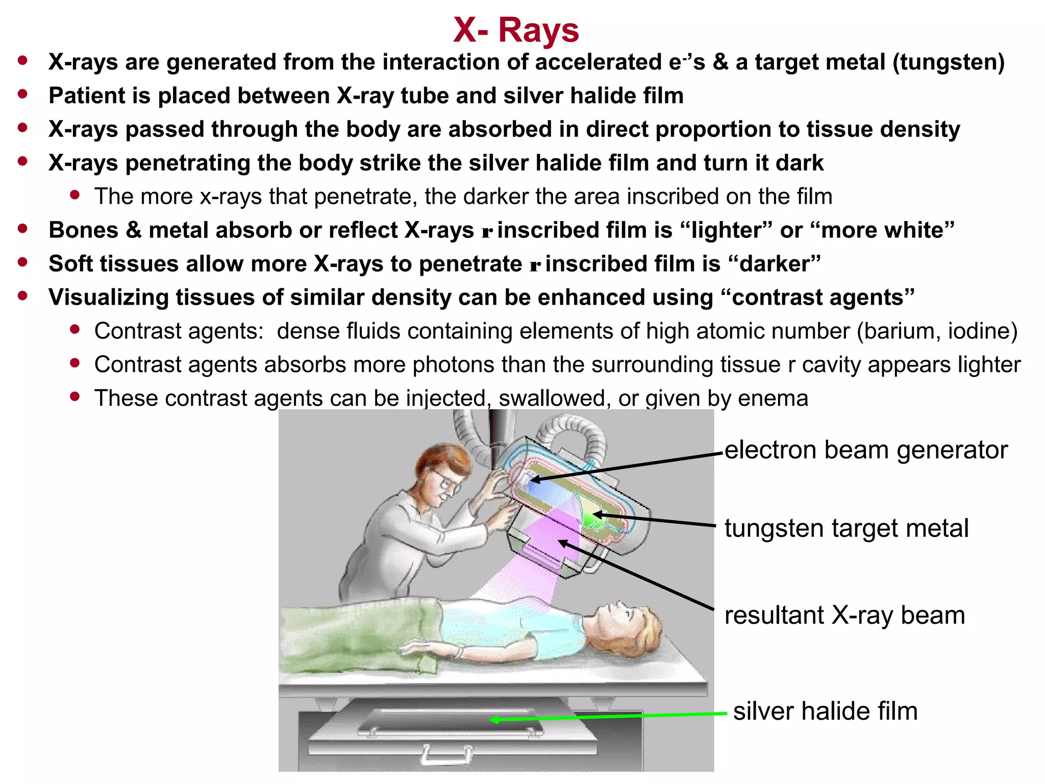
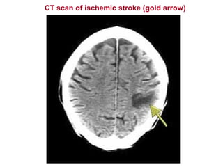
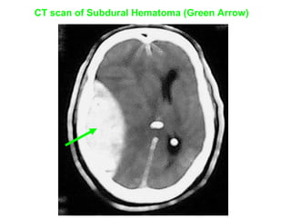
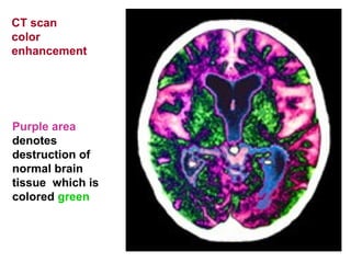
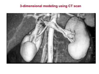
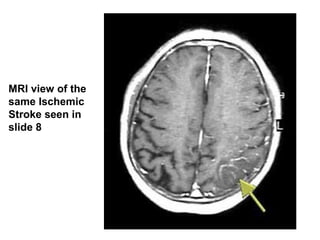
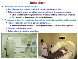
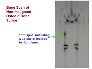
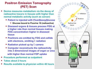
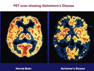

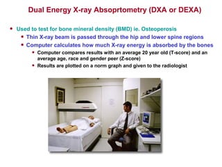


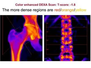

X-rays are generated when accelerated electrons interact with a metal target, producing X-ray beams that are passed through the body and absorbed at different rates by tissues depending on their density. The X-rays that pass through the body expose silver halide film, creating an image where bone and other dense tissues appear lighter since they absorb more X-rays. Contrast agents can enhance the visualization of similar tissues. CT scans use X-rays and a computer to generate cross-sectional images of the body, while MRI uses magnetic fields and radio waves to produce detailed images of soft tissues without radiation exposure. Bone scans and PET scans use radioactive tracers to detect bone abnormalities and areas of increased metabolic activity. DEXA scans measure bone


![Patient care [autosaved]](https://cdn.slidesharecdn.com/ss_thumbnails/patientcareautosaved-150405120334-conversion-gate01-thumbnail.jpg?width=640&height=640&fit=bounds)


