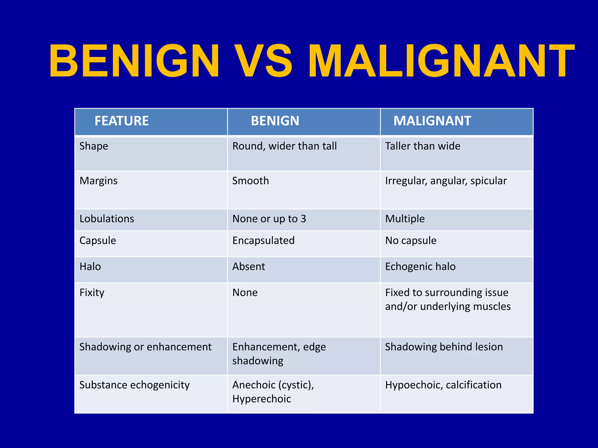
RADIOLOGICAL APPROACH FOR
MALIGNANT BREAST LESIONS
DR. ANJUM MEHDI
MBBS, DMRD, FCPS (Radiology)
Associate Professor Radiology
Punjab Medical College, Faisalabad.
Breast Cancer
• Incidence = 90 / 100 000.
• 10 % of women will have a Breast Cancer
• Treatment efficient for local disease
• 5 % of Women will die
Risk Factors Before Menopause
>>>
Tall & Slim
Border Line Lesions
> 1 Abortion
Long Study
Contraceptive Pill > 5 years very early
Short Menstrual Cycles (<26 days)
Psychological Factors
>
>
>
>
>
?
?
?
?
>>>
>>>
Nulliparous < 25 years
First menstruation <13 years
Alcoholism
Family History
History of pulmonary TB
Risk Factors after Menopause
Nulliparous < 25 years >>>
Menopause > 52 years >>>
First menstrual cycle < 13 years >>>
Obesity >>>
Alcoholism >
High social class >
Urban Habitat >
Family History >
History of pulmonary TB ?
Celibate ?
Predisposing Genes of Breast and
Ovary cancer
Breast Cancer Ovarian Cancer
BRCA1 9 q 34
(Japan)BRCA 2
BRCA 3 (8p)
Androgen-R
FNA
Magnification
Core Biopsy SurgeryFallow-up
SpotUltrasound
Decision
Micro calcifications Stellate
Lesions
Mass
Screening Mammography
Diagnostic Stratégie
Breast Cancers
Tubular
Invasive
Ductal
64%
Invasive
Lobular
Mucinous
DCIS
11%
Papillary
Medullary
TNM Classification
Tumor
• T0 : No Clinical Sign
•T is : Carcinoma in situ
• T1 < 2 cm
•T1a < 0,5 cm
•T1b > 0,5 cm &< 1 cm
•T1c > 0,5 cm &< 2 cm
• T2 : 2 to 5 cm
• T3 > 5 cm
• T4 : Skin or Pectoral Adherence
•T4a : extension to the Wall
•T4b : Edema, ulceration
•T4c : Inflammatory Carcinoma
Nodes
Non palpable : N0
Mobiles : N1
Fixed : N2
Ipsilateral or
Susclavian : N3
Metastasis
Absent : M0
Present : M1
p
Nodes
pN0 : No invasion
•pN1 : Axillary extent mobile
• pN1b: Metastasis >2 mm
• pN1a: Micro metastasis
•pN2 : Axillary extent fixed
•pN3 : Internal Mammary Extent
Pathologic
Classification
p TNM
STAGES
0 I II III IV
T is N0 M0 T1 N0 M0
Tn Nn M1
T0 N1 M0
T1 N1 M0
T2 N0 M0
T2 N1 M0
T3 N0 M0
A B A B
T0 N2 M0
T1 N2 M0
T2 N2 M0
T3N1&2M0
T4 Nn M0
Mortality of Breast Cancer
0
20
40
Deathfor100000
Breast
Uterus
Lung Ovaries
1975 1995 2015
Women 35 - 64 years
BREAST ANATOMY
Breast tissue is composed
of 3 layers.
1. Premammary.
2. Mammary.
3. Retromammory.
 Breast contains 15-18
lobes,
 Each lobe contain 20-40
lobules.
Cooper’s ligament are
fibrous bands that course
between the superficial
fascia and the deep fascia.
BREAST ANATOMY
 Basic functional unit of breast is a
TDLU/lobule.
 TDLU consist of 10 -100 acini that
drain into the terminal duct.
 The terminal duct drains into 
larger ducts main duct of the lobe
 nipple.
 The terminal ductal lobular unit is
an important structure because
most invasive cancers arise from
the TDLU.
It also is the site of origin of ductal
carcinoma in situ (DCIS), lobular
carcinoma in situ, fibroadenoma
and fibrocystic disease.
BENIGN VS MALIGNANT
Feature Benign Malignant
Shape Round, wider than tall Taller than wide
Margins Smooth Irregular, angular, spicular
Lobulations None or up to 3 Multiple
Capsule Encapsulated No capsule
Halo Absent Echogenic halo
Fixity None Fixed to surrounding issue
and/or underlying muscles
Shadowing or enhancement Enhancement, edge
shadowing
Shadowing behind lesion
Substance echogenicity Anechoic (cystic), Hyperechoic Hypoechoic, calcification
FEATURE BENIGN MALIGNANT
Shape Round, wider than tall Taller than wide
Margins Smooth Irregular, angular, spicular
Lobulations None or up to 3 Multiple
Capsule Encapsulated No capsule
Halo Absent Echogenic halo
Fixity None Fixed to surrounding issue
and/or underlying muscles
Shadowing or enhancement Enhancement, edge
shadowing
Shadowing behind lesion
Substance echogenicity Anechoic (cystic),
Hyperechoic
Hypoechoic, calcification
ANGULAR MARGINS
 Indicative of invasion.
 The angles of lesion
margins can be acute, right
angle or obtuse.
 A single angle of any type
on the surface should be
considered suspicious.
 Angles on the surface of the
lesion occur in regions of
low resistance to
invasion(fatty tissue).
MARKEDLY HYPOECHOIC
 Marked
hypoechogenicity of a
solid nodule (compared
with fat) is a suspicious
sonographic finding for
malignancy.
AXILLARY LYMPHADENOPATHY
• √Normal lymph nodes have a hypoechoic cortex with a thickness up
to 2 mm, and a fatty hyperechoic central hilum.
• Normal nodes can be very large but almost entirely made up of fat
with a thin rim of hypoechoic cortex.
• Measurement of nodal length is therefore useless in predicting nodal
infiltration by tumour.
• √ Normal nodes are typically oval in shape. Metastatic nodes are
frequently round rather than oval (l:s axis ratio < 2),they show either
concentric or eccentric cortical thickening of >2 mm with
concomitant narrowing of the hilum.
• √ Some of the proposed criteria for malignant lymph nodes have
included size greater than 2 cm, round or irregular shape and
absence of a fatty hilum.
AXILLARY
LYMPHADENOPATHY
Normal lymph node. On sonography, features include an
ovoid shape and thin cortex (arrowhead), well-defined
margins, and a preserved fatty hilum (arrow).
AXILLARY LYMPHADENOPATHY
Suspicious sonographic characteristics of lymph nodes. Nodes A and B show cortical thickening
with compression and displacement of the central fatty hilum. Nodes C and D demonstrate focal
eccentric cortical thickening. Nodes E and F are completely replaced with loss of hilum. Note the
rounded configuration of nodes B and F. The ratio of the long-to-short axis is less than 2. Node G
is hypervascular with a nonhilar blood flow pattern.
BREAST IMAGING REPORTING
AND DATA SYSTEM (BIRADS)
classification of breast lesions
is an attempt to standardize the
reading and reporting of
mammograms
0 Category
additional evaluation (e.g., magnification or
spot compression view, old films for
comparison, or ultrasound ) needed
1 Category
breasts are symmetric and normal, annual
screening is recommended
2 Category
benign lesions (stable mass, simple cyst,
calcified fibroadenoma, scattered benign
calcifications, normal lymph node) annual
screening is recommended
• 67 Year old women
with left
cephalocaudal
mammogram
Simple Cyst
3 Category
probably benign lesion (multiple rounded
densities, round calcifications, circumscribed
mass on a first mammogram) short interval
follow up suggested (6 months)
Fibroadenomas
4 Category
suspicious lesion(10-30% chance of
malignancy) biopsy recommended
• 38 year-old woman with a
palpable mass; Right MLO view
(a) demonstrates a high density
irregular mass with indistinct
margins in the upper aspect of
the right breast. Also present is
adenopathy in the axilla.
Ultrasound (b) demonstrates
the palpable mass to be
hypoechoic with irregular
margins. Pathology: Invasive
ductal carcinoma.
5 Category
malignant lesion (spiculated lesion- 90%
chance of malignancy) appropriate action
(e.g.,biopsy, excision)should be taken.
• 43 year-old woman for
screening mammography.
Right craniocaudal views
show a classical appearing
carcinoma, which is a high
density mass with spiculated
margins, and
microcalcifications.
Pathology: Invasive ductal
and lobular carcinoma.
Infiltrating ductal carcinoma
• 71 year old women
had a firm mass in
the left breast
• Cephalocaudal view
of the left breast
Calcifications on mammography
• Microcalcifications less than 0.5mm
• Not specific to carcinoma
• Is seen in 30-40% of carcinoma on
mammography
• Macrocalcifications more than 0.5mm
• May be found in carcinoma
PROBABLY BENIGN
• Widespread – all one/both breasts.
• Macrocalcification of one size.
• Symmetrical distribution.
• Widely separated opacities.
• Superficial distribution.
• Normal parenchyma.
POSSIBLY MALIGNANT
Biopsy indicated
• Microcalcification – particularly segmental,
cluster distribution (> 5 particles in 1.0 cm3
space; of these 30% will be malignant).
• Mixture of sizes and shapes – linear,
branching, punctate.
• Associated suspicious soft-tissue opacity.
• Microcalcification eccentrically located in soft-
tissue mass.
• Deterioration on serial mammography.
PLEOMORPHIC
CALCIFICATIONS
DUCTOGRAPHY
• Galactography, or ductography, is a
mammographic technique that involves
injection of a contrast agent (dye) into a milk
duct. This study may be useful in the
evaluation of unilateral spontaneous nipple
discharge that is bloody. (Nipple discharge
that is milky, yellow, or green is rarely
associated with breast cancers.)
Ninety-degree mediolateral ductogram shows a filling defect (arrows) within
dilated ducts, which represents a papilloma.
Carcinoma. Craniocaudal
ductogram shows large filling
defects near the nipple
(arrow). Sanguineous
spontaneous nipple
discharge prompted
acquisition of a diagnostic
ductogram. The standard
central duct excision would
have resulted in excision of
tissue within a cone limited
by the white lines and the
nipple. Note the filling
defects outside the margin
of the standard excision
(arrowheads). Histologic
analysis demonstrated
extensive ductal carcinoma
in situ (DCIS) involving much
of the area opacified with
ductography.
Where do we stand today
in breast imaging?
Breast
Imaging
Mammography Breast Ultrasound Breast MRI
Lymphoscintigraphy
Pre-surgical marking of sentinal lymph node
• Performed for invasive cancers
• Tc-99 SC injected at tumor site or peri-areolar
• Static planner images or CT-SPECT to identify the
sentinal lymph node
– LN marked on skin
– Surgeons explore with Gamma probe during surgery
RadioGraphics 2004; 24:121–145
Ultrasound Elastography
• Uses assessing inherent tissue elasticity to find
cancers
• Cancers that are harder than breast tissues /
benign lesions show different values
• Cancers usually measures larger on elastograms
compared to grey-scale
Standardization is currently a real problem
Elasticity Scores
Figure 1: Images present general appearance of lesions for elasticity scores of (a) 1,
(b) 2, (c) 3, (d) 4, and (e) 5. Black circle indicates outline of hypoechoic lesion (ie,
border between lesion and surrounding breast tissue) on B-mode images.
Elastography
RADIOLOGY: VOLUME 239: NUMBER 2 2006
Breast specific gamma Imaging (BSGI)
• High sensitivity (>90%), specificity (>45%)
• Intravenous Tc-99 Sestamibi scintigraphy
• Comparable images to mammography
• Not affected by breast density
• High dose of radiation (20-30 X mammography)
• Limitations with biopsy capabilities
BSGI
Positron Emission Mammography (PEM) –
F-18 FDG
• Sensitivity and specificity similar to MRI
• > 90% sensitivity for DCIS
• Is not affected by the breast density or hormonal
status
• Newer agents more specific to DNA synthesis
• High dose of radiation
• Current limitation for biopsy
Positron Emission Mammography
Positron Emission Mammography (PEM)
Digital Tomosynthesis
(3D-Mammography)
• Digital mammography and computed
tomography
• 3-D image slices through the breast
• Initial research suggest lesser recall and
finding more cancer
• Hybrid units with Tomo + functional imaging
• Time consuming (scanning and reading)
• Not significantly better for calcifications
Digital Tomosynthesis
(3D-Mammography)
Low dose breast CT scan
• Still in early stages of research
• Contrast enhanced CT improves detection
of benign and malignant breast masses
Prionas et al. ‘Radiology’Sept. 2010
Low dose breast CT scan
Calcifications Enhancing mass
Contrast enhanced ultrasound
CEUS
• Not FDA approved in USA for clinical use
• Assesses enhancement suggesting neovascularity
in cancer
• Analysis of time-intensity curves or enhancement
pattern (peak%, Time to peak, Mean transit time)
• Significant overlap between benign and malignant
• Peripheral enhancement seen with malignancy
but low sensitivity (39.5%) and high specificity
(98%)
Homogeneous and peripheral enhancement
Invasive cancers
Breast MRI
INDICATIONS
• High risk for breast cancer: personal or strong family history (especially
premenopausal cancer in first degree relative - mother, sister or daughter)
• Breast cancer gene present
• Prior to breast cancer conservation surgery to look for occult breast cancer
in either breast
• Problem-solving for breast diagnosis
• Breast implants with a question of leak
• Technique of choice in the differentiation between postoperative scarring
and local recurrence
• Differentiation of axillary recurrence and brachial plexopathy post
radiotherapy
A: Fibroadenoma
B: Invasive Lobular
Carcinoma
Five minutes after contrast injection: Subtracted images (only the cancer is visible):
Controlled Movement means that Subtraction can be used.
MRI Spectroscopy
• Information on intracellular metabolites
• Increased Choline peak between the water and
lipid peaks in cancers
• Has shown high sensitivity (83%) and specificity
(85%)
• Utility may improve with 3T MR
RADIOGRAPHICS;27:1213-1229 2007
MRI Spectroscopy
Image-localized magnetic resonance spectroscopy (MRS) of a
breast tumor. Left panel is an MRI image with tumor voxel
(square) selected. Right panel is the corresponding 1H MRS
spectrum with the tCho resonance at 3.2 ppm
Breast MRI enhancement curves
Following administration of Gadolinium there
can be three possible enhancement kinetic
curves for a lesion on breast MRI.
• type I curve: progressive enhancement pattern
– typically shows a continuous increase in signal intensity throughout
time
– usually considered benign with only a small proportion of (~9%) of
malignant lesions having this pattern
• type II curve: plateau pattern
– initial uptake followed by the plateau phase towards the latter part of
the study
– considered concerning for malignancy
• type III curve: washout pattern
– has a relatively rapid uptake shows reduction in enhancement towards
the latter part of the study
– considered strongly suggestive of malignancy
Breast MRI enhancement curves
RECOMMENDATIONS FOR SCREENING
• AMERICAN CANCER SOCIETY
• AMERICAN COLLEGE OF RADIOLOGY
WOMEN AT NORMAL RISK SHOULD BEGIN
ANNUAL BREAST CANCER SCREENING AT
STARTING AT AGE 40
Team-work
Radiologist
PathologistBreast Surgeon
Survival of Invasive Ductal
Carcinoma / Grade
0 1 2 3 4 5 6 7 8 9 10 11 12
0
0.1
0.2
0.3
0.4
0.5
0.6
0.7
0.8
0.9
1
Grade 1
Grade 2
Grade 3
Survival of Invasive Ductal Carcinoma
Related to the Size
0 2 4 6 8 10 12
0
0.5
1 <0.9 mm
1-14 mm
15-19 mm
20-29 mm
30-49 mm
> 50 mm
0 1
2 3
4 5 6
7 8
9 10
11 12
0
0.1
0.2
0.3
0.4
0.5
0.6
0.7
0.8
0.9
1
Grade 1
Grade 2
Grade 3
Survival of Invasive Ductal
Carcinoma / Grade & Size
0 2 4 6 8 10 12
1
2
3
0
0.2
0.4
0.6
0.8
1
30-49 mm15-19 mm
BRCA-1 (chromosome 17), BRCA-2
(chromosome 13), BRCA3 (chromosome 11),
and BRCA4 (chromosome 13) are tumor
suppressor genes. Mutations in these genes
are associated with an increased risk of
hereditary (familial), early onset (pre-
menopausal) bilateral breast or ovarian cancer.
Hereditary breast cancer accounts for 5% of all
breast cancer. The BRCA mutation confers a
50% - 85% lifetime risk of developing breast
cancer, as compared to 10% for the general
population. The mutation confers a 15% - 40%
lifetime risk for ovarian cancer, as compared to
1.5% in the general population.

Radiological approach for malignant breast lesions

  • 2.
    RADIOLOGICAL APPROACH FOR MALIGNANTBREAST LESIONS DR. ANJUM MEHDI MBBS, DMRD, FCPS (Radiology) Associate Professor Radiology Punjab Medical College, Faisalabad.
  • 3.
    Breast Cancer • Incidence= 90 / 100 000. • 10 % of women will have a Breast Cancer • Treatment efficient for local disease • 5 % of Women will die
  • 4.
    Risk Factors BeforeMenopause >>> Tall & Slim Border Line Lesions > 1 Abortion Long Study Contraceptive Pill > 5 years very early Short Menstrual Cycles (<26 days) Psychological Factors > > > > > ? ? ? ? >>> >>> Nulliparous < 25 years First menstruation <13 years Alcoholism Family History History of pulmonary TB
  • 5.
    Risk Factors afterMenopause Nulliparous < 25 years >>> Menopause > 52 years >>> First menstrual cycle < 13 years >>> Obesity >>> Alcoholism > High social class > Urban Habitat > Family History > History of pulmonary TB ? Celibate ?
  • 6.
    Predisposing Genes ofBreast and Ovary cancer Breast Cancer Ovarian Cancer BRCA1 9 q 34 (Japan)BRCA 2 BRCA 3 (8p) Androgen-R
  • 7.
    FNA Magnification Core Biopsy SurgeryFallow-up SpotUltrasound Decision Microcalcifications Stellate Lesions Mass Screening Mammography Diagnostic Stratégie
  • 8.
  • 9.
    TNM Classification Tumor • T0: No Clinical Sign •T is : Carcinoma in situ • T1 < 2 cm •T1a < 0,5 cm •T1b > 0,5 cm &< 1 cm •T1c > 0,5 cm &< 2 cm • T2 : 2 to 5 cm • T3 > 5 cm • T4 : Skin or Pectoral Adherence •T4a : extension to the Wall •T4b : Edema, ulceration •T4c : Inflammatory Carcinoma Nodes Non palpable : N0 Mobiles : N1 Fixed : N2 Ipsilateral or Susclavian : N3 Metastasis Absent : M0 Present : M1 p Nodes pN0 : No invasion •pN1 : Axillary extent mobile • pN1b: Metastasis >2 mm • pN1a: Micro metastasis •pN2 : Axillary extent fixed •pN3 : Internal Mammary Extent Pathologic Classification p TNM
  • 10.
    STAGES 0 I IIIII IV T is N0 M0 T1 N0 M0 Tn Nn M1 T0 N1 M0 T1 N1 M0 T2 N0 M0 T2 N1 M0 T3 N0 M0 A B A B T0 N2 M0 T1 N2 M0 T2 N2 M0 T3N1&2M0 T4 Nn M0
  • 11.
    Mortality of BreastCancer 0 20 40 Deathfor100000 Breast Uterus Lung Ovaries 1975 1995 2015 Women 35 - 64 years
  • 12.
    BREAST ANATOMY Breast tissueis composed of 3 layers. 1. Premammary. 2. Mammary. 3. Retromammory.  Breast contains 15-18 lobes,  Each lobe contain 20-40 lobules. Cooper’s ligament are fibrous bands that course between the superficial fascia and the deep fascia.
  • 13.
    BREAST ANATOMY  Basicfunctional unit of breast is a TDLU/lobule.  TDLU consist of 10 -100 acini that drain into the terminal duct.  The terminal duct drains into  larger ducts main duct of the lobe  nipple.  The terminal ductal lobular unit is an important structure because most invasive cancers arise from the TDLU. It also is the site of origin of ductal carcinoma in situ (DCIS), lobular carcinoma in situ, fibroadenoma and fibrocystic disease.
  • 14.
    BENIGN VS MALIGNANT FeatureBenign Malignant Shape Round, wider than tall Taller than wide Margins Smooth Irregular, angular, spicular Lobulations None or up to 3 Multiple Capsule Encapsulated No capsule Halo Absent Echogenic halo Fixity None Fixed to surrounding issue and/or underlying muscles Shadowing or enhancement Enhancement, edge shadowing Shadowing behind lesion Substance echogenicity Anechoic (cystic), Hyperechoic Hypoechoic, calcification FEATURE BENIGN MALIGNANT Shape Round, wider than tall Taller than wide Margins Smooth Irregular, angular, spicular Lobulations None or up to 3 Multiple Capsule Encapsulated No capsule Halo Absent Echogenic halo Fixity None Fixed to surrounding issue and/or underlying muscles Shadowing or enhancement Enhancement, edge shadowing Shadowing behind lesion Substance echogenicity Anechoic (cystic), Hyperechoic Hypoechoic, calcification
  • 15.
    ANGULAR MARGINS  Indicativeof invasion.  The angles of lesion margins can be acute, right angle or obtuse.  A single angle of any type on the surface should be considered suspicious.  Angles on the surface of the lesion occur in regions of low resistance to invasion(fatty tissue).
  • 16.
    MARKEDLY HYPOECHOIC  Marked hypoechogenicityof a solid nodule (compared with fat) is a suspicious sonographic finding for malignancy.
  • 17.
    AXILLARY LYMPHADENOPATHY • √Normallymph nodes have a hypoechoic cortex with a thickness up to 2 mm, and a fatty hyperechoic central hilum. • Normal nodes can be very large but almost entirely made up of fat with a thin rim of hypoechoic cortex. • Measurement of nodal length is therefore useless in predicting nodal infiltration by tumour. • √ Normal nodes are typically oval in shape. Metastatic nodes are frequently round rather than oval (l:s axis ratio < 2),they show either concentric or eccentric cortical thickening of >2 mm with concomitant narrowing of the hilum. • √ Some of the proposed criteria for malignant lymph nodes have included size greater than 2 cm, round or irregular shape and absence of a fatty hilum.
  • 18.
    AXILLARY LYMPHADENOPATHY Normal lymph node.On sonography, features include an ovoid shape and thin cortex (arrowhead), well-defined margins, and a preserved fatty hilum (arrow).
  • 19.
    AXILLARY LYMPHADENOPATHY Suspicious sonographiccharacteristics of lymph nodes. Nodes A and B show cortical thickening with compression and displacement of the central fatty hilum. Nodes C and D demonstrate focal eccentric cortical thickening. Nodes E and F are completely replaced with loss of hilum. Note the rounded configuration of nodes B and F. The ratio of the long-to-short axis is less than 2. Node G is hypervascular with a nonhilar blood flow pattern.
  • 20.
    BREAST IMAGING REPORTING ANDDATA SYSTEM (BIRADS) classification of breast lesions is an attempt to standardize the reading and reporting of mammograms
  • 21.
    0 Category additional evaluation(e.g., magnification or spot compression view, old films for comparison, or ultrasound ) needed
  • 22.
    1 Category breasts aresymmetric and normal, annual screening is recommended
  • 24.
    2 Category benign lesions(stable mass, simple cyst, calcified fibroadenoma, scattered benign calcifications, normal lymph node) annual screening is recommended
  • 25.
    • 67 Yearold women with left cephalocaudal mammogram Simple Cyst
  • 26.
    3 Category probably benignlesion (multiple rounded densities, round calcifications, circumscribed mass on a first mammogram) short interval follow up suggested (6 months)
  • 27.
  • 28.
    4 Category suspicious lesion(10-30%chance of malignancy) biopsy recommended
  • 29.
    • 38 year-oldwoman with a palpable mass; Right MLO view (a) demonstrates a high density irregular mass with indistinct margins in the upper aspect of the right breast. Also present is adenopathy in the axilla. Ultrasound (b) demonstrates the palpable mass to be hypoechoic with irregular margins. Pathology: Invasive ductal carcinoma.
  • 30.
    5 Category malignant lesion(spiculated lesion- 90% chance of malignancy) appropriate action (e.g.,biopsy, excision)should be taken.
  • 31.
    • 43 year-oldwoman for screening mammography. Right craniocaudal views show a classical appearing carcinoma, which is a high density mass with spiculated margins, and microcalcifications. Pathology: Invasive ductal and lobular carcinoma.
  • 32.
    Infiltrating ductal carcinoma •71 year old women had a firm mass in the left breast • Cephalocaudal view of the left breast
  • 33.
    Calcifications on mammography •Microcalcifications less than 0.5mm • Not specific to carcinoma • Is seen in 30-40% of carcinoma on mammography • Macrocalcifications more than 0.5mm • May be found in carcinoma
  • 34.
    PROBABLY BENIGN • Widespread– all one/both breasts. • Macrocalcification of one size. • Symmetrical distribution. • Widely separated opacities. • Superficial distribution. • Normal parenchyma.
  • 35.
    POSSIBLY MALIGNANT Biopsy indicated •Microcalcification – particularly segmental, cluster distribution (> 5 particles in 1.0 cm3 space; of these 30% will be malignant). • Mixture of sizes and shapes – linear, branching, punctate. • Associated suspicious soft-tissue opacity. • Microcalcification eccentrically located in soft- tissue mass. • Deterioration on serial mammography.
  • 36.
  • 37.
    DUCTOGRAPHY • Galactography, orductography, is a mammographic technique that involves injection of a contrast agent (dye) into a milk duct. This study may be useful in the evaluation of unilateral spontaneous nipple discharge that is bloody. (Nipple discharge that is milky, yellow, or green is rarely associated with breast cancers.)
  • 39.
    Ninety-degree mediolateral ductogramshows a filling defect (arrows) within dilated ducts, which represents a papilloma.
  • 40.
    Carcinoma. Craniocaudal ductogram showslarge filling defects near the nipple (arrow). Sanguineous spontaneous nipple discharge prompted acquisition of a diagnostic ductogram. The standard central duct excision would have resulted in excision of tissue within a cone limited by the white lines and the nipple. Note the filling defects outside the margin of the standard excision (arrowheads). Histologic analysis demonstrated extensive ductal carcinoma in situ (DCIS) involving much of the area opacified with ductography.
  • 41.
    Where do westand today in breast imaging?
  • 42.
  • 43.
    Lymphoscintigraphy Pre-surgical marking ofsentinal lymph node • Performed for invasive cancers • Tc-99 SC injected at tumor site or peri-areolar • Static planner images or CT-SPECT to identify the sentinal lymph node – LN marked on skin – Surgeons explore with Gamma probe during surgery
  • 44.
  • 45.
  • 46.
    Ultrasound Elastography • Usesassessing inherent tissue elasticity to find cancers • Cancers that are harder than breast tissues / benign lesions show different values • Cancers usually measures larger on elastograms compared to grey-scale Standardization is currently a real problem
  • 47.
    Elasticity Scores Figure 1:Images present general appearance of lesions for elasticity scores of (a) 1, (b) 2, (c) 3, (d) 4, and (e) 5. Black circle indicates outline of hypoechoic lesion (ie, border between lesion and surrounding breast tissue) on B-mode images.
  • 48.
  • 49.
    Breast specific gammaImaging (BSGI) • High sensitivity (>90%), specificity (>45%) • Intravenous Tc-99 Sestamibi scintigraphy • Comparable images to mammography • Not affected by breast density • High dose of radiation (20-30 X mammography) • Limitations with biopsy capabilities
  • 50.
  • 51.
    Positron Emission Mammography(PEM) – F-18 FDG • Sensitivity and specificity similar to MRI • > 90% sensitivity for DCIS • Is not affected by the breast density or hormonal status • Newer agents more specific to DNA synthesis • High dose of radiation • Current limitation for biopsy
  • 53.
  • 54.
  • 55.
    Digital Tomosynthesis (3D-Mammography) • Digitalmammography and computed tomography • 3-D image slices through the breast • Initial research suggest lesser recall and finding more cancer • Hybrid units with Tomo + functional imaging • Time consuming (scanning and reading) • Not significantly better for calcifications
  • 56.
  • 57.
    Low dose breastCT scan • Still in early stages of research • Contrast enhanced CT improves detection of benign and malignant breast masses Prionas et al. ‘Radiology’Sept. 2010
  • 58.
  • 59.
    Low dose breastCT scan Calcifications Enhancing mass
  • 60.
    Contrast enhanced ultrasound CEUS •Not FDA approved in USA for clinical use • Assesses enhancement suggesting neovascularity in cancer • Analysis of time-intensity curves or enhancement pattern (peak%, Time to peak, Mean transit time) • Significant overlap between benign and malignant • Peripheral enhancement seen with malignancy but low sensitivity (39.5%) and high specificity (98%)
  • 61.
    Homogeneous and peripheralenhancement Invasive cancers
  • 62.
  • 63.
    INDICATIONS • High riskfor breast cancer: personal or strong family history (especially premenopausal cancer in first degree relative - mother, sister or daughter) • Breast cancer gene present • Prior to breast cancer conservation surgery to look for occult breast cancer in either breast • Problem-solving for breast diagnosis • Breast implants with a question of leak • Technique of choice in the differentiation between postoperative scarring and local recurrence • Differentiation of axillary recurrence and brachial plexopathy post radiotherapy
  • 64.
    A: Fibroadenoma B: InvasiveLobular Carcinoma Five minutes after contrast injection: Subtracted images (only the cancer is visible): Controlled Movement means that Subtraction can be used.
  • 65.
    MRI Spectroscopy • Informationon intracellular metabolites • Increased Choline peak between the water and lipid peaks in cancers • Has shown high sensitivity (83%) and specificity (85%) • Utility may improve with 3T MR RADIOGRAPHICS;27:1213-1229 2007
  • 66.
    MRI Spectroscopy Image-localized magneticresonance spectroscopy (MRS) of a breast tumor. Left panel is an MRI image with tumor voxel (square) selected. Right panel is the corresponding 1H MRS spectrum with the tCho resonance at 3.2 ppm
  • 67.
    Breast MRI enhancementcurves Following administration of Gadolinium there can be three possible enhancement kinetic curves for a lesion on breast MRI. • type I curve: progressive enhancement pattern – typically shows a continuous increase in signal intensity throughout time – usually considered benign with only a small proportion of (~9%) of malignant lesions having this pattern • type II curve: plateau pattern – initial uptake followed by the plateau phase towards the latter part of the study – considered concerning for malignancy • type III curve: washout pattern – has a relatively rapid uptake shows reduction in enhancement towards the latter part of the study – considered strongly suggestive of malignancy
  • 68.
  • 69.
    RECOMMENDATIONS FOR SCREENING •AMERICAN CANCER SOCIETY • AMERICAN COLLEGE OF RADIOLOGY WOMEN AT NORMAL RISK SHOULD BEGIN ANNUAL BREAST CANCER SCREENING AT STARTING AT AGE 40
  • 70.
  • 72.
    Survival of InvasiveDuctal Carcinoma / Grade 0 1 2 3 4 5 6 7 8 9 10 11 12 0 0.1 0.2 0.3 0.4 0.5 0.6 0.7 0.8 0.9 1 Grade 1 Grade 2 Grade 3
  • 73.
    Survival of InvasiveDuctal Carcinoma Related to the Size 0 2 4 6 8 10 12 0 0.5 1 <0.9 mm 1-14 mm 15-19 mm 20-29 mm 30-49 mm > 50 mm
  • 74.
    0 1 2 3 45 6 7 8 9 10 11 12 0 0.1 0.2 0.3 0.4 0.5 0.6 0.7 0.8 0.9 1 Grade 1 Grade 2 Grade 3 Survival of Invasive Ductal Carcinoma / Grade & Size 0 2 4 6 8 10 12 1 2 3 0 0.2 0.4 0.6 0.8 1 30-49 mm15-19 mm
  • 75.
    BRCA-1 (chromosome 17),BRCA-2 (chromosome 13), BRCA3 (chromosome 11), and BRCA4 (chromosome 13) are tumor suppressor genes. Mutations in these genes are associated with an increased risk of hereditary (familial), early onset (pre- menopausal) bilateral breast or ovarian cancer. Hereditary breast cancer accounts for 5% of all breast cancer. The BRCA mutation confers a 50% - 85% lifetime risk of developing breast cancer, as compared to 10% for the general population. The mutation confers a 15% - 40% lifetime risk for ovarian cancer, as compared to 1.5% in the general population.