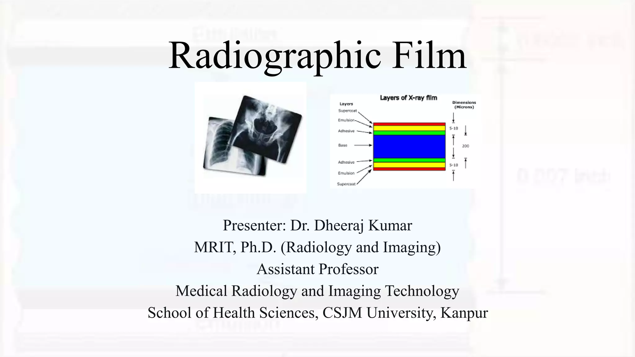
SlideShare a Scribd company logo
Radiographic Film
Presenter: Dr. Dheeraj Kumar
MRIT, Ph.D. (Radiology and Imaging)
Assistant Professor
Medical Radiology and Imaging Technology
School of Health Sciences, CSJM University, Kanpur
Overview of Radiographic Film
• Radiographic film is a light-sensitive material used in medical imaging to record X-ray images. It
acts as a medium to capture X-rays that pass through the patient's body, resulting in an image that
helps diagnose various medical conditions.
• The history of radiographic film dates back to the early 20th century when it revolutionized the
field of radiology. Today, it remains an essential tool in medical imaging, despite the advancements
in digital technology.
• Radiographic film is utilized in various imaging modalities, including conventional radiography,
fluoroscopy, and mammography. Its versatility and ease of use make it a preferred choice in many
clinical settings.
History
• Discovery of X-rays: In 1895, Wilhelm Conrad Roentgen, a German physicist, accidentally
discovered X-rays while experimenting with cathode-ray tubes. He noticed that a mysterious,
invisible radiation could penetrate solid objects and produce images on photographic plates. This
ground breaking discovery laid the foundation for the field of radiology and medical imaging.
• Early Photographic Plates: Following Roentgen's discovery, early attempts at medical imaging
involved using photographic plates to capture X-ray images. These plates were sensitive to X-rays
but required long exposure times, making them impractical for clinical use.
History continue….
• Introduction of Glass Plate Radiographs: By the late 19th century, glass plate
radiographs became more widely used. The X-ray image was captured on glass plates
coated with photographic emulsion. These plates provided better image quality than early
photographic plates but still required lengthy exposure times.
• Flexible Celluloid Films: In 1896, Thomas Edison and Clarence Dally introduced the
idea of using flexible celluloid film for radiography. They coated celluloid sheets with a
photosensitive emulsion, leading to the creation of flexible X-ray films. However, the
quality of these early films was limited, and they were highly flammable.
History continue….
• Introduction of X-ray Film Screens: In the early 20th century, intensifying screens were
introduced to reduce exposure times during X-ray imaging. Intensifying screens contained
fluorescent materials that converted X-rays into visible light, which in turn exposed the
X-ray film. This significantly reduced patient radiation dose and improved image quality.
• Single-Emulsion Radiographic Film: In 1918, the single-emulsion radiographic film
was developed by using a single layer of photosensitive emulsion on one side of the film
base. This film offered improved sensitivity and image quality, making it a standard
choice for medical imaging for several decades.
History continue….
• Double-Emulsion Radiographic Film: In the 1940s, double-emulsion radiographic film
was introduced, which contained two layers of photosensitive emulsion on opposite sides
of the film base. This advancement further improved image quality and reduced the need
for retakes.
• Development of Screen-Film Systems: Throughout the mid-20th century, screen-film
systems became the dominant method of radiographic imaging. These systems combined
the use of intensifying screens with double-emulsion radiographic film, drastically
reducing exposure times and radiation dose while providing high-quality images.
History continue….
• Introduction of Computed Radiography (CR): In the 1980s, computed radiography
(CR) revolutionized medical imaging by replacing traditional film-based radiography
with digital imaging technology. CR systems used photo stimulable phosphor plates to
capture X-ray images digitally, eliminating the need for film processing.
• Transition to Digital Radiography (DR): In the late 20th century and early 21st century,
digital radiography (DR) systems gained popularity, directly capturing X-ray images
using solid-state detectors. DR offered faster image acquisition, better image
manipulation, and improved workflow efficiency.
Emergence of Digital Imaging
• In recent years, advancements in digital technology have continued to shape the field of radiology.
Digital imaging technologies, such as picture archiving and communication systems (PACS) and
teleradiology, have transformed the way radiologists view, store, and share medical images.
• Radiographic film is gradually being replaced by digital imaging systems, which offer numerous
advantages in terms of image quality, accessibility, and integration with other medical systems.
Despite this transition, the rich history of radiographic film remains an integral part of the
evolution of medical imaging and continues to be appreciated for its contribution to the field of
radiology.
Types of Radiographic Film
• There are different types of
radiographic films available,
each with distinct characteristics
and applications. Let's explore
the main types:
Screen-Film Radiography
• Traditional radiographic film combined
with intensifying screens.
• Intensifying screens convert X-rays into
visible light, enhancing image formation.
• Two types: single-emulsion (used for
general radiography) and double-emulsion
(used for higher resolution imaging).
Digital Radiography (DR)
• Replaces traditional film with digital
sensors to capture X-ray images
directly.
• Two main types: Direct DR (uses
amorphous selenium or other
materials) and Indirect DR (uses a
scintillator to convert X-rays to
visible light).
Computed Radiography (CR)
• Utilizes photostimulable phosphor plates to
record X-ray images.
• These plates store energy when exposed to X-
rays and release it when scanned with a laser to
create the digital image.
• Each type of radiographic film has its
advantages and limitations, and understanding
their differences is essential for choosing the
appropriate imaging method for specific clinical
scenarios.
Structure of Radiographic Film
• Radiographic films consist of several layers
that work together to produce a high-quality
image. The primary components include:
• Emulsion Layer:
• Contains light-sensitive silver halide crystals
suspended in gelatin.
• These crystals react to X-rays, capturing the
image information.
Structure of Radiographic Film Continue….
• Base:
• Provides support and stability to the emulsion layer.
• Usually made of polyester, ensuring the film's durability.
• Protective Layer:
• Guards the emulsion from damage during handling and processing.
• Enhances the film's resistance to scratches and chemical reactions.
• The combination of these layers ensures efficient image formation and protects the film
from external factors that might compromise image quality.
Screen-Film Radiography
• Screen-film radiography is a widely used imaging technique that incorporates intensifying
screens to enhance image capture. The process involves the following steps:
• X-ray Interaction:
• X-rays pass through the patient's body, interacting with tissues and creating a latent image on the
radiographic film's emulsion.
• Intensifying Screens:
• Intensifying screens, placed on both sides of the radiographic film, convert X-rays into visible light.
• This light exposes the emulsion, creating a visible image on the film.
Screen-Film Radiography Continue…
• Image Formation:
• The combination of X-ray interactions and light
emission forms the final radiographic image.
• The resulting film can be developed and viewed for
diagnostic purposes.
• Screen-film radiography remains essential in
many clinical scenarios, but it has limitations,
such as lower image resolution compared to
digital methods.
Digital Radiography (DR)
• Digital radiography represents a
significant advancement in medical
imaging technology. Unlike
screen-film radiography, DR
directly captures X-ray images
using digital sensors. Let's explore
the two main types of DR:
Direct DR
• Utilizes solid-state detectors (e.g.,
amorphous selenium) to directly
convert X-rays into electrical
signals.
• These signals are processed and
converted into digital images
without the need for intensifying
screens.
Indirect DR
• Employs scintillator materials (e.g.,
cesium iodide) to convert X-rays into
visible light.
• The light is then detected by an
amorphous silicon photodiode array,
which converts it into electrical signals
for image formation.
• DR offers numerous
advantages, including rapid
image acquisition, dose
reduction, and the ability to
post-process images for better
visualization.
Computed Radiography (CR)
• Computed Radiography (CR) is another digital imaging method that utilizes
photostimulable phosphor plates. Here's how it works:
• X-ray Exposure:
• X-rays expose the phosphor plates, causing them to store energy in the form of a latent image.
• Image Readout:
• The phosphor plates are scanned with a laser, stimulating the release of stored energy as visible
light.
• Image Formation:
• The emitted light is detected and converted into a digital image that can be displayed on a computer.
CR offers flexibility and the ability to retrofit existing conventional
radiography systems for digital imaging.
Image Quality and Artifacts
• Image quality is crucial in radiology, as it directly impacts diagnostic
accuracy. Several factors influence image quality, including:
• Spatial Resolution:
• The ability of the system to distinguish fine details and structures in the image.
• Higher spatial resolution results in sharper images and improved diagnostic
capabilities.
Image Quality and Artifacts Continue….
• Image Contrast:
• The difference in brightness between different tissues and structures in the image.
• Optimal contrast allows for better visualization and differentiation of anatomical features.
• Image Noise:
• Random fluctuations in pixel intensity, reducing image clarity and detail.
• Image noise should be minimized to achieve high-quality radiographs.
• Artifacts can compromise image quality and lead to misinterpretation of results. Common
Artifacts include motion blur, grid lines, and processing Artifacts. Identifying and mitigating
Artifacts are crucial for accurate diagnoses.
Handling and Storage of Radiographic Films
• Proper handling and storage of radiographic films are essential to maintain image quality and
prolong their lifespan. Follow these guidelines:
• Film Handling:
• Always handle films with clean, dry hands to avoid contamination.
• Avoid bending or folding the film to prevent damage.
• Film Storage:
• Store films in a cool, dry environment away from direct sunlight.
• Use protective sleeves or boxes to shield films from dust and scratches.
• Labelling and organizing films are vital to ensure easy retrieval and proper patient documentation.
Film Processing
• In traditional screen-film radiography, film processing is a critical step to produce high-quality images.
The process involves several stages:
• Development:
• Immersing the exposed film in a chemical developer to convert the latent image into a visible image.
• Fixation:
• Removing unexposed silver halide crystals from the film using a chemical fixer.
• Washing:
• Rinsing the film in water to remove residual chemicals.
• Drying:
• Allowing the film to dry completely before viewing or archiving.
Radiographic Film Analysis
• As radiology students, the ability to analysis radiographic images is vital for accurate diagnosis.
Consider the following criteria during image evaluation:
• Image Clarity:
• Assess the sharpness and detail of anatomical structures.
• Image Density:
• Evaluate the brightness and darkness of tissues in the image.
• Positioning:
• Check for proper patient positioning and alignment to the X-ray beam.
• Artifacts:
• Identify and analyse any Artifacts that may affect image quality.
Transition to Digital Imaging
• With the rapid advancement of digital imaging technology, the healthcare industry is
transitioning from traditional radiographic film to digital systems. This shift offers
numerous benefits, including:
• Faster Image Acquisition:
• Digital systems provide real-time image capture, reducing patient waiting times.
• Enhanced Image Manipulation:
• Digital images can be post-processed to optimize visualization.
• Lower Radiation Dose:
• Some digital systems use lower X-ray doses, contributing to patient safety.
Future Trends in Radiographic Film
Technology
• The future of radiographic film technology is promising, with ongoing research
and development in the field. Some potential trends include:
• Advanced Digital Detectors:
• Continual improvements in solid-state detectors for higher image resolution.
• Artificial Intelligence (AI) Integration:
• AI algorithms may assist in image analysis and diagnosis.
• Portable and Wireless Solutions:
• Compact and wireless digital imaging devices for greater convenience.
Conclusion
• In conclusion, radiographic film remains a cornerstone of diagnostic
radiology. Understanding the different types of radiographic film, their
structures, and their applications will serve us well in our future careers.
• As radiology students, developing our image analysis skills and embracing
digital imaging technologies will enable us to provide accurate and efficient
diagnoses to improve patient care.
References
• Smith, J. (2020). Fundamentals of Radiography. Springer International
Publishing.
• Bushong, S. C. (2017). Radiologic Science for Technologists. Elsevier.
• Image critique in radiography - A practical guide. (2016).
Radiography, 22(1), e1-e7.
Questions and Answers
• Now, the floor is open for questions. Feel free to ask any queries you may
have about radiographic film, digital imaging, or any related topics.
Thank You

More Related Content

What's hot

Computed Radiography and digital radiography
Computed Radiography and  digital radiographyComputed Radiography and  digital radiography
Computed Radiography and digital radiography
Durga Singh
 
Soft tissue radiography.pptx
Soft tissue radiography.pptxSoft tissue radiography.pptx
Soft tissue radiography.pptx
Ritupanta1
 
Beam restriction
Beam restrictionBeam restriction
Beam restriction
aslam bs
 
Quality assurance
Quality  assuranceQuality  assurance
Quality assurance
Khursheed Ganie
 
Faults in x ray tube and its care
Faults in x ray tube and its careFaults in x ray tube and its care
Faults in x ray tube and its care
SaruGosain
 
Production of xray
Production of xrayProduction of xray
Production of xray
Prajwith Rai
 
X ray generator new.ppt
X ray generator new.pptX ray generator new.ppt
X ray generator new.ppt
jayapandiyan Paraman
 
X-Ray Properties.pptx
X-Ray Properties.pptxX-Ray Properties.pptx
X-Ray Properties.pptx
Dr. Dheeraj Kumar
 
Production of x rays
Production of x rays Production of x rays
Production of x rays
VIneeth C
 
soft tissue radiography
soft tissue radiographysoft tissue radiography
soft tissue radiography
Dinesh Darshana
 
Production of x rays & generators
Production of x rays & generatorsProduction of x rays & generators
Production of x rays & generators
Rakesh Ca
 
Faults in xray tube by Sandesh magar
Faults in xray tube by Sandesh magarFaults in xray tube by Sandesh magar
Faults in xray tube by Sandesh magar
SandeshMagar4
 
Filters
FiltersFilters
Filters
aslam bs
 
Beam restricted device and filter used in x ray
Beam restricted device and filter used in x rayBeam restricted device and filter used in x ray
Beam restricted device and filter used in x ray
SushilPattar
 
Factors affecting x-ray beam quality and quantity
Factors affecting x-ray beam quality and quantityFactors affecting x-ray beam quality and quantity
Factors affecting x-ray beam quality and quantity
ravi christian
 
Mobile & portable radiography
Mobile & portable radiographyMobile & portable radiography
Mobile & portable radiography
Priyanka Parimala
 
ICRP-International commission on Radiation Protection
ICRP-International commission on Radiation ProtectionICRP-International commission on Radiation Protection
ICRP-International commission on Radiation Protection
Chandan Prasad
 
MACRORADIOGRAPHY.pptx
MACRORADIOGRAPHY.pptxMACRORADIOGRAPHY.pptx
MACRORADIOGRAPHY.pptx
ThejaTej6
 
Fluroscopy
Fluroscopy Fluroscopy
Fluroscopy
Rakesh Ca
 
X rays generators and transformers
X rays   generators  and transformersX rays   generators  and transformers
X rays generators and transformers
shariq ahmad shah
 

What's hot (20)

Computed Radiography and digital radiography
Computed Radiography and  digital radiographyComputed Radiography and  digital radiography
Computed Radiography and digital radiography
 
Soft tissue radiography.pptx
Soft tissue radiography.pptxSoft tissue radiography.pptx
Soft tissue radiography.pptx
 
Beam restriction
Beam restrictionBeam restriction
Beam restriction
 
Quality assurance
Quality  assuranceQuality  assurance
Quality assurance
 
Faults in x ray tube and its care
Faults in x ray tube and its careFaults in x ray tube and its care
Faults in x ray tube and its care
 
Production of xray
Production of xrayProduction of xray
Production of xray
 
X ray generator new.ppt
X ray generator new.pptX ray generator new.ppt
X ray generator new.ppt
 
X-Ray Properties.pptx
X-Ray Properties.pptxX-Ray Properties.pptx
X-Ray Properties.pptx
 
Production of x rays
Production of x rays Production of x rays
Production of x rays
 
soft tissue radiography
soft tissue radiographysoft tissue radiography
soft tissue radiography
 
Production of x rays & generators
Production of x rays & generatorsProduction of x rays & generators
Production of x rays & generators
 
Faults in xray tube by Sandesh magar
Faults in xray tube by Sandesh magarFaults in xray tube by Sandesh magar
Faults in xray tube by Sandesh magar
 
Filters
FiltersFilters
Filters
 
Beam restricted device and filter used in x ray
Beam restricted device and filter used in x rayBeam restricted device and filter used in x ray
Beam restricted device and filter used in x ray
 
Factors affecting x-ray beam quality and quantity
Factors affecting x-ray beam quality and quantityFactors affecting x-ray beam quality and quantity
Factors affecting x-ray beam quality and quantity
 
Mobile & portable radiography
Mobile & portable radiographyMobile & portable radiography
Mobile & portable radiography
 
ICRP-International commission on Radiation Protection
ICRP-International commission on Radiation ProtectionICRP-International commission on Radiation Protection
ICRP-International commission on Radiation Protection
 
MACRORADIOGRAPHY.pptx
MACRORADIOGRAPHY.pptxMACRORADIOGRAPHY.pptx
MACRORADIOGRAPHY.pptx
 
Fluroscopy
Fluroscopy Fluroscopy
Fluroscopy
 
X rays generators and transformers
X rays   generators  and transformersX rays   generators  and transformers
X rays generators and transformers
 

Similar to Radiographic Film.pptx

3-MEDICAL-IMAGING…………. ……………………………….
3-MEDICAL-IMAGING………….     ……………………………….3-MEDICAL-IMAGING………….     ……………………………….
3-MEDICAL-IMAGING…………. ……………………………….
hdhdufyfuei78
 
X-ray Machine
X-ray Machine X-ray Machine
X-ray Machine
SamyaQadi
 
Ndtm 5
Ndtm 5Ndtm 5
Digital Radiography
Digital RadiographyDigital Radiography
Digital Radiography
Upakar Paudel
 
nasif radiology
nasif radiologynasif radiology
nasif radiology
Mohamed Gad
 
Image receptors for radiology seminar
Image receptors   for radiology seminarImage receptors   for radiology seminar
Image receptors for radiology seminar
Puneet Choudhary
 
Image receptors/ dental crown & bridge courses
Image receptors/ dental crown & bridge coursesImage receptors/ dental crown & bridge courses
Image receptors/ dental crown & bridge courses
Indian dental academy
 
digital imaging ppt.ppt
digital imaging ppt.pptdigital imaging ppt.ppt
digital imaging ppt.ppt
vishalmehrotra10
 
Radiographic Considerations in Implants
Radiographic Considerations in ImplantsRadiographic Considerations in Implants
Radiographic Considerations in Implants
Dr.Richa Sahai
 
radiographs basics
radiographs basicsradiographs basics
radiographs basics
Neha Sharma
 
Basics of imaging by mohamed abou el ghar
Basics of imaging by mohamed abou el gharBasics of imaging by mohamed abou el ghar
Basics of imaging by mohamed abou el ghar
FarragBahbah
 
X ray machine l
X ray machine lX ray machine l
X ray machine l
DiNa Amin
 
Digital radiography.. an update
Digital radiography.. an updateDigital radiography.. an update
Digital radiography.. an update
Shivam Batra
 
General x-ray machine and fluoroscopy
General x-ray machine and fluoroscopyGeneral x-ray machine and fluoroscopy
General x-ray machine and fluoroscopy
Osama Albedaiwi
 
Medical x ray image sensors
Medical x ray image sensorsMedical x ray image sensors
Medical x ray image sensors
Islam Kotb Ismail
 
DIGITAL IMAGING
DIGITAL IMAGINGDIGITAL IMAGING
DIGITAL IMAGING
SharmaRajan4
 
Radiographic system clinical engineering
Radiographic system clinical engineeringRadiographic system clinical engineering
Radiographic system clinical engineering
HasibulHasanHredoy
 
Introduction to Digital Dentistry
Introduction to Digital DentistryIntroduction to Digital Dentistry
Introduction to Digital Dentistry
Ibrahim Tayyan
 
intensifying screens and grids.pptx
intensifying screens and grids.pptxintensifying screens and grids.pptx
intensifying screens and grids.pptx
ChandiniDabbiru1
 
Orthodontic radiograph..
Orthodontic radiograph.. Orthodontic radiograph..
Orthodontic radiograph..
nehal albelasy
 

Similar to Radiographic Film.pptx (20)

3-MEDICAL-IMAGING…………. ……………………………….
3-MEDICAL-IMAGING………….     ……………………………….3-MEDICAL-IMAGING………….     ……………………………….
3-MEDICAL-IMAGING…………. ……………………………….
 
X-ray Machine
X-ray Machine X-ray Machine
X-ray Machine
 
Ndtm 5
Ndtm 5Ndtm 5
Ndtm 5
 
Digital Radiography
Digital RadiographyDigital Radiography
Digital Radiography
 
nasif radiology
nasif radiologynasif radiology
nasif radiology
 
Image receptors for radiology seminar
Image receptors   for radiology seminarImage receptors   for radiology seminar
Image receptors for radiology seminar
 
Image receptors/ dental crown & bridge courses
Image receptors/ dental crown & bridge coursesImage receptors/ dental crown & bridge courses
Image receptors/ dental crown & bridge courses
 
digital imaging ppt.ppt
digital imaging ppt.pptdigital imaging ppt.ppt
digital imaging ppt.ppt
 
Radiographic Considerations in Implants
Radiographic Considerations in ImplantsRadiographic Considerations in Implants
Radiographic Considerations in Implants
 
radiographs basics
radiographs basicsradiographs basics
radiographs basics
 
Basics of imaging by mohamed abou el ghar
Basics of imaging by mohamed abou el gharBasics of imaging by mohamed abou el ghar
Basics of imaging by mohamed abou el ghar
 
X ray machine l
X ray machine lX ray machine l
X ray machine l
 
Digital radiography.. an update
Digital radiography.. an updateDigital radiography.. an update
Digital radiography.. an update
 
General x-ray machine and fluoroscopy
General x-ray machine and fluoroscopyGeneral x-ray machine and fluoroscopy
General x-ray machine and fluoroscopy
 
Medical x ray image sensors
Medical x ray image sensorsMedical x ray image sensors
Medical x ray image sensors
 
DIGITAL IMAGING
DIGITAL IMAGINGDIGITAL IMAGING
DIGITAL IMAGING
 
Radiographic system clinical engineering
Radiographic system clinical engineeringRadiographic system clinical engineering
Radiographic system clinical engineering
 
Introduction to Digital Dentistry
Introduction to Digital DentistryIntroduction to Digital Dentistry
Introduction to Digital Dentistry
 
intensifying screens and grids.pptx
intensifying screens and grids.pptxintensifying screens and grids.pptx
intensifying screens and grids.pptx
 
Orthodontic radiograph..
Orthodontic radiograph.. Orthodontic radiograph..
Orthodontic radiograph..
 

More from Dr. Dheeraj Kumar

Ultrasound Image Artifacts and Their Remedies.pptx
Ultrasound Image Artifacts and Their Remedies.pptxUltrasound Image Artifacts and Their Remedies.pptx
Ultrasound Image Artifacts and Their Remedies.pptx
Dr. Dheeraj Kumar
 
MRI Artifacts and Their Remedies/Corrections.pptx
MRI Artifacts and Their Remedies/Corrections.pptxMRI Artifacts and Their Remedies/Corrections.pptx
MRI Artifacts and Their Remedies/Corrections.pptx
Dr. Dheeraj Kumar
 
Measurement of Radiation and Dosimetric Procedure.pptx
Measurement of Radiation and Dosimetric Procedure.pptxMeasurement of Radiation and Dosimetric Procedure.pptx
Measurement of Radiation and Dosimetric Procedure.pptx
Dr. Dheeraj Kumar
 
Radiation Dosimetry Parameters and Isodose Curves.pptx
Radiation Dosimetry Parameters and Isodose Curves.pptxRadiation Dosimetry Parameters and Isodose Curves.pptx
Radiation Dosimetry Parameters and Isodose Curves.pptx
Dr. Dheeraj Kumar
 
Culture and Health Disorders Social change.pptx
Culture and Health Disorders Social change.pptxCulture and Health Disorders Social change.pptx
Culture and Health Disorders Social change.pptx
Dr. Dheeraj Kumar
 
Units of Radiation Measurements, Quality Specification, Half-Value Thickness,...
Units of Radiation Measurements, Quality Specification, Half-Value Thickness,...Units of Radiation Measurements, Quality Specification, Half-Value Thickness,...
Units of Radiation Measurements, Quality Specification, Half-Value Thickness,...
Dr. Dheeraj Kumar
 
X-Ray Beam Restrictors/Beam Modifying devices.pptx
X-Ray Beam Restrictors/Beam Modifying devices.pptxX-Ray Beam Restrictors/Beam Modifying devices.pptx
X-Ray Beam Restrictors/Beam Modifying devices.pptx
Dr. Dheeraj Kumar
 
Range of Secondary Electrons and Electron Build-Up: Impact on Scatter in Homo...
Range of Secondary Electrons and Electron Build-Up: Impact on Scatter in Homo...Range of Secondary Electrons and Electron Build-Up: Impact on Scatter in Homo...
Range of Secondary Electrons and Electron Build-Up: Impact on Scatter in Homo...
Dr. Dheeraj Kumar
 
Transmission of X-ray through body tissues linear energy transfer..pptx
Transmission of X-ray through body tissues linear energy transfer..pptxTransmission of X-ray through body tissues linear energy transfer..pptx
Transmission of X-ray through body tissues linear energy transfer..pptx
Dr. Dheeraj Kumar
 
X-ray Production A Journey Through History and the X-ray Tube.pptx
X-ray Production A Journey Through History and the X-ray Tube.pptxX-ray Production A Journey Through History and the X-ray Tube.pptx
X-ray Production A Journey Through History and the X-ray Tube.pptx
Dr. Dheeraj Kumar
 
Population of India and Family welfare programme.pptx
Population of India and Family welfare programme.pptxPopulation of India and Family welfare programme.pptx
Population of India and Family welfare programme.pptx
Dr. Dheeraj Kumar
 
Artificial Radionuclide Generators in Medicine Applications in Radiotherapy.pptx
Artificial Radionuclide Generators in Medicine Applications in Radiotherapy.pptxArtificial Radionuclide Generators in Medicine Applications in Radiotherapy.pptx
Artificial Radionuclide Generators in Medicine Applications in Radiotherapy.pptx
Dr. Dheeraj Kumar
 
Effects of variation of tube voltage current, filtration..pptx
Effects of variation of tube voltage current, filtration..pptxEffects of variation of tube voltage current, filtration..pptx
Effects of variation of tube voltage current, filtration..pptx
Dr. Dheeraj Kumar
 
Radioactivity spectrum of diagnostic imaging and therapy X ray..pptx
Radioactivity spectrum of diagnostic imaging and therapy X ray..pptxRadioactivity spectrum of diagnostic imaging and therapy X ray..pptx
Radioactivity spectrum of diagnostic imaging and therapy X ray..pptx
Dr. Dheeraj Kumar
 
Atomic structure as applied to generation of X-rays.pptx
Atomic structure as applied to generation of X-rays.pptxAtomic structure as applied to generation of X-rays.pptx
Atomic structure as applied to generation of X-rays.pptx
Dr. Dheeraj Kumar
 
Radiation and Spectra.pptx
Radiation and Spectra.pptxRadiation and Spectra.pptx
Radiation and Spectra.pptx
Dr. Dheeraj Kumar
 
Guidelines for X-Ray Equipments.pptx
Guidelines for X-Ray Equipments.pptxGuidelines for X-Ray Equipments.pptx
Guidelines for X-Ray Equipments.pptx
Dr. Dheeraj Kumar
 
Clinical Applications and Procedure of MRI.pptx
Clinical Applications and Procedure of MRI.pptxClinical Applications and Procedure of MRI.pptx
Clinical Applications and Procedure of MRI.pptx
Dr. Dheeraj Kumar
 
Physical Principle of MRI.pptx
Physical Principle of MRI.pptxPhysical Principle of MRI.pptx
Physical Principle of MRI.pptx
Dr. Dheeraj Kumar
 
Tomography.pptx
Tomography.pptxTomography.pptx
Tomography.pptx
Dr. Dheeraj Kumar
 

More from Dr. Dheeraj Kumar (20)

Ultrasound Image Artifacts and Their Remedies.pptx
Ultrasound Image Artifacts and Their Remedies.pptxUltrasound Image Artifacts and Their Remedies.pptx
Ultrasound Image Artifacts and Their Remedies.pptx
 
MRI Artifacts and Their Remedies/Corrections.pptx
MRI Artifacts and Their Remedies/Corrections.pptxMRI Artifacts and Their Remedies/Corrections.pptx
MRI Artifacts and Their Remedies/Corrections.pptx
 
Measurement of Radiation and Dosimetric Procedure.pptx
Measurement of Radiation and Dosimetric Procedure.pptxMeasurement of Radiation and Dosimetric Procedure.pptx
Measurement of Radiation and Dosimetric Procedure.pptx
 
Radiation Dosimetry Parameters and Isodose Curves.pptx
Radiation Dosimetry Parameters and Isodose Curves.pptxRadiation Dosimetry Parameters and Isodose Curves.pptx
Radiation Dosimetry Parameters and Isodose Curves.pptx
 
Culture and Health Disorders Social change.pptx
Culture and Health Disorders Social change.pptxCulture and Health Disorders Social change.pptx
Culture and Health Disorders Social change.pptx
 
Units of Radiation Measurements, Quality Specification, Half-Value Thickness,...
Units of Radiation Measurements, Quality Specification, Half-Value Thickness,...Units of Radiation Measurements, Quality Specification, Half-Value Thickness,...
Units of Radiation Measurements, Quality Specification, Half-Value Thickness,...
 
X-Ray Beam Restrictors/Beam Modifying devices.pptx
X-Ray Beam Restrictors/Beam Modifying devices.pptxX-Ray Beam Restrictors/Beam Modifying devices.pptx
X-Ray Beam Restrictors/Beam Modifying devices.pptx
 
Range of Secondary Electrons and Electron Build-Up: Impact on Scatter in Homo...
Range of Secondary Electrons and Electron Build-Up: Impact on Scatter in Homo...Range of Secondary Electrons and Electron Build-Up: Impact on Scatter in Homo...
Range of Secondary Electrons and Electron Build-Up: Impact on Scatter in Homo...
 
Transmission of X-ray through body tissues linear energy transfer..pptx
Transmission of X-ray through body tissues linear energy transfer..pptxTransmission of X-ray through body tissues linear energy transfer..pptx
Transmission of X-ray through body tissues linear energy transfer..pptx
 
X-ray Production A Journey Through History and the X-ray Tube.pptx
X-ray Production A Journey Through History and the X-ray Tube.pptxX-ray Production A Journey Through History and the X-ray Tube.pptx
X-ray Production A Journey Through History and the X-ray Tube.pptx
 
Population of India and Family welfare programme.pptx
Population of India and Family welfare programme.pptxPopulation of India and Family welfare programme.pptx
Population of India and Family welfare programme.pptx
 
Artificial Radionuclide Generators in Medicine Applications in Radiotherapy.pptx
Artificial Radionuclide Generators in Medicine Applications in Radiotherapy.pptxArtificial Radionuclide Generators in Medicine Applications in Radiotherapy.pptx
Artificial Radionuclide Generators in Medicine Applications in Radiotherapy.pptx
 
Effects of variation of tube voltage current, filtration..pptx
Effects of variation of tube voltage current, filtration..pptxEffects of variation of tube voltage current, filtration..pptx
Effects of variation of tube voltage current, filtration..pptx
 
Radioactivity spectrum of diagnostic imaging and therapy X ray..pptx
Radioactivity spectrum of diagnostic imaging and therapy X ray..pptxRadioactivity spectrum of diagnostic imaging and therapy X ray..pptx
Radioactivity spectrum of diagnostic imaging and therapy X ray..pptx
 
Atomic structure as applied to generation of X-rays.pptx
Atomic structure as applied to generation of X-rays.pptxAtomic structure as applied to generation of X-rays.pptx
Atomic structure as applied to generation of X-rays.pptx
 
Radiation and Spectra.pptx
Radiation and Spectra.pptxRadiation and Spectra.pptx
Radiation and Spectra.pptx
 
Guidelines for X-Ray Equipments.pptx
Guidelines for X-Ray Equipments.pptxGuidelines for X-Ray Equipments.pptx
Guidelines for X-Ray Equipments.pptx
 
Clinical Applications and Procedure of MRI.pptx
Clinical Applications and Procedure of MRI.pptxClinical Applications and Procedure of MRI.pptx
Clinical Applications and Procedure of MRI.pptx
 
Physical Principle of MRI.pptx
Physical Principle of MRI.pptxPhysical Principle of MRI.pptx
Physical Principle of MRI.pptx
 
Tomography.pptx
Tomography.pptxTomography.pptx
Tomography.pptx
 

Recently uploaded

Part II - Body Grief: Losing parts of ourselves and our identity before, duri...
Part II - Body Grief: Losing parts of ourselves and our identity before, duri...Part II - Body Grief: Losing parts of ourselves and our identity before, duri...
Part II - Body Grief: Losing parts of ourselves and our identity before, duri...
bkling
 
Local Advanced Lung Cancer: Artificial Intelligence, Synergetics, Complex Sys...
Local Advanced Lung Cancer: Artificial Intelligence, Synergetics, Complex Sys...Local Advanced Lung Cancer: Artificial Intelligence, Synergetics, Complex Sys...
Local Advanced Lung Cancer: Artificial Intelligence, Synergetics, Complex Sys...
Oleg Kshivets
 
TEST BANK For Community Health Nursing A Canadian Perspective, 5th Edition by...
TEST BANK For Community Health Nursing A Canadian Perspective, 5th Edition by...TEST BANK For Community Health Nursing A Canadian Perspective, 5th Edition by...
TEST BANK For Community Health Nursing A Canadian Perspective, 5th Edition by...
Donc Test
 
Role of Mukta Pishti in the Management of Hyperthyroidism
Role of Mukta Pishti in the Management of HyperthyroidismRole of Mukta Pishti in the Management of Hyperthyroidism
Role of Mukta Pishti in the Management of Hyperthyroidism
Dr. Jyothirmai Paindla
 
Hemodialysis: Chapter 4, Dialysate Circuit - Dr.Gawad
Hemodialysis: Chapter 4, Dialysate Circuit - Dr.GawadHemodialysis: Chapter 4, Dialysate Circuit - Dr.Gawad
Hemodialysis: Chapter 4, Dialysate Circuit - Dr.Gawad
NephroTube - Dr.Gawad
 
#cALL# #gIRLS# In Dehradun ꧁❤8107221448❤꧂#cALL# #gIRLS# Service In Dehradun W...
#cALL# #gIRLS# In Dehradun ꧁❤8107221448❤꧂#cALL# #gIRLS# Service In Dehradun W...#cALL# #gIRLS# In Dehradun ꧁❤8107221448❤꧂#cALL# #gIRLS# Service In Dehradun W...
#cALL# #gIRLS# In Dehradun ꧁❤8107221448❤꧂#cALL# #gIRLS# Service In Dehradun W...
chandankumarsmartiso
 
Journal Article Review on Rasamanikya
Journal Article Review on RasamanikyaJournal Article Review on Rasamanikya
Journal Article Review on Rasamanikya
Dr. Jyothirmai Paindla
 
Top 10 Best Ayurvedic Kidney Stone Syrups in India
Top 10 Best Ayurvedic Kidney Stone Syrups in IndiaTop 10 Best Ayurvedic Kidney Stone Syrups in India
Top 10 Best Ayurvedic Kidney Stone Syrups in India
Swastik Ayurveda
 
Novas diretrizes da OMS para os cuidados perinatais de mais qualidade
Novas diretrizes da OMS para os cuidados perinatais de mais qualidadeNovas diretrizes da OMS para os cuidados perinatais de mais qualidade
Novas diretrizes da OMS para os cuidados perinatais de mais qualidade
Prof. Marcus Renato de Carvalho
 
Muscles of Mastication by Dr. Rabia Inam Gandapore.pptx
Muscles of Mastication by Dr. Rabia Inam Gandapore.pptxMuscles of Mastication by Dr. Rabia Inam Gandapore.pptx
Muscles of Mastication by Dr. Rabia Inam Gandapore.pptx
Dr. Rabia Inam Gandapore
 
Adhd Medication Shortage Uk - trinexpharmacy.com
Adhd Medication Shortage Uk - trinexpharmacy.comAdhd Medication Shortage Uk - trinexpharmacy.com
Adhd Medication Shortage Uk - trinexpharmacy.com
reignlana06
 
Does Over-Masturbation Contribute to Chronic Prostatitis.pptx
Does Over-Masturbation Contribute to Chronic Prostatitis.pptxDoes Over-Masturbation Contribute to Chronic Prostatitis.pptx
Does Over-Masturbation Contribute to Chronic Prostatitis.pptx
walterHu5
 
ABDOMINAL TRAUMA in pediatrics part one.
ABDOMINAL TRAUMA in pediatrics part one.ABDOMINAL TRAUMA in pediatrics part one.
ABDOMINAL TRAUMA in pediatrics part one.
drhasanrajab
 
CHEMOTHERAPY_RDP_CHAPTER 1_ANTI TB DRUGS.pdf
CHEMOTHERAPY_RDP_CHAPTER 1_ANTI TB DRUGS.pdfCHEMOTHERAPY_RDP_CHAPTER 1_ANTI TB DRUGS.pdf
CHEMOTHERAPY_RDP_CHAPTER 1_ANTI TB DRUGS.pdf
rishi2789
 
THERAPEUTIC ANTISENSE MOLECULES .pptx
THERAPEUTIC ANTISENSE MOLECULES    .pptxTHERAPEUTIC ANTISENSE MOLECULES    .pptx
THERAPEUTIC ANTISENSE MOLECULES .pptx
70KRISHPATEL
 
CHEMOTHERAPY_RDP_CHAPTER 3_ANTIFUNGAL AGENT.pdf
CHEMOTHERAPY_RDP_CHAPTER 3_ANTIFUNGAL AGENT.pdfCHEMOTHERAPY_RDP_CHAPTER 3_ANTIFUNGAL AGENT.pdf
CHEMOTHERAPY_RDP_CHAPTER 3_ANTIFUNGAL AGENT.pdf
rishi2789
 
Best Ayurvedic medicine for Gas and Indigestion
Best Ayurvedic medicine for Gas and IndigestionBest Ayurvedic medicine for Gas and Indigestion
Best Ayurvedic medicine for Gas and Indigestion
Swastik Ayurveda
 
A Classical Text Review on Basavarajeeyam
A Classical Text Review on BasavarajeeyamA Classical Text Review on Basavarajeeyam
A Classical Text Review on Basavarajeeyam
Dr. Jyothirmai Paindla
 
Efficacy of Avartana Sneha in Ayurveda
Efficacy of Avartana Sneha in AyurvedaEfficacy of Avartana Sneha in Ayurveda
Efficacy of Avartana Sneha in Ayurveda
Dr. Jyothirmai Paindla
 
Phone Us ❤8107221448❤ #ℂall #gIRLS In Dehradun By Dehradun @ℂall @Girls Hotel...
Phone Us ❤8107221448❤ #ℂall #gIRLS In Dehradun By Dehradun @ℂall @Girls Hotel...Phone Us ❤8107221448❤ #ℂall #gIRLS In Dehradun By Dehradun @ℂall @Girls Hotel...
Phone Us ❤8107221448❤ #ℂall #gIRLS In Dehradun By Dehradun @ℂall @Girls Hotel...
chandankumarsmartiso
 

Recently uploaded (20)

Part II - Body Grief: Losing parts of ourselves and our identity before, duri...
Part II - Body Grief: Losing parts of ourselves and our identity before, duri...Part II - Body Grief: Losing parts of ourselves and our identity before, duri...
Part II - Body Grief: Losing parts of ourselves and our identity before, duri...
 
Local Advanced Lung Cancer: Artificial Intelligence, Synergetics, Complex Sys...
Local Advanced Lung Cancer: Artificial Intelligence, Synergetics, Complex Sys...Local Advanced Lung Cancer: Artificial Intelligence, Synergetics, Complex Sys...
Local Advanced Lung Cancer: Artificial Intelligence, Synergetics, Complex Sys...
 
TEST BANK For Community Health Nursing A Canadian Perspective, 5th Edition by...
TEST BANK For Community Health Nursing A Canadian Perspective, 5th Edition by...TEST BANK For Community Health Nursing A Canadian Perspective, 5th Edition by...
TEST BANK For Community Health Nursing A Canadian Perspective, 5th Edition by...
 
Role of Mukta Pishti in the Management of Hyperthyroidism
Role of Mukta Pishti in the Management of HyperthyroidismRole of Mukta Pishti in the Management of Hyperthyroidism
Role of Mukta Pishti in the Management of Hyperthyroidism
 
Hemodialysis: Chapter 4, Dialysate Circuit - Dr.Gawad
Hemodialysis: Chapter 4, Dialysate Circuit - Dr.GawadHemodialysis: Chapter 4, Dialysate Circuit - Dr.Gawad
Hemodialysis: Chapter 4, Dialysate Circuit - Dr.Gawad
 
#cALL# #gIRLS# In Dehradun ꧁❤8107221448❤꧂#cALL# #gIRLS# Service In Dehradun W...
#cALL# #gIRLS# In Dehradun ꧁❤8107221448❤꧂#cALL# #gIRLS# Service In Dehradun W...#cALL# #gIRLS# In Dehradun ꧁❤8107221448❤꧂#cALL# #gIRLS# Service In Dehradun W...
#cALL# #gIRLS# In Dehradun ꧁❤8107221448❤꧂#cALL# #gIRLS# Service In Dehradun W...
 
Journal Article Review on Rasamanikya
Journal Article Review on RasamanikyaJournal Article Review on Rasamanikya
Journal Article Review on Rasamanikya
 
Top 10 Best Ayurvedic Kidney Stone Syrups in India
Top 10 Best Ayurvedic Kidney Stone Syrups in IndiaTop 10 Best Ayurvedic Kidney Stone Syrups in India
Top 10 Best Ayurvedic Kidney Stone Syrups in India
 
Novas diretrizes da OMS para os cuidados perinatais de mais qualidade
Novas diretrizes da OMS para os cuidados perinatais de mais qualidadeNovas diretrizes da OMS para os cuidados perinatais de mais qualidade
Novas diretrizes da OMS para os cuidados perinatais de mais qualidade
 
Muscles of Mastication by Dr. Rabia Inam Gandapore.pptx
Muscles of Mastication by Dr. Rabia Inam Gandapore.pptxMuscles of Mastication by Dr. Rabia Inam Gandapore.pptx
Muscles of Mastication by Dr. Rabia Inam Gandapore.pptx
 
Adhd Medication Shortage Uk - trinexpharmacy.com
Adhd Medication Shortage Uk - trinexpharmacy.comAdhd Medication Shortage Uk - trinexpharmacy.com
Adhd Medication Shortage Uk - trinexpharmacy.com
 
Does Over-Masturbation Contribute to Chronic Prostatitis.pptx
Does Over-Masturbation Contribute to Chronic Prostatitis.pptxDoes Over-Masturbation Contribute to Chronic Prostatitis.pptx
Does Over-Masturbation Contribute to Chronic Prostatitis.pptx
 
ABDOMINAL TRAUMA in pediatrics part one.
ABDOMINAL TRAUMA in pediatrics part one.ABDOMINAL TRAUMA in pediatrics part one.
ABDOMINAL TRAUMA in pediatrics part one.
 
CHEMOTHERAPY_RDP_CHAPTER 1_ANTI TB DRUGS.pdf
CHEMOTHERAPY_RDP_CHAPTER 1_ANTI TB DRUGS.pdfCHEMOTHERAPY_RDP_CHAPTER 1_ANTI TB DRUGS.pdf
CHEMOTHERAPY_RDP_CHAPTER 1_ANTI TB DRUGS.pdf
 
THERAPEUTIC ANTISENSE MOLECULES .pptx
THERAPEUTIC ANTISENSE MOLECULES    .pptxTHERAPEUTIC ANTISENSE MOLECULES    .pptx
THERAPEUTIC ANTISENSE MOLECULES .pptx
 
CHEMOTHERAPY_RDP_CHAPTER 3_ANTIFUNGAL AGENT.pdf
CHEMOTHERAPY_RDP_CHAPTER 3_ANTIFUNGAL AGENT.pdfCHEMOTHERAPY_RDP_CHAPTER 3_ANTIFUNGAL AGENT.pdf
CHEMOTHERAPY_RDP_CHAPTER 3_ANTIFUNGAL AGENT.pdf
 
Best Ayurvedic medicine for Gas and Indigestion
Best Ayurvedic medicine for Gas and IndigestionBest Ayurvedic medicine for Gas and Indigestion
Best Ayurvedic medicine for Gas and Indigestion
 
A Classical Text Review on Basavarajeeyam
A Classical Text Review on BasavarajeeyamA Classical Text Review on Basavarajeeyam
A Classical Text Review on Basavarajeeyam
 
Efficacy of Avartana Sneha in Ayurveda
Efficacy of Avartana Sneha in AyurvedaEfficacy of Avartana Sneha in Ayurveda
Efficacy of Avartana Sneha in Ayurveda
 
Phone Us ❤8107221448❤ #ℂall #gIRLS In Dehradun By Dehradun @ℂall @Girls Hotel...
Phone Us ❤8107221448❤ #ℂall #gIRLS In Dehradun By Dehradun @ℂall @Girls Hotel...Phone Us ❤8107221448❤ #ℂall #gIRLS In Dehradun By Dehradun @ℂall @Girls Hotel...
Phone Us ❤8107221448❤ #ℂall #gIRLS In Dehradun By Dehradun @ℂall @Girls Hotel...
 

Radiographic Film.pptx

  • 1. Radiographic Film Presenter: Dr. Dheeraj Kumar MRIT, Ph.D. (Radiology and Imaging) Assistant Professor Medical Radiology and Imaging Technology School of Health Sciences, CSJM University, Kanpur
  • 2. Overview of Radiographic Film • Radiographic film is a light-sensitive material used in medical imaging to record X-ray images. It acts as a medium to capture X-rays that pass through the patient's body, resulting in an image that helps diagnose various medical conditions. • The history of radiographic film dates back to the early 20th century when it revolutionized the field of radiology. Today, it remains an essential tool in medical imaging, despite the advancements in digital technology. • Radiographic film is utilized in various imaging modalities, including conventional radiography, fluoroscopy, and mammography. Its versatility and ease of use make it a preferred choice in many clinical settings.
  • 3. History • Discovery of X-rays: In 1895, Wilhelm Conrad Roentgen, a German physicist, accidentally discovered X-rays while experimenting with cathode-ray tubes. He noticed that a mysterious, invisible radiation could penetrate solid objects and produce images on photographic plates. This ground breaking discovery laid the foundation for the field of radiology and medical imaging. • Early Photographic Plates: Following Roentgen's discovery, early attempts at medical imaging involved using photographic plates to capture X-ray images. These plates were sensitive to X-rays but required long exposure times, making them impractical for clinical use.
  • 4. History continue…. • Introduction of Glass Plate Radiographs: By the late 19th century, glass plate radiographs became more widely used. The X-ray image was captured on glass plates coated with photographic emulsion. These plates provided better image quality than early photographic plates but still required lengthy exposure times. • Flexible Celluloid Films: In 1896, Thomas Edison and Clarence Dally introduced the idea of using flexible celluloid film for radiography. They coated celluloid sheets with a photosensitive emulsion, leading to the creation of flexible X-ray films. However, the quality of these early films was limited, and they were highly flammable.
  • 5. History continue…. • Introduction of X-ray Film Screens: In the early 20th century, intensifying screens were introduced to reduce exposure times during X-ray imaging. Intensifying screens contained fluorescent materials that converted X-rays into visible light, which in turn exposed the X-ray film. This significantly reduced patient radiation dose and improved image quality. • Single-Emulsion Radiographic Film: In 1918, the single-emulsion radiographic film was developed by using a single layer of photosensitive emulsion on one side of the film base. This film offered improved sensitivity and image quality, making it a standard choice for medical imaging for several decades.
  • 6. History continue…. • Double-Emulsion Radiographic Film: In the 1940s, double-emulsion radiographic film was introduced, which contained two layers of photosensitive emulsion on opposite sides of the film base. This advancement further improved image quality and reduced the need for retakes. • Development of Screen-Film Systems: Throughout the mid-20th century, screen-film systems became the dominant method of radiographic imaging. These systems combined the use of intensifying screens with double-emulsion radiographic film, drastically reducing exposure times and radiation dose while providing high-quality images.
  • 7. History continue…. • Introduction of Computed Radiography (CR): In the 1980s, computed radiography (CR) revolutionized medical imaging by replacing traditional film-based radiography with digital imaging technology. CR systems used photo stimulable phosphor plates to capture X-ray images digitally, eliminating the need for film processing. • Transition to Digital Radiography (DR): In the late 20th century and early 21st century, digital radiography (DR) systems gained popularity, directly capturing X-ray images using solid-state detectors. DR offered faster image acquisition, better image manipulation, and improved workflow efficiency.
  • 8. Emergence of Digital Imaging • In recent years, advancements in digital technology have continued to shape the field of radiology. Digital imaging technologies, such as picture archiving and communication systems (PACS) and teleradiology, have transformed the way radiologists view, store, and share medical images. • Radiographic film is gradually being replaced by digital imaging systems, which offer numerous advantages in terms of image quality, accessibility, and integration with other medical systems. Despite this transition, the rich history of radiographic film remains an integral part of the evolution of medical imaging and continues to be appreciated for its contribution to the field of radiology.
  • 9. Types of Radiographic Film • There are different types of radiographic films available, each with distinct characteristics and applications. Let's explore the main types:
  • 10. Screen-Film Radiography • Traditional radiographic film combined with intensifying screens. • Intensifying screens convert X-rays into visible light, enhancing image formation. • Two types: single-emulsion (used for general radiography) and double-emulsion (used for higher resolution imaging).
  • 11. Digital Radiography (DR) • Replaces traditional film with digital sensors to capture X-ray images directly. • Two main types: Direct DR (uses amorphous selenium or other materials) and Indirect DR (uses a scintillator to convert X-rays to visible light).
  • 12. Computed Radiography (CR) • Utilizes photostimulable phosphor plates to record X-ray images. • These plates store energy when exposed to X- rays and release it when scanned with a laser to create the digital image. • Each type of radiographic film has its advantages and limitations, and understanding their differences is essential for choosing the appropriate imaging method for specific clinical scenarios.
  • 13. Structure of Radiographic Film • Radiographic films consist of several layers that work together to produce a high-quality image. The primary components include: • Emulsion Layer: • Contains light-sensitive silver halide crystals suspended in gelatin. • These crystals react to X-rays, capturing the image information.
  • 14. Structure of Radiographic Film Continue…. • Base: • Provides support and stability to the emulsion layer. • Usually made of polyester, ensuring the film's durability. • Protective Layer: • Guards the emulsion from damage during handling and processing. • Enhances the film's resistance to scratches and chemical reactions. • The combination of these layers ensures efficient image formation and protects the film from external factors that might compromise image quality.
  • 15. Screen-Film Radiography • Screen-film radiography is a widely used imaging technique that incorporates intensifying screens to enhance image capture. The process involves the following steps: • X-ray Interaction: • X-rays pass through the patient's body, interacting with tissues and creating a latent image on the radiographic film's emulsion. • Intensifying Screens: • Intensifying screens, placed on both sides of the radiographic film, convert X-rays into visible light. • This light exposes the emulsion, creating a visible image on the film.
  • 16. Screen-Film Radiography Continue… • Image Formation: • The combination of X-ray interactions and light emission forms the final radiographic image. • The resulting film can be developed and viewed for diagnostic purposes. • Screen-film radiography remains essential in many clinical scenarios, but it has limitations, such as lower image resolution compared to digital methods.
  • 17. Digital Radiography (DR) • Digital radiography represents a significant advancement in medical imaging technology. Unlike screen-film radiography, DR directly captures X-ray images using digital sensors. Let's explore the two main types of DR:
  • 18. Direct DR • Utilizes solid-state detectors (e.g., amorphous selenium) to directly convert X-rays into electrical signals. • These signals are processed and converted into digital images without the need for intensifying screens.
  • 19. Indirect DR • Employs scintillator materials (e.g., cesium iodide) to convert X-rays into visible light. • The light is then detected by an amorphous silicon photodiode array, which converts it into electrical signals for image formation.
  • 20. • DR offers numerous advantages, including rapid image acquisition, dose reduction, and the ability to post-process images for better visualization.
  • 21. Computed Radiography (CR) • Computed Radiography (CR) is another digital imaging method that utilizes photostimulable phosphor plates. Here's how it works: • X-ray Exposure: • X-rays expose the phosphor plates, causing them to store energy in the form of a latent image. • Image Readout: • The phosphor plates are scanned with a laser, stimulating the release of stored energy as visible light. • Image Formation: • The emitted light is detected and converted into a digital image that can be displayed on a computer.
  • 22. CR offers flexibility and the ability to retrofit existing conventional radiography systems for digital imaging.
  • 23. Image Quality and Artifacts • Image quality is crucial in radiology, as it directly impacts diagnostic accuracy. Several factors influence image quality, including: • Spatial Resolution: • The ability of the system to distinguish fine details and structures in the image. • Higher spatial resolution results in sharper images and improved diagnostic capabilities.
  • 24. Image Quality and Artifacts Continue…. • Image Contrast: • The difference in brightness between different tissues and structures in the image. • Optimal contrast allows for better visualization and differentiation of anatomical features. • Image Noise: • Random fluctuations in pixel intensity, reducing image clarity and detail. • Image noise should be minimized to achieve high-quality radiographs. • Artifacts can compromise image quality and lead to misinterpretation of results. Common Artifacts include motion blur, grid lines, and processing Artifacts. Identifying and mitigating Artifacts are crucial for accurate diagnoses.
  • 25. Handling and Storage of Radiographic Films • Proper handling and storage of radiographic films are essential to maintain image quality and prolong their lifespan. Follow these guidelines: • Film Handling: • Always handle films with clean, dry hands to avoid contamination. • Avoid bending or folding the film to prevent damage. • Film Storage: • Store films in a cool, dry environment away from direct sunlight. • Use protective sleeves or boxes to shield films from dust and scratches. • Labelling and organizing films are vital to ensure easy retrieval and proper patient documentation.
  • 26. Film Processing • In traditional screen-film radiography, film processing is a critical step to produce high-quality images. The process involves several stages: • Development: • Immersing the exposed film in a chemical developer to convert the latent image into a visible image. • Fixation: • Removing unexposed silver halide crystals from the film using a chemical fixer. • Washing: • Rinsing the film in water to remove residual chemicals. • Drying: • Allowing the film to dry completely before viewing or archiving.
  • 27.
  • 28. Radiographic Film Analysis • As radiology students, the ability to analysis radiographic images is vital for accurate diagnosis. Consider the following criteria during image evaluation: • Image Clarity: • Assess the sharpness and detail of anatomical structures. • Image Density: • Evaluate the brightness and darkness of tissues in the image. • Positioning: • Check for proper patient positioning and alignment to the X-ray beam. • Artifacts: • Identify and analyse any Artifacts that may affect image quality.
  • 29. Transition to Digital Imaging • With the rapid advancement of digital imaging technology, the healthcare industry is transitioning from traditional radiographic film to digital systems. This shift offers numerous benefits, including: • Faster Image Acquisition: • Digital systems provide real-time image capture, reducing patient waiting times. • Enhanced Image Manipulation: • Digital images can be post-processed to optimize visualization. • Lower Radiation Dose: • Some digital systems use lower X-ray doses, contributing to patient safety.
  • 30. Future Trends in Radiographic Film Technology • The future of radiographic film technology is promising, with ongoing research and development in the field. Some potential trends include: • Advanced Digital Detectors: • Continual improvements in solid-state detectors for higher image resolution. • Artificial Intelligence (AI) Integration: • AI algorithms may assist in image analysis and diagnosis. • Portable and Wireless Solutions: • Compact and wireless digital imaging devices for greater convenience.
  • 31. Conclusion • In conclusion, radiographic film remains a cornerstone of diagnostic radiology. Understanding the different types of radiographic film, their structures, and their applications will serve us well in our future careers. • As radiology students, developing our image analysis skills and embracing digital imaging technologies will enable us to provide accurate and efficient diagnoses to improve patient care.
  • 32. References • Smith, J. (2020). Fundamentals of Radiography. Springer International Publishing. • Bushong, S. C. (2017). Radiologic Science for Technologists. Elsevier. • Image critique in radiography - A practical guide. (2016). Radiography, 22(1), e1-e7.
  • 33. Questions and Answers • Now, the floor is open for questions. Feel free to ask any queries you may have about radiographic film, digital imaging, or any related topics.