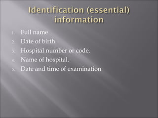
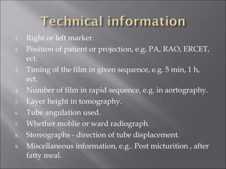
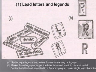
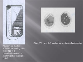
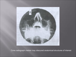
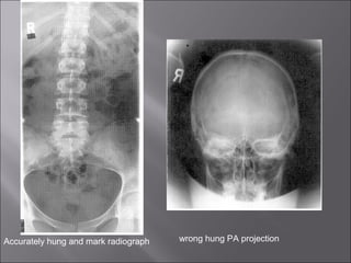
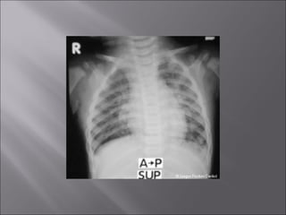
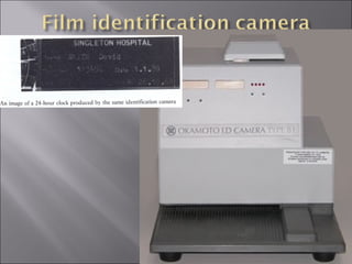
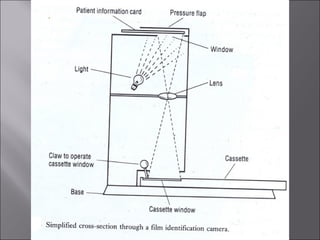
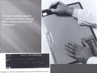
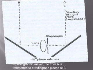
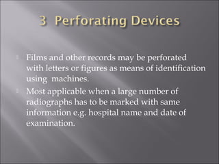
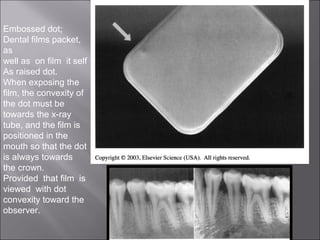
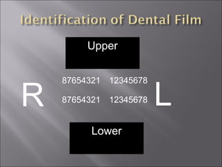
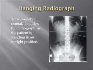
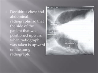
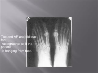
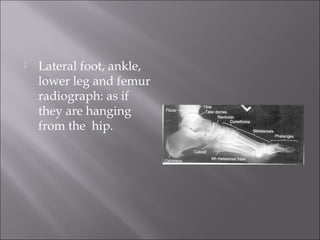
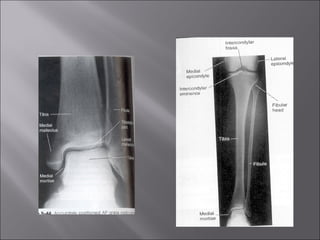
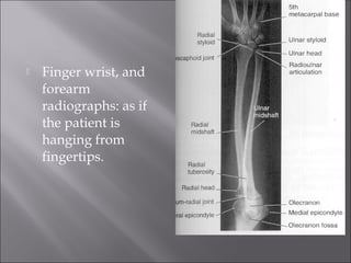
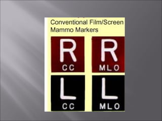


What is required in a radiograph in addition
to adequate demonstration of a part examined?



Very simply the answer is identification.


Any radiograph whatsoever should
included on it, preferably in
indelible form the following
information;
1.
2.
3.
4.
5.

Full name
Date of birth.
Hospital number or code.
Name of hospital.
Date and time of examination
1.
2.

3.

4.
5.
6.
7.
8.
9.

Right or left marker.
Position of patient or projection, e.g. PA, RAO, ERCET,
ect.
Timing of the film in given sequence, e.g. 5 min, 1 h,
ect.
Number of film in rapid sequence, e.g. in aortography.
Layer height in tomography.
Tube angulation used.
Whether moblie or ward radiograph.
Stereographs - direction of tube displacement.
Miscellaneous information, e.g.. Post micturition , after
fatty meal.
2.

Readable when the radiograph is viewed from
correct aspect.
Not superimposed on any important anatomy.

3.

Included within the collimated area.

1.
1.

Radiographer’s or technologist’s identity.

2.

The particular cassette or screen used.
1.
2.
3.

Opaque letters and legends.
Perforating devices.
Actinic marker.
(1) Lead letters and legends

(a) Radiopaque legends and letters for use in marking radiograph
(b) Marker for radiographs upper the letter is incised in a thin piece of metal.
Centre the letter lead, mounted in a Perspex plaque. Lower single lead character.
.Right (R) and left marker for anatomical orientation
Anatomical marker
suitable for placing over
the edge of a
cassette to record on
the film either the right
or left.
Cone radiograph marker may obscured anatomical structures of interest
Accurately hung and mark radiograph

wrong hung PA projection
The character should not be placed where to obscure
a feature of diagnostic importance.
 If the irradiated field is limited by a cone or
collimator its useless to place a marker close to the
border of the cassette as it will receive no exposure.

(a) Contact printing.
(b) photographic marker (using simple lens
system).


Work either like direct printing boxes or like
simple camera and light is used to affect the
film.
An actinic marker of printing-box type.
A Kodak X-omatic cassette
showing the widow through
Which patient detail may be
Photograph.
A photographic maker, the from A is
transferred to a radiograph placed at B





Permanent identification.
Is economical in time.
Shows information neatly and uniformly.
Reduces the likelihood of error




Films and other records may be perforated
with letters or figures as means of identification
using machines.
Most applicable when a large number of
radiographs has to be marked with same
information e.g. hospital name and date of
examination.
Embossed dot;
Dental films packet,
as
well as on film it self
As raised dot.
When exposing the
film, the convexity of
the dot must be
towards the x-ray
tube, and the film is
positioned in the
mouth so that the dot
is always towards
the crown.
Provided that film is
viewed with dot
convexity toward the
observer.
Upper

R

87654321

12345678

87654321

12345678

Lower

L
Features of good illuminator are;
(1)Light of even intensity.
(2)Light should as white as possible.
(3)Minimal heat given off by the light source.
(4)A facility for varying the brightness.
(5)A high intensity light (spot light)
incorporated.
(6)Transparent film grips, so that
identification is always visible.


Torso, vertebral,
cranial, shoulder,
hip radiograph: as if
the patient is
standing in an
upright position.


Decubitus chest and
abdominal
radiographs: so that
the side of the
patient that was
positioned upward
when radiograph
was taken is upward
on the hung
radiograph.
Toe and AP and oblique
foot
radiographs: as if the
patient
is hanging from toes.


Lateral foot, ankle,
lower leg and femur
radiograph: as if
they are hanging
from the hip.


Finger wrist, and
forearm
radiographs: as if
the patient is
hanging from
fingertips.


Elbow and humerus
radiograph: as if
they are hanging
from the patients
shoulder.
The presentation of radiograph

The presentation of radiograph

  • 1.
     What is requiredin a radiograph in addition to adequate demonstration of a part examined?  Very simply the answer is identification.
  • 2.
     Any radiograph whatsoevershould included on it, preferably in indelible form the following information;
  • 3.
    1. 2. 3. 4. 5. Full name Date ofbirth. Hospital number or code. Name of hospital. Date and time of examination
  • 4.
    1. 2. 3. 4. 5. 6. 7. 8. 9. Right or leftmarker. Position of patient or projection, e.g. PA, RAO, ERCET, ect. Timing of the film in given sequence, e.g. 5 min, 1 h, ect. Number of film in rapid sequence, e.g. in aortography. Layer height in tomography. Tube angulation used. Whether moblie or ward radiograph. Stereographs - direction of tube displacement. Miscellaneous information, e.g.. Post micturition , after fatty meal.
  • 5.
    2. Readable when theradiograph is viewed from correct aspect. Not superimposed on any important anatomy. 3. Included within the collimated area. 1.
  • 6.
    1. Radiographer’s or technologist’sidentity. 2. The particular cassette or screen used.
  • 7.
    1. 2. 3. Opaque letters andlegends. Perforating devices. Actinic marker.
  • 8.
    (1) Lead lettersand legends (a) Radiopaque legends and letters for use in marking radiograph (b) Marker for radiographs upper the letter is incised in a thin piece of metal. Centre the letter lead, mounted in a Perspex plaque. Lower single lead character.
  • 9.
    .Right (R) andleft marker for anatomical orientation Anatomical marker suitable for placing over the edge of a cassette to record on the film either the right or left.
  • 10.
    Cone radiograph markermay obscured anatomical structures of interest
  • 11.
    Accurately hung andmark radiograph wrong hung PA projection
  • 15.
    The character shouldnot be placed where to obscure a feature of diagnostic importance.  If the irradiated field is limited by a cone or collimator its useless to place a marker close to the border of the cassette as it will receive no exposure. 
  • 16.
    (a) Contact printing. (b)photographic marker (using simple lens system).  Work either like direct printing boxes or like simple camera and light is used to affect the film.
  • 17.
    An actinic markerof printing-box type.
  • 20.
    A Kodak X-omaticcassette showing the widow through Which patient detail may be Photograph.
  • 21.
    A photographic maker,the from A is transferred to a radiograph placed at B
  • 22.
        Permanent identification. Is economicalin time. Shows information neatly and uniformly. Reduces the likelihood of error
  • 23.
      Films and otherrecords may be perforated with letters or figures as means of identification using machines. Most applicable when a large number of radiographs has to be marked with same information e.g. hospital name and date of examination.
  • 24.
    Embossed dot; Dental filmspacket, as well as on film it self As raised dot. When exposing the film, the convexity of the dot must be towards the x-ray tube, and the film is positioned in the mouth so that the dot is always towards the crown. Provided that film is viewed with dot convexity toward the observer.
  • 25.
  • 26.
    Features of goodilluminator are; (1)Light of even intensity. (2)Light should as white as possible. (3)Minimal heat given off by the light source. (4)A facility for varying the brightness. (5)A high intensity light (spot light) incorporated. (6)Transparent film grips, so that identification is always visible.
  • 27.
     Torso, vertebral, cranial, shoulder, hipradiograph: as if the patient is standing in an upright position.
  • 29.
     Decubitus chest and abdominal radiographs:so that the side of the patient that was positioned upward when radiograph was taken is upward on the hung radiograph.
  • 30.
    Toe and APand oblique foot radiographs: as if the patient is hanging from toes.
  • 31.
     Lateral foot, ankle, lowerleg and femur radiograph: as if they are hanging from the hip.
  • 33.
     Finger wrist, and forearm radiographs:as if the patient is hanging from fingertips.
  • 34.
     Elbow and humerus radiograph:as if they are hanging from the patients shoulder.