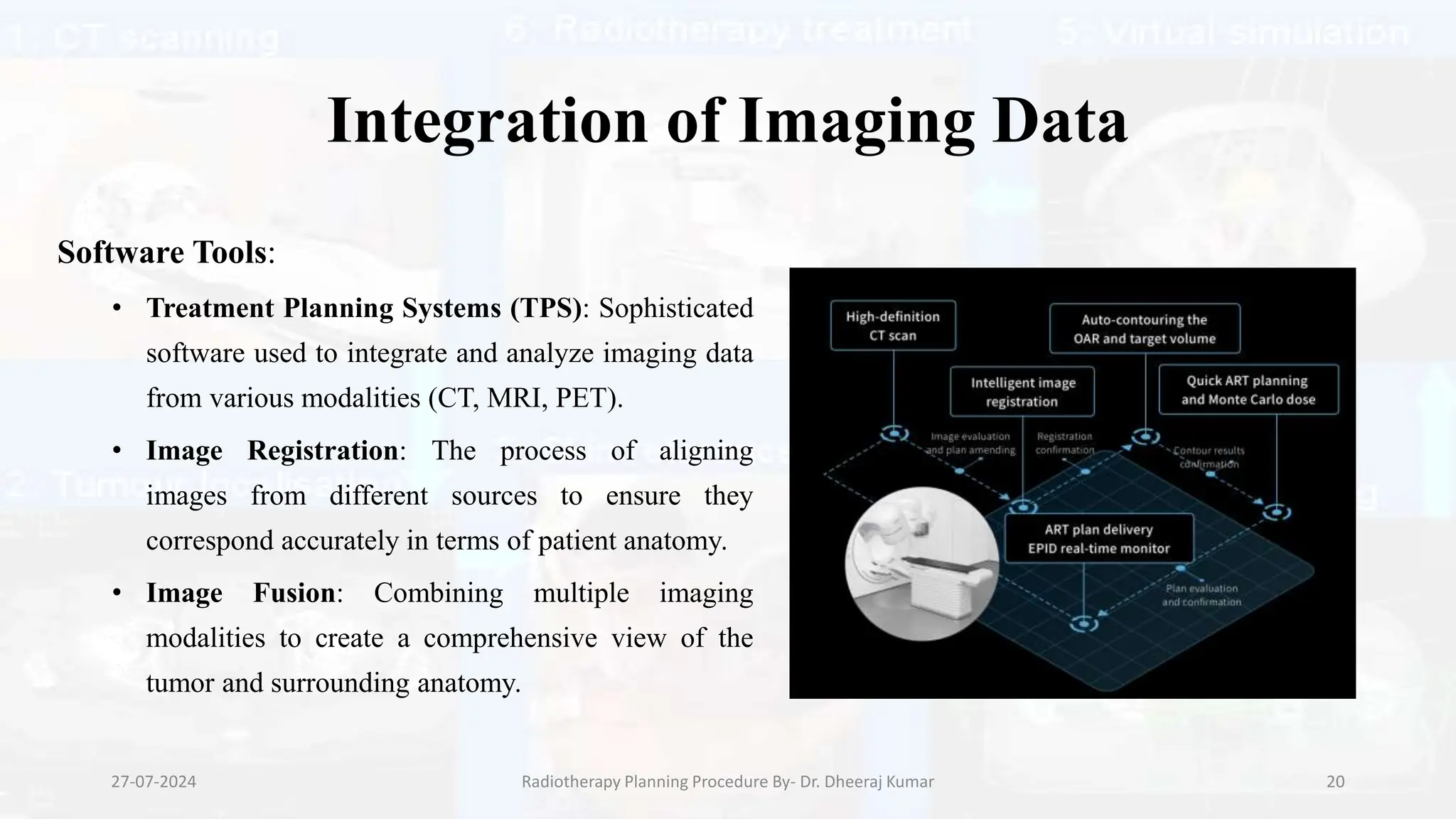
Radiotherapy Planning
Procedure
Presenter: Dheeraj Kumar
MRIT, Ph.D. (Radiology and Imaging)
Assistant Professor
Medical Radiology and Imaging Technology
School of Health Sciences, CSJM University, Kanpur
Introduction to Radiotherapy Planning
Definition:
• Radiotherapy planning is a multi-step process used to design and optimize a
treatment regimen that delivers precise doses of radiation to cancerous tissues
while minimizing damage to surrounding healthy tissues.
27-07-2024 Radiotherapy Planning Procedure By- Dr. Dheeraj Kumar 2
• Objective: The primary goal of radiotherapy planning is to ensure the
accurate delivery of the therapeutic dose to the tumor. This involves
detailed mapping and calculation to achieve maximum tumor control with
minimal side effects.
• Importance: Proper planning is crucial for the success of radiotherapy as it
directly impacts treatment efficacy and patient safety. Advances in imaging
and computer technology have significantly improved the precision and
effectiveness of radiotherapy planning.
27-07-2024 Radiotherapy Planning Procedure By- Dr. Dheeraj Kumar 3
Stages of Radiotherapy Planning
1.Patient Preparation
2.Imaging for Localization
3.Contouring
4.Dose Calculation
5.Plan Evaluation and Verification
27-07-2024 Radiotherapy Planning Procedure By- Dr. Dheeraj Kumar 4
Patient Preparation
Simulation:
• Simulation is a critical component
of patient preparation. During
simulation, the patient is positioned
as they will be during actual
treatment. This session is used to
gather imaging data and establish
reproducible patient positioning.
27-07-2024 Radiotherapy Planning Procedure By- Dr. Dheeraj Kumar 5
Immobilization Devices:
• Various devices are used to immobilize
the patient to ensure that they remain in
the same position for each treatment
session. Examples include:
• Masks: For head and neck treatments.
• Body Molds: For body positioning.
• Vacuum Cushions: Custom-fitted cushions
that mold to the patient's body.
27-07-2024 Radiotherapy Planning Procedure By- Dr. Dheeraj Kumar 6
Marking:
• Temporary or permanent marks are
made on the patient’s skin to assist in
accurate positioning. These marks are
aligned with external lasers in the
treatment room to ensure correct setup.
27-07-2024 Radiotherapy Planning Procedure By- Dr. Dheeraj Kumar 7
Imaging for Localization
Conventional Radiography:
• Uses X-rays to create 2D images of the
body.
• Important for initial localization and for
simple cases where detailed 3D imaging
may not be required.
• Quick and readily available but limited in
providing comprehensive spatial
information.
27-07-2024 Radiotherapy Planning Procedure By- Dr. Dheeraj Kumar 8
Simulator Imaging:
• Simulation machines are specifically designed to
replicate the geometry of the treatment unit.
• 2D Imaging: Simulator can produce 2D images similar
to conventional radiographs.
• 3D Imaging: Advanced simulators provide 3D imaging
capabilities, which offer more detailed information about
the tumor and surrounding anatomy.
• Provides the basis for defining treatment fields and
planning the appropriate radiation beams.
27-07-2024 Radiotherapy Planning Procedure By- Dr. Dheeraj Kumar 9
Role of Imaging:
• Essential for accurately identifying the
tumor's location and extent.
• Helps in visualizing the relationship
between the tumor and critical organs or
structures.
• Provides data necessary for contouring
and treatment planning.
27-07-2024 Radiotherapy Planning Procedure By- Dr. Dheeraj Kumar 10
Tumor Localization
Purpose:
• The main aim of tumor localization
is to accurately identify the tumor's
position and boundaries within the
body. This precision is vital to ensure
the radiation dose is delivered
effectively to the tumor while sparing
healthy tissues.
27-07-2024 Radiotherapy Planning Procedure By- Dr. Dheeraj Kumar 11
Techniques
• Conventional Radiography
• Simulator Imaging
27-07-2024 Radiotherapy Planning Procedure By- Dr. Dheeraj Kumar 12
Conventional Radiographic Method
Procedure:
• Patient Positioning: The patient is
positioned in a specific posture to expose
the area of interest.
• X-ray Imaging: X-ray images are taken
to visualize the tumor and surrounding
structures.
• Marking: Physical marks are placed on
the patient’s skin to guide treatment.
27-07-2024 Radiotherapy Planning Procedure By- Dr. Dheeraj Kumar 13
Advantages:
• Simplicity: Easy to perform and widely available.
• Speed: Quick process, suitable for initial assessments and less complex cases.
Limitations:
• 2D Imaging: Provides only two-dimensional images, which may not be
sufficient for complex cases.
• Accuracy: Less precise compared to advanced imaging techniques,
potentially leading to less accurate treatment planning.
27-07-2024 Radiotherapy Planning Procedure By- Dr. Dheeraj Kumar 14
Simulator Imaging
Procedure:
• Simulation Setup: The patient is positioned on a
simulation machine that replicates the actual
treatment setup.
• 2D and 3D Imaging: The machine captures both
2D and 3D images to provide comprehensive
visualization of the tumor and surrounding
anatomy.
• Data Collection: The images collected are used
to define the treatment fields and plan the
radiation beams.
27-07-2024 Radiotherapy Planning Procedure By- Dr. Dheeraj Kumar 15
Advantages:
• Accuracy: Provides detailed 3D images, improving the precision of tumor
localization.
• Detailed Visualization: Allows for better differentiation between the tumor and
surrounding tissues.
Limitations:
• Complexity: More complex and time-consuming than conventional radiography.
• Resource Intensive: Requires specialized equipment and trained personnel.
27-07-2024 Radiotherapy Planning Procedure By- Dr. Dheeraj Kumar 16
Target Volume Measurement
Gross Tumor Volume (GTV):
• The visible or palpable extent of the tumor.
• Includes only the tumor itself, not accounting for microscopic spread.
Clinical Target Volume (CTV):
• GTV plus any areas of suspected microscopic disease.
• Accounts for potential spread of cancer cells that are not visible on
imaging.
Planning Target Volume (PTV):
• CTV plus a margin to accommodate patient movement, variations in daily
setup, and organ motion.
• Ensures that the entire tumor receives the prescribed dose even if there are
minor changes in positioning.
27-07-2024 Radiotherapy Planning Procedure By- Dr. Dheeraj Kumar 17
Measurement Techniques
Conventional Radiography:
• Method: Uses 2D X-ray images to
estimate the tumor's size and location.
• Advantages: Quick and simple,
providing a basic outline of the tumor.
• Limitations: Less precise for internal
tumors, limited by its 2D nature.
27-07-2024 Radiotherapy Planning Procedure By- Dr. Dheeraj Kumar 18
Simulator Imaging:
• Method: Employs simulation machines to produce
detailed 3D images.
• Advantages:
• Precision: Offers more accurate delineation of GTV,
CTV, and PTV.
• Comprehensive Visualization: Enables better planning
by providing a clear view of the tumor and its
relationship with surrounding tissues.
• Limitations: Requires more time and resources,
including specialized equipment and trained
personnel.
27-07-2024 Radiotherapy Planning Procedure By- Dr. Dheeraj Kumar 19
Integration of Imaging Data
Software Tools:
• Treatment Planning Systems (TPS): Sophisticated
software used to integrate and analyze imaging data
from various modalities (CT, MRI, PET).
• Image Registration: The process of aligning
images from different sources to ensure they
correspond accurately in terms of patient anatomy.
• Image Fusion: Combining multiple imaging
modalities to create a comprehensive view of the
tumor and surrounding anatomy.
27-07-2024 Radiotherapy Planning Procedure By- Dr. Dheeraj Kumar 20
Fusion of Images
CT and MRI: CT provides detailed anatomical information,
while MRI offers superior soft tissue contrast. Fusing these
images enhances tumor delineation.
CT and PET: PET provides metabolic information, helping to
identify active tumor regions. Fusing PET with CT enhances
the ability to target metabolically active tumor areas.
Benefits:
• Improved Accuracy: Combining anatomical and functional
information leads to more precise tumor localization and
contouring.
• Enhanced Treatment Planning: A comprehensive view of the
tumor and surrounding structures allows for better planning of
radiation dose distribution.
27-07-2024 Radiotherapy Planning Procedure By- Dr. Dheeraj Kumar 21
Workflow
Acquisition: Obtain high-quality images from various modalities.
Registration and Fusion: Align and combine images to create a unified dataset.
Contouring: Define the tumor and critical structures on the fused images.
Planning: Use the integrated data for dose calculation and optimization.
27-07-2024 Radiotherapy Planning Procedure By- Dr. Dheeraj Kumar 22
Dose Calculation
Treatment Planning System (TPS):
• Function: TPS is a specialized software that
calculates the optimal radiation dose
distribution based on the tumor and
surrounding anatomy.
• Algorithms: Utilizes advanced algorithms to
simulate how radiation interacts with tissues.
Common algorithms include
convolution/superposition and Monte Carlo
simulations.
27-07-2024 Radiotherapy Planning Procedure By- Dr. Dheeraj Kumar 23
Factors Considered:
• Tumor Characteristics: Size, shape, and location of the tumor.
• Surrounding Anatomy: Proximity of critical organs and structures.
• Radiation Modality: Type of radiation used (e.g., photons, electrons,
protons).
• Beam Geometry: Angles, shapes, and energies of the radiation beams.
27-07-2024 Radiotherapy Planning Procedure By- Dr. Dheeraj Kumar 24
Optimization:
• Objective: Maximize dose to the tumor
(GTV, CTV, PTV) while minimizing
exposure to healthy tissues.
• Techniques:
• Intensity-Modulated Radiotherapy (IMRT):
Varies the intensity of radiation beams to
conform to the tumor shape.
• Volumetric Modulated Arc Therapy (VMAT):
Delivers radiation dose continuously as the
machine rotates around the patient.
27-07-2024 Radiotherapy Planning Procedure By- Dr. Dheeraj Kumar 25
Plan Evaluation and Verification
Plan Evaluation:
• Dosimetric Analysis: Reviewing dose-
volume histograms (DVHs) to ensure the
prescribed dose covers the target volumes
while sparing critical structures.
• Quality Assurance (QA): Verifying the
plan meets clinical goals and institutional
protocols.
27-07-2024 Radiotherapy Planning Procedure By- Dr. Dheeraj Kumar 26
Verification Techniques:
• Imaging:
• On-Board Imaging (OBI): Uses imaging systems
attached to the treatment machine to verify patient
positioning before and during treatment.
• Cone Beam CT (CBCT): Provides 3D imaging
capabilities for verifying patient setup and anatomy.
• Portal Dosimetry: Uses electronic portal
imaging devices (EPIDs) to verify the radiation
dose delivered matches the planned dose.
27-07-2024 Radiotherapy Planning Procedure By- Dr. Dheeraj Kumar 27
Adaptive Radiotherapy:
• Concept: Adjusting the treatment plan based on changes in the patient's
anatomy or tumor size during the course of treatment.
• Implementation: Regular imaging and re-planning as necessary to account for
anatomical changes.
27-07-2024 Radiotherapy Planning Procedure By- Dr. Dheeraj Kumar 28
Quality Assurance
Importance:
• QA is crucial to ensure the safety, accuracy, and effectiveness of radiotherapy treatments. It helps
prevent errors and ensures that the treatment delivered matches the treatment plan.
Procedures:
• Machine Calibration: Regular calibration of treatment machines to ensure accurate dose delivery.
• Dosimetry Checks: Routine checks using phantoms and dosimeters to verify dose distributions.
• Patient-Specific QA: Verification of each patient's treatment plan before the first treatment
session.
27-07-2024 Radiotherapy Planning Procedure By- Dr. Dheeraj Kumar 29
Daily QA:
• Checks: Daily checks of machine performance, including beam output, alignment,
and mechanical functionality.
• Documentation: Detailed records of QA procedures and results for traceability and
compliance.
Regulatory Compliance:
• Standards and Guidelines: Adherence to standards set by organizations such as the
American Association of Physicists in Medicine (AAPM) and the International
Atomic Energy Agency (IAEA).
• Audits and Inspections: Regular audits by regulatory bodies to ensure compliance
with safety and quality standards.
27-07-2024 Radiotherapy Planning Procedure By- Dr. Dheeraj Kumar 30
References
• "The Physics of Radiation Therapy"Authors: Faiz M. Khan, John P. Gibbon - Publisher: Lippincott Williams &
Wilkins, 2014
• "Principles and Practice of Radiation Oncology"Authors: Carlos A. Perez, Luther W. Brady- Publisher: Lippincott
Williams & Wilkins, 2013
• "Radiation Oncology: A Question-Based Review"Authors: Borislav Hristov, Steven H. Lin, John P.
Christodouleas- Publisher: Lippincott Williams & Wilkins, 2014
• "Radiotherapy in Practice: Imaging"Authors : Peter Hoskin, Patricia Price - Publisher: Oxford University Press,
2021
• "Image-Guided and Adaptive Radiation Therapy"Editors: Robert D. Timmerman, Lei Xing Publisher: Lippincott
Williams & Wilkins, 2012
27-07-2024 Radiotherapy Planning Procedure By- Dr. Dheeraj Kumar 31
THANK YOU
27-07-2024 Radiotherapy Planning Procedure By- Dr. Dheeraj Kumar 32

Radiotherapy Treatment Planning Procedure.pptx

  • 1.
    Radiotherapy Planning Procedure Presenter: DheerajKumar MRIT, Ph.D. (Radiology and Imaging) Assistant Professor Medical Radiology and Imaging Technology School of Health Sciences, CSJM University, Kanpur
  • 2.
    Introduction to RadiotherapyPlanning Definition: • Radiotherapy planning is a multi-step process used to design and optimize a treatment regimen that delivers precise doses of radiation to cancerous tissues while minimizing damage to surrounding healthy tissues. 27-07-2024 Radiotherapy Planning Procedure By- Dr. Dheeraj Kumar 2
  • 3.
    • Objective: Theprimary goal of radiotherapy planning is to ensure the accurate delivery of the therapeutic dose to the tumor. This involves detailed mapping and calculation to achieve maximum tumor control with minimal side effects. • Importance: Proper planning is crucial for the success of radiotherapy as it directly impacts treatment efficacy and patient safety. Advances in imaging and computer technology have significantly improved the precision and effectiveness of radiotherapy planning. 27-07-2024 Radiotherapy Planning Procedure By- Dr. Dheeraj Kumar 3
  • 4.
    Stages of RadiotherapyPlanning 1.Patient Preparation 2.Imaging for Localization 3.Contouring 4.Dose Calculation 5.Plan Evaluation and Verification 27-07-2024 Radiotherapy Planning Procedure By- Dr. Dheeraj Kumar 4
  • 5.
    Patient Preparation Simulation: • Simulationis a critical component of patient preparation. During simulation, the patient is positioned as they will be during actual treatment. This session is used to gather imaging data and establish reproducible patient positioning. 27-07-2024 Radiotherapy Planning Procedure By- Dr. Dheeraj Kumar 5
  • 6.
    Immobilization Devices: • Variousdevices are used to immobilize the patient to ensure that they remain in the same position for each treatment session. Examples include: • Masks: For head and neck treatments. • Body Molds: For body positioning. • Vacuum Cushions: Custom-fitted cushions that mold to the patient's body. 27-07-2024 Radiotherapy Planning Procedure By- Dr. Dheeraj Kumar 6
  • 7.
    Marking: • Temporary orpermanent marks are made on the patient’s skin to assist in accurate positioning. These marks are aligned with external lasers in the treatment room to ensure correct setup. 27-07-2024 Radiotherapy Planning Procedure By- Dr. Dheeraj Kumar 7
  • 8.
    Imaging for Localization ConventionalRadiography: • Uses X-rays to create 2D images of the body. • Important for initial localization and for simple cases where detailed 3D imaging may not be required. • Quick and readily available but limited in providing comprehensive spatial information. 27-07-2024 Radiotherapy Planning Procedure By- Dr. Dheeraj Kumar 8
  • 9.
    Simulator Imaging: • Simulationmachines are specifically designed to replicate the geometry of the treatment unit. • 2D Imaging: Simulator can produce 2D images similar to conventional radiographs. • 3D Imaging: Advanced simulators provide 3D imaging capabilities, which offer more detailed information about the tumor and surrounding anatomy. • Provides the basis for defining treatment fields and planning the appropriate radiation beams. 27-07-2024 Radiotherapy Planning Procedure By- Dr. Dheeraj Kumar 9
  • 10.
    Role of Imaging: •Essential for accurately identifying the tumor's location and extent. • Helps in visualizing the relationship between the tumor and critical organs or structures. • Provides data necessary for contouring and treatment planning. 27-07-2024 Radiotherapy Planning Procedure By- Dr. Dheeraj Kumar 10
  • 11.
    Tumor Localization Purpose: • Themain aim of tumor localization is to accurately identify the tumor's position and boundaries within the body. This precision is vital to ensure the radiation dose is delivered effectively to the tumor while sparing healthy tissues. 27-07-2024 Radiotherapy Planning Procedure By- Dr. Dheeraj Kumar 11
  • 12.
    Techniques • Conventional Radiography •Simulator Imaging 27-07-2024 Radiotherapy Planning Procedure By- Dr. Dheeraj Kumar 12
  • 13.
    Conventional Radiographic Method Procedure: •Patient Positioning: The patient is positioned in a specific posture to expose the area of interest. • X-ray Imaging: X-ray images are taken to visualize the tumor and surrounding structures. • Marking: Physical marks are placed on the patient’s skin to guide treatment. 27-07-2024 Radiotherapy Planning Procedure By- Dr. Dheeraj Kumar 13
  • 14.
    Advantages: • Simplicity: Easyto perform and widely available. • Speed: Quick process, suitable for initial assessments and less complex cases. Limitations: • 2D Imaging: Provides only two-dimensional images, which may not be sufficient for complex cases. • Accuracy: Less precise compared to advanced imaging techniques, potentially leading to less accurate treatment planning. 27-07-2024 Radiotherapy Planning Procedure By- Dr. Dheeraj Kumar 14
  • 15.
    Simulator Imaging Procedure: • SimulationSetup: The patient is positioned on a simulation machine that replicates the actual treatment setup. • 2D and 3D Imaging: The machine captures both 2D and 3D images to provide comprehensive visualization of the tumor and surrounding anatomy. • Data Collection: The images collected are used to define the treatment fields and plan the radiation beams. 27-07-2024 Radiotherapy Planning Procedure By- Dr. Dheeraj Kumar 15
  • 16.
    Advantages: • Accuracy: Providesdetailed 3D images, improving the precision of tumor localization. • Detailed Visualization: Allows for better differentiation between the tumor and surrounding tissues. Limitations: • Complexity: More complex and time-consuming than conventional radiography. • Resource Intensive: Requires specialized equipment and trained personnel. 27-07-2024 Radiotherapy Planning Procedure By- Dr. Dheeraj Kumar 16
  • 17.
    Target Volume Measurement GrossTumor Volume (GTV): • The visible or palpable extent of the tumor. • Includes only the tumor itself, not accounting for microscopic spread. Clinical Target Volume (CTV): • GTV plus any areas of suspected microscopic disease. • Accounts for potential spread of cancer cells that are not visible on imaging. Planning Target Volume (PTV): • CTV plus a margin to accommodate patient movement, variations in daily setup, and organ motion. • Ensures that the entire tumor receives the prescribed dose even if there are minor changes in positioning. 27-07-2024 Radiotherapy Planning Procedure By- Dr. Dheeraj Kumar 17
  • 18.
    Measurement Techniques Conventional Radiography: •Method: Uses 2D X-ray images to estimate the tumor's size and location. • Advantages: Quick and simple, providing a basic outline of the tumor. • Limitations: Less precise for internal tumors, limited by its 2D nature. 27-07-2024 Radiotherapy Planning Procedure By- Dr. Dheeraj Kumar 18
  • 19.
    Simulator Imaging: • Method:Employs simulation machines to produce detailed 3D images. • Advantages: • Precision: Offers more accurate delineation of GTV, CTV, and PTV. • Comprehensive Visualization: Enables better planning by providing a clear view of the tumor and its relationship with surrounding tissues. • Limitations: Requires more time and resources, including specialized equipment and trained personnel. 27-07-2024 Radiotherapy Planning Procedure By- Dr. Dheeraj Kumar 19
  • 20.
    Integration of ImagingData Software Tools: • Treatment Planning Systems (TPS): Sophisticated software used to integrate and analyze imaging data from various modalities (CT, MRI, PET). • Image Registration: The process of aligning images from different sources to ensure they correspond accurately in terms of patient anatomy. • Image Fusion: Combining multiple imaging modalities to create a comprehensive view of the tumor and surrounding anatomy. 27-07-2024 Radiotherapy Planning Procedure By- Dr. Dheeraj Kumar 20
  • 21.
    Fusion of Images CTand MRI: CT provides detailed anatomical information, while MRI offers superior soft tissue contrast. Fusing these images enhances tumor delineation. CT and PET: PET provides metabolic information, helping to identify active tumor regions. Fusing PET with CT enhances the ability to target metabolically active tumor areas. Benefits: • Improved Accuracy: Combining anatomical and functional information leads to more precise tumor localization and contouring. • Enhanced Treatment Planning: A comprehensive view of the tumor and surrounding structures allows for better planning of radiation dose distribution. 27-07-2024 Radiotherapy Planning Procedure By- Dr. Dheeraj Kumar 21
  • 22.
    Workflow Acquisition: Obtain high-qualityimages from various modalities. Registration and Fusion: Align and combine images to create a unified dataset. Contouring: Define the tumor and critical structures on the fused images. Planning: Use the integrated data for dose calculation and optimization. 27-07-2024 Radiotherapy Planning Procedure By- Dr. Dheeraj Kumar 22
  • 23.
    Dose Calculation Treatment PlanningSystem (TPS): • Function: TPS is a specialized software that calculates the optimal radiation dose distribution based on the tumor and surrounding anatomy. • Algorithms: Utilizes advanced algorithms to simulate how radiation interacts with tissues. Common algorithms include convolution/superposition and Monte Carlo simulations. 27-07-2024 Radiotherapy Planning Procedure By- Dr. Dheeraj Kumar 23
  • 24.
    Factors Considered: • TumorCharacteristics: Size, shape, and location of the tumor. • Surrounding Anatomy: Proximity of critical organs and structures. • Radiation Modality: Type of radiation used (e.g., photons, electrons, protons). • Beam Geometry: Angles, shapes, and energies of the radiation beams. 27-07-2024 Radiotherapy Planning Procedure By- Dr. Dheeraj Kumar 24
  • 25.
    Optimization: • Objective: Maximizedose to the tumor (GTV, CTV, PTV) while minimizing exposure to healthy tissues. • Techniques: • Intensity-Modulated Radiotherapy (IMRT): Varies the intensity of radiation beams to conform to the tumor shape. • Volumetric Modulated Arc Therapy (VMAT): Delivers radiation dose continuously as the machine rotates around the patient. 27-07-2024 Radiotherapy Planning Procedure By- Dr. Dheeraj Kumar 25
  • 26.
    Plan Evaluation andVerification Plan Evaluation: • Dosimetric Analysis: Reviewing dose- volume histograms (DVHs) to ensure the prescribed dose covers the target volumes while sparing critical structures. • Quality Assurance (QA): Verifying the plan meets clinical goals and institutional protocols. 27-07-2024 Radiotherapy Planning Procedure By- Dr. Dheeraj Kumar 26
  • 27.
    Verification Techniques: • Imaging: •On-Board Imaging (OBI): Uses imaging systems attached to the treatment machine to verify patient positioning before and during treatment. • Cone Beam CT (CBCT): Provides 3D imaging capabilities for verifying patient setup and anatomy. • Portal Dosimetry: Uses electronic portal imaging devices (EPIDs) to verify the radiation dose delivered matches the planned dose. 27-07-2024 Radiotherapy Planning Procedure By- Dr. Dheeraj Kumar 27
  • 28.
    Adaptive Radiotherapy: • Concept:Adjusting the treatment plan based on changes in the patient's anatomy or tumor size during the course of treatment. • Implementation: Regular imaging and re-planning as necessary to account for anatomical changes. 27-07-2024 Radiotherapy Planning Procedure By- Dr. Dheeraj Kumar 28
  • 29.
    Quality Assurance Importance: • QAis crucial to ensure the safety, accuracy, and effectiveness of radiotherapy treatments. It helps prevent errors and ensures that the treatment delivered matches the treatment plan. Procedures: • Machine Calibration: Regular calibration of treatment machines to ensure accurate dose delivery. • Dosimetry Checks: Routine checks using phantoms and dosimeters to verify dose distributions. • Patient-Specific QA: Verification of each patient's treatment plan before the first treatment session. 27-07-2024 Radiotherapy Planning Procedure By- Dr. Dheeraj Kumar 29
  • 30.
    Daily QA: • Checks:Daily checks of machine performance, including beam output, alignment, and mechanical functionality. • Documentation: Detailed records of QA procedures and results for traceability and compliance. Regulatory Compliance: • Standards and Guidelines: Adherence to standards set by organizations such as the American Association of Physicists in Medicine (AAPM) and the International Atomic Energy Agency (IAEA). • Audits and Inspections: Regular audits by regulatory bodies to ensure compliance with safety and quality standards. 27-07-2024 Radiotherapy Planning Procedure By- Dr. Dheeraj Kumar 30
  • 31.
    References • "The Physicsof Radiation Therapy"Authors: Faiz M. Khan, John P. Gibbon - Publisher: Lippincott Williams & Wilkins, 2014 • "Principles and Practice of Radiation Oncology"Authors: Carlos A. Perez, Luther W. Brady- Publisher: Lippincott Williams & Wilkins, 2013 • "Radiation Oncology: A Question-Based Review"Authors: Borislav Hristov, Steven H. Lin, John P. Christodouleas- Publisher: Lippincott Williams & Wilkins, 2014 • "Radiotherapy in Practice: Imaging"Authors : Peter Hoskin, Patricia Price - Publisher: Oxford University Press, 2021 • "Image-Guided and Adaptive Radiation Therapy"Editors: Robert D. Timmerman, Lei Xing Publisher: Lippincott Williams & Wilkins, 2012 27-07-2024 Radiotherapy Planning Procedure By- Dr. Dheeraj Kumar 31
  • 32.
    THANK YOU 27-07-2024 RadiotherapyPlanning Procedure By- Dr. Dheeraj Kumar 32