Download as PDF, PPTX
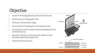
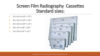
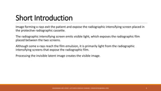
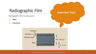


The document provides an overview of screen film radiography, detailing topics such as standard cassette sizes, radiographic film construction, and the process of forming a latent image. It emphasizes the interaction of x-rays with a radiographic intensifying screen and film, creating a visible image through processing. Additionally, the document covers the components and handling of radiographic film to maintain image quality.


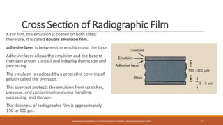
![Hypothalamus short ppt by Dr. Neha [PT].pptx](https://cdn.slidesharecdn.com/ss_thumbnails/hypothalamusbydr-260124145759-b9f94a93-thumbnail.jpg?width=640&height=640&fit=bounds)


