A brief historyon Radiography
1895: Discovery of X-rays by Wilhelm Röntgen.
Early X-ray images were captured on glass plates.
1896: First commercial X-ray film developed by George Eastman
(Kodak).
1913: Kodak introduces the first flexible X-ray film.
Advances in film materials, faster exposure times, and better image
quality over decades.
Film-based radiography became the standard for medical and
dental imaging.
Early 1980s: Development of digital radiography.
Key shift from film to digital imaging in the 1990s due to
advancements in computer technology.
Uses digital sensors instead of traditional film.
4.
History of Dentalradiography
Dr Otto Walkhoff, a German scientist, is credited with creating the
first dental radiograph in 1896.
It required a 25 minutes exposure time, but thankfully, he used
himself as the subject.
C. Edmond Kells, a dentist from New Orleans, created some of the
first practical applications.
He also did a fair bit towards the understanding of the damage
done by X-rays to human tissue, losing an arm and eventually dying
from the effects of radiation.
7.
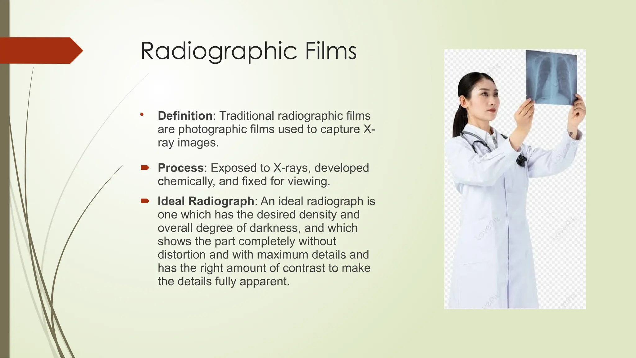
Radiographic Films
Definition:Traditional radiographic films
are photographic films used to capture X-
ray images.
Process: Exposed to X-rays, developed
chemically, and fixed for viewing.
Ideal Radiograph: An ideal radiograph is
one which has the desired density and
overall degree of darkness, and which
shows the part completely without
distortion and with maximum details and
has the right amount of contrast to make
the details fully apparent.
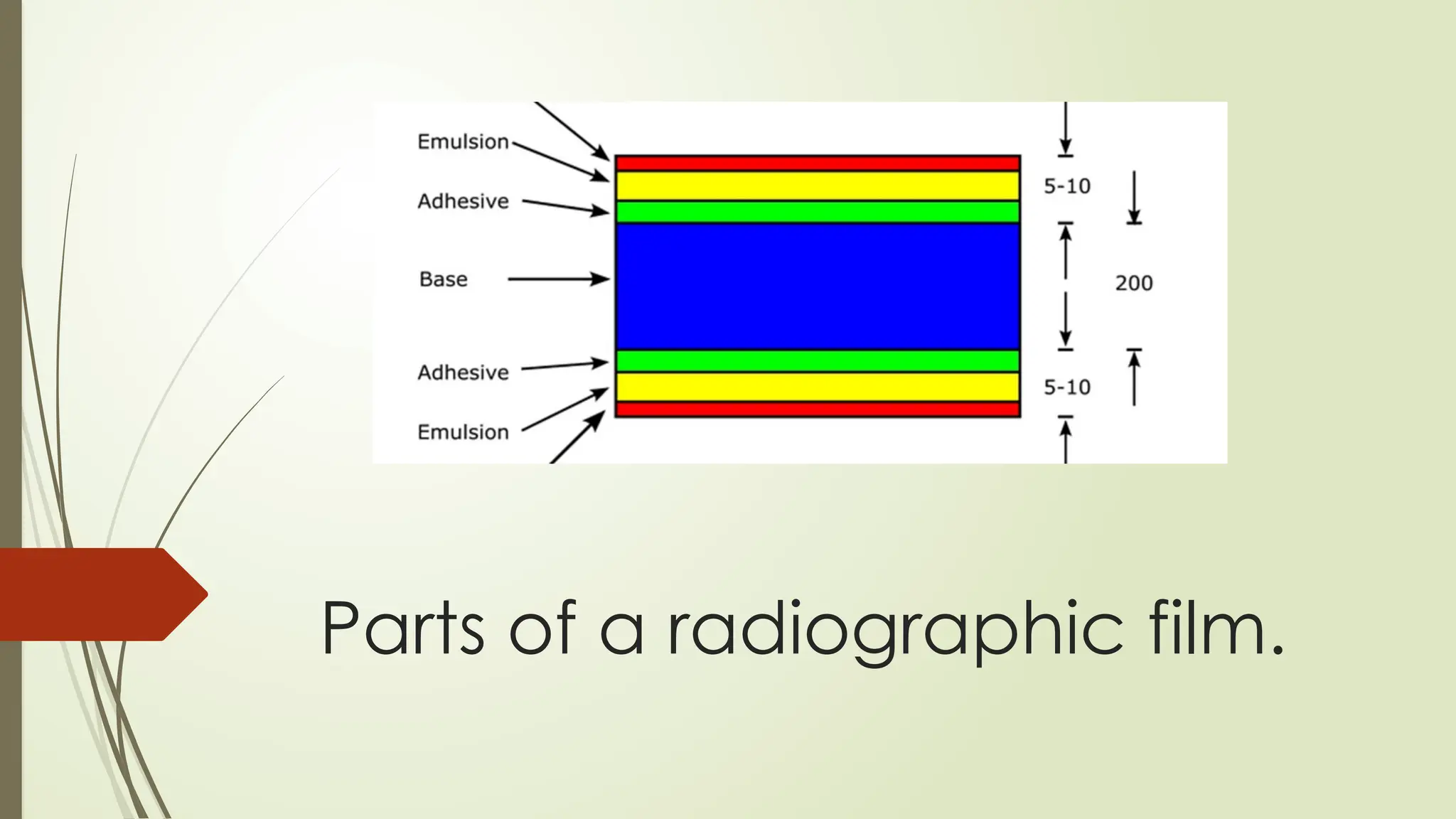
1. BaseLayer: Usually made from polyester or cellulose
acetate.
Properties: Provides strength and flexibility to the film.
Transparent, allowing light from the exposed areas to pass
through.
2. Emulsion Layer: A gelatin layer containing silver halide
crystals
Properties: The emulsion layer is sensitive to radiation (X-rays or
light), which allows the film to record the image.
The silver halide crystals undergo a chemical reaction when
exposed to radiation, forming a latent image.
3. Adhesive Layer: Gelatin is used to hold the silver halide
crystals in place.
Properties: It allows for the proper diffusion of chemicals during
processing.
4. Protective Coating: A thin, clear layer of protective material,
often made of a polymer.
Properties: Protects the emulsion layer from scratches, dirt, and
other physical damage.
Ensures the integrity and longevity of the film.
10.
Digital Radiography
DigitalRadiography is a modern imaging technique used in
medical diagnostics, which captures X-ray images digitally rather
than using traditional photographic film.
1. Direct Digital Radiography: Uses a flat-panel detector or a
charge-coupled device to capture the X-ray image directly.
The X-ray photons are converted into electrical signals, which are
then processed by a computer to produce the digital image.
2. Computed Radiography: Uses a photostimulable phosphor (PSP)
plate to capture the X-ray image, which is then processed in a
scanner.
The PSP plate absorbs the X-ray energy and stores it. After exposure,
the plate is scanned by a laser to release the stored image data,
which is then digitized and processed.
12.
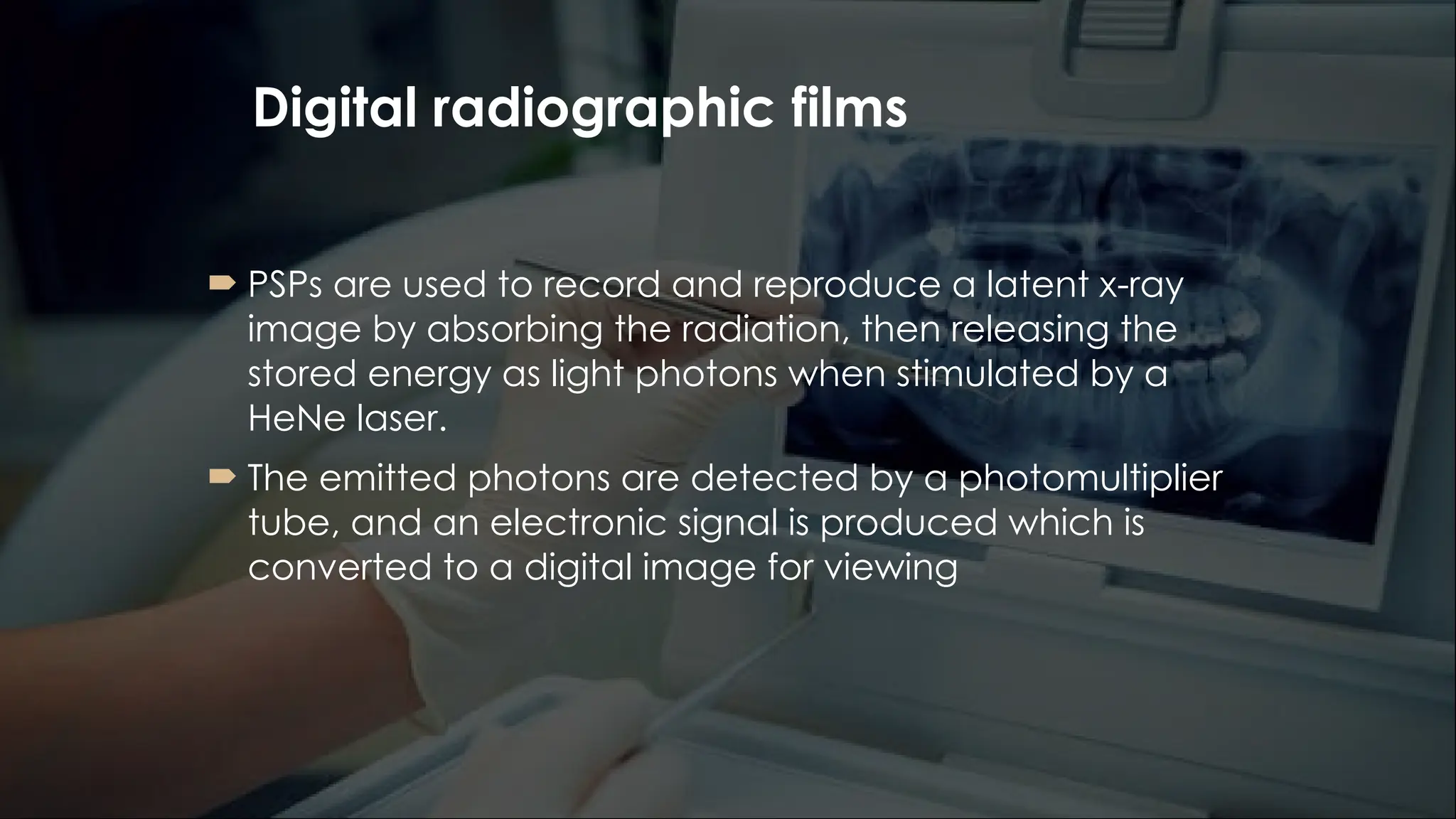
Digital radiographic films
PSPs are used to record and reproduce a latent x-ray
image by absorbing the radiation, then releasing the
stored energy as light photons when stimulated by a
HeNe laser.
The emitted photons are detected by a photomultiplier
tube, and an electronic signal is produced which is
converted to a digital image for viewing
13.
Parts of PSP
1. Phosphor Material: Often made from rare earth
elements like barium fluorohalide doped with
europium or other materials with the ability to absorb
X-rays or light and store the energy in the form of
trapped electrons.
It absorbs and stores energy when exposed to
radiation. The stored energy is released as light when
the phosphor is subsequently exposed to a stimulating
light source.
2. Protective Layer: A protective layer is often applied
on top of the phosphor material to shield it from
environmental damage and physical wear. It helps
improve the durability and longevity of the PSP.
3. Supporting layer: The phosphor layer is usually
applied to a flexible or rigid substrate material, which
gives the PSP its structural integrity and ensures it is
easy to handle and fit into imaging systems.
14.
Comparison of Radiographicfilms and
Digital films
Radiographic films
Image quality is good but can
degrade overtime
Exposure time is longer
Cost is lower initially but ongoing
expenses for films and chemicals
Physical storage required
Processing requires chemical
development
More wastage from chemicals
and films
Digital films
Superior, with adjustable
contrast/brightness
Shorter, with reduced radiation
Higher initial cost but no
film/chemical costs
Digital storage, easy to backup
Immediate results, no chemicals
required
Less wastage
15.
Digital Radiography isthe Future
digital radiographic films offer high image quality, faster processing
times, lower radiation doses, and enhanced capabilities for image
manipulation and storage.
Digital radiography has revolutionized medical imaging, providing
faster, more accurate diagnostics while improving patient care and
workflow efficiency
Digital detectors are replacing films to provide images instantly with
no additional film processing. They are also being supplemented
with digital light scanners that create 3-D images of tooth and gum
surfaces. These innovations have led to improved and quicker
diagnosis.
Advancements in sensor technology and integration of AI will lead
to better image quality, enhanced diagnosis and detailed image
analysis, making them an essential tool in modern medical imaging.


![general_and_extraoral_examination[1]__-__Read-Only n.pptx](https://cdn.slidesharecdn.com/ss_thumbnails/generalandextraoralexamination1-read-onlyn-250216162540-10c854ca-thumbnail.jpg?width=640&height=640&fit=bounds)


