Downloaded 340 times

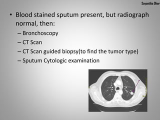

Lung cancer is characterized by uncontrolled cell growth in lung tissues. Worldwide, it is the leading cause of cancer death in men and women, responsible for 1.3 million deaths annually. The main causes are smoking and exposure to radon, asbestos, viruses and other particulates. Symptoms include coughing, shortness of breath, and weight loss. Diagnosis involves imaging tests and biopsies. Treatment depends on cancer type and stage but may include surgery, chemotherapy, and radiation therapy.


![PERI-PROSTHETIC FRACTURE NAIL-PLATE CONSTRUCT [NPC].pptx](https://cdn.slidesharecdn.com/ss_thumbnails/drarunkumardrmohamedashrafperiprostheticfrasturenail-plateconstructnpc-260209164459-7e9d15a1-thumbnail.jpg?width=640&height=640&fit=bounds)


