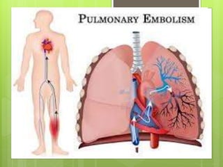
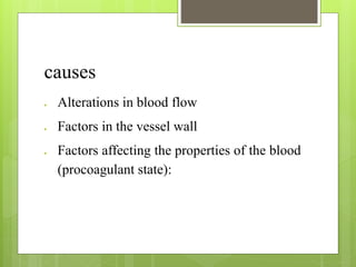
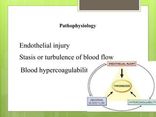
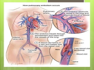
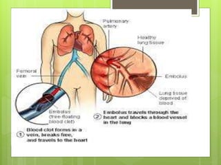
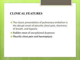
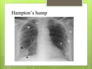
BHAGYASREE N P
2ND MSC NURSING
GOVT CON
ALAPPUZHA
Definition
 Pulmonary embolus (PE) refers to obstruction of
the pulmonary artery or one of its branches by
material (eg, thrombus, tumor, air, or fat) that
originated elsewhere in the body.
causes
 Alterations in blood flow
 Factors in the vessel wall
 Factors affecting the properties of the blood
(procoagulant state):
Pathophysiology
Endothelial injury
Stasis or turbulence of blood flow
Blood hypercoagulability
CLINICAL FEATURES
 The classic presentation of pulmonary embolism is
the abrupt onset of pleuritic chest pain, shortness
of breath, and hypoxia
 Sudden onset of unexplained dyspnoea
 Pleuritic chest pain and haemoptysis
Small/medium pulmonary embolism
 pleuritic chest pain and breathlessness
 Haemoptysis occurs in 30%
 On examination,
 tachypnoeic with a localized pleural rub and often
coarse crackles
 exudative pleural effusion
Massive pulmonary embolism
 central chest pain
 Syncope
 On examination,
 tachypnoeic
 tachycardia with hypotension
 The jugular venous pressure (JVP) is raised
Multiple recurrent pulmonary emboli
 breathlessness, often over weeks or months
 weakness, syncope on exertion and occasionally
angina
 On examination,
 signs of right ventricular overload with a right
ventricular heave and loud pulmonary second
sound
Revised Geneva score for the clinical prediction of a pulmonary embolism
Risk factors
Score
 Age >65 years
+1
 Previous deep venous thrombosis or pulmonary
embolism
+3
 Surgery or fracture within 1month
+2
 Active malignancy
+2
Symptoms
 Unilateral leg pain
+3
 Haemoptysis
+2
Clinical signs
Heart rate (b.p.m.):
 75-94 +3
 >95 +5
 Pain on leg deep vein palpation and unilateral oedema
+4
Clinical probability
 Low 0-3
 Intermediate 4-10
 High >11
INVESTIGATIONS
 D-dimer and other circulating markers
 Ischemia-Modified Albumin levels
 White Blood Cell Count
 Arterial blood gases
 Troponin levels
 Brain Natriuretic Peptide
Pulmonary angiography
V/Q scan
Chest x-ray
Signs with relative high specificity but low
sensitivity for acute pulmonary embolism:
 Decreased vascularity in the peripheral lung
(Westermark sign).
 Enlargement of the central pulmonary artery
(Fleischner sign).
 Pleural based areas of increased opacity (Hampton
hump).
 Hemidiaphragm elevation
Westermark sign
Hampton’s hump
MANAGEMENT
 low-dose anticoagulation with standard UFH,
LMWH, or fondaparinux
 heparin 5000 U given subcutaneously every 12
hours
 Anticoagulation together with pneumatic
compression
 Oxygen should be given to all hypoxaemic patients
Treatment Acute management
 high-flow oxygen (60-100%)
 intravenous fluids and inotropic agents
 Fibrinolytic therapy
 Surgical embolectomy
Anticoagulation and thrombolysis
Anticoagulation
 Unfractionated heparin
 Low-molecular-weight heparin
 Factor Xa Inhibitors
 Fondaparinux
Warfarin
Thrombolytic agents
 Alteplase
 Reteplase
 Urokinase
 Streptokinase
Vena Cava Filters
Complications
Sudden cardiac death
Obstructive shock
Pulseless electrical activity
trial or ventricular arrhythmias
Secondary pulmonary arterial hypertension
Cor pulmonale

 Severe hypoxemia
 Right-to-left intracardiac shunt
 Lung infarction
 Pleural effusion
 Paradoxical embolism
 Heparin-induced thrombocytopenia
Thrombophlebitis

Pulmonary embolism

  • 1.
    BHAGYASREE N P 2NDMSC NURSING GOVT CON ALAPPUZHA
  • 2.
    Definition  Pulmonary embolus(PE) refers to obstruction of the pulmonary artery or one of its branches by material (eg, thrombus, tumor, air, or fat) that originated elsewhere in the body.
  • 4.
    causes  Alterations inblood flow  Factors in the vessel wall  Factors affecting the properties of the blood (procoagulant state):
  • 5.
    Pathophysiology Endothelial injury Stasis orturbulence of blood flow Blood hypercoagulability
  • 8.
    CLINICAL FEATURES  Theclassic presentation of pulmonary embolism is the abrupt onset of pleuritic chest pain, shortness of breath, and hypoxia  Sudden onset of unexplained dyspnoea  Pleuritic chest pain and haemoptysis
  • 9.
    Small/medium pulmonary embolism pleuritic chest pain and breathlessness  Haemoptysis occurs in 30%  On examination,  tachypnoeic with a localized pleural rub and often coarse crackles  exudative pleural effusion
  • 10.
    Massive pulmonary embolism central chest pain  Syncope  On examination,  tachypnoeic  tachycardia with hypotension  The jugular venous pressure (JVP) is raised
  • 11.
    Multiple recurrent pulmonaryemboli  breathlessness, often over weeks or months  weakness, syncope on exertion and occasionally angina  On examination,  signs of right ventricular overload with a right ventricular heave and loud pulmonary second sound
  • 12.
    Revised Geneva scorefor the clinical prediction of a pulmonary embolism Risk factors Score  Age >65 years +1  Previous deep venous thrombosis or pulmonary embolism +3  Surgery or fracture within 1month +2  Active malignancy +2
  • 13.
    Symptoms  Unilateral legpain +3  Haemoptysis +2
  • 14.
    Clinical signs Heart rate(b.p.m.):  75-94 +3  >95 +5  Pain on leg deep vein palpation and unilateral oedema +4
  • 15.
    Clinical probability  Low0-3  Intermediate 4-10  High >11
  • 16.
    INVESTIGATIONS  D-dimer andother circulating markers  Ischemia-Modified Albumin levels  White Blood Cell Count  Arterial blood gases  Troponin levels  Brain Natriuretic Peptide
  • 17.
  • 18.
  • 19.
    Chest x-ray Signs withrelative high specificity but low sensitivity for acute pulmonary embolism:  Decreased vascularity in the peripheral lung (Westermark sign).  Enlargement of the central pulmonary artery (Fleischner sign).  Pleural based areas of increased opacity (Hampton hump).  Hemidiaphragm elevation
  • 20.
  • 22.
  • 23.
    MANAGEMENT  low-dose anticoagulationwith standard UFH, LMWH, or fondaparinux  heparin 5000 U given subcutaneously every 12 hours  Anticoagulation together with pneumatic compression  Oxygen should be given to all hypoxaemic patients
  • 24.
    Treatment Acute management high-flow oxygen (60-100%)  intravenous fluids and inotropic agents  Fibrinolytic therapy  Surgical embolectomy
  • 25.
    Anticoagulation and thrombolysis Anticoagulation Unfractionated heparin  Low-molecular-weight heparin  Factor Xa Inhibitors  Fondaparinux Warfarin
  • 26.
    Thrombolytic agents  Alteplase Reteplase  Urokinase  Streptokinase
  • 27.
  • 29.
    Complications Sudden cardiac death Obstructiveshock Pulseless electrical activity trial or ventricular arrhythmias Secondary pulmonary arterial hypertension Cor pulmonale 
  • 30.
     Severe hypoxemia Right-to-left intracardiac shunt  Lung infarction  Pleural effusion  Paradoxical embolism  Heparin-induced thrombocytopenia Thrombophlebitis