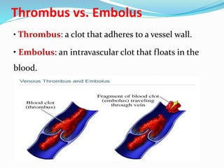
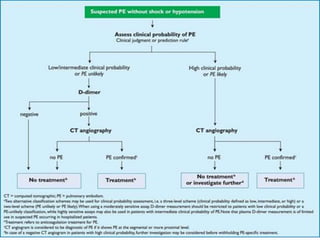
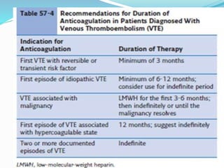
Prepared by
Dr/ Doaa Gadallah Hassanien
Lecturer of chest diseases
Hemostasis
• prevention of blood loss.
• Hemostasis is the process of forming clots in
the walls of damaged blood vessels and
preventing blood loss, while maintaining blood
in a fluid state within the vascular system.
• Physiologic
Thrombosis:
• The formation of an unwanted clot within a
blood vessel.
• The most common abnormality of
hemostasis.
• Pathologic
Thrombus vs. Embolus
• Thrombus: a clot that adheres to a vessel wall.
• Embolus: an intravascular clot that floats in the
blood.
Pulmonary embolism (PE) is a blockage
of pulmonary artery by a substance that has moved
from elsewhere in the body through the
bloodstream. Most pulmonary emboli are multiple,
more commonly in the lower lobes.
Venous thromboembolism (VTE) encompasses
deep vein thrombosis (DVT) and pulmonary
embolism (PE).
A saddle pulmonary embolism is described
as a clot located in the main pulmonary artery
that traverses the right and left pulmonary
arteries.
Lobar, segmental, and subsegmental PEs:
are clots located in the branches of the pulmonary
artery corresponding to the anatomical lung
segment.
Pulmonary Infarction : The pathological changes
which develop in the lung as a result of pulmonary
embolism.
Epidemiology
.PE is the third most common cause of death among
hospitalized patients.
.PE is present in 60-80% of patients with DVT.
.Acute PE is the most serious clinical presentation of VTE.
.The epidemiology of PE is difficult to determine because it
may remain asymptomatic, or its diagnosis may be an
incidental finding, in some cases, the first presentation of PE
may be sudden death.
.The incidence of PE also is age-dependent,
with increasing incidence of death with
advancing age
The percentages of the different subtypes
of PE:
Massive PE: 5-10%
Sub massive PE: 20-25%
Low-risk PE: ~70%
Sources of emboli
(80%–95%) of PE occur as a result of thrombus originating in
the lower extremity: deep veins of the calf , the popliteal and
femoral veins.
Other sources: -the pelvic veins (pregnancy, pelvic infection,
prostate disease, or recent pelvic surgery)
-Emboli may also originate from upper extremity thrombosis
associated with central venous catheters or intravascular
cardiac devices
-the renal veins
Pathogenesis
Thrombus often begins at a site where blood flow is
turbulent, such as at a venous bifurcation, or behind
a venous valve. When thrombus propagation
exceeds the rate of thrombus organization and
adherence to the endothelium, part or all of
thrombus may break away and migrate via the
venous system to the lungs
Classification
I. Thrombotic: The most common.
II. Non-thrombotic PE (NTPE)
- Embolization to the pulmonary circulation
of different cell types (adipocytes,
hematopoietic, amniotic or tumor), bacteria,
fungi, foreign material or gas.
Etiology
Three primary influences predispose a patient
to thrombus formation; these form the so-
called Virchow triad, which consists of the
following:
.Endothelial injury
.Stasis or turbulence of blood flow
.Blood hypercoagulability
Risk factors for venous
thromboembolism
INHERITED RISK FACTORS:
.Factor V Leiden mutation
.Protein C deficiency
.Protein S deficiency
.Antithrombin deficiency
.Prothrombin gene mutation
.Anticardiolipin antibodies
.Lupus anticoagulant
Risk factors for venous
thromboembolism
Major risk factors (relative
risk 5–20):
Surgery*
• Major abdominal/pelvic
surgery
• Hip/knee replacement
• Postoperative intensive care
Obstetrics
• Late pregnancy
•Caesarian section
• Puerperium
Lower limb problems
• Fracture
•Varicose veins
Malignancy
• Abdominal/pelvic
•metastatic
Reduced mobility
• Hospitalization
•Institutional care
Miscellaneous
• Previous proven VTE
Risk factors for venous
thromboembolism
Minor risk factors (relative
risk 2–4):
Cardiovascular
• Congenital heart disease
• Congestive cardiac failure
• Hypertension
• Superficial venous
thrombosis
• Indwelling central vein
catheter
Estrogens
•Oral contraceptive
• Hormone replacement
therapy
Miscellaneous
• COPD
• Occult malignancy
• Long distance sedentary travel
• Obesity
Clinical classification of pulmonary
embolism severity
Diagnosis
‘confirmed PE’ is defined as a probability of
PE high enough to indicate the need for PE-
specific treatment
‘excluded PE’ as a probability of PE low
enough to justify withholding PE-specific
treatment.
Clinical presentation
PE
confir
med
signs
96%Tachypnea (respiratory rate
>16/min)
58%Rales
53%Accentuated second heart sound
44%Tachycardia (heart rate
>100/min)
43%Fever (temperature >37.8°C
32%Clinical signs and symptoms
suggesting thrombophlebitis
24%Lower extremity edema
23%Cardiac murmur
19%Cyanosis
Differential Diagnosis for Acute
Pulmonary Embolism
.Pneumonia or bronchitis
.Asthma or exacerbation of chronic obstructive lung disease
.Pleuritis .Pericarditis/cardiac tamponed
.Pneumothorax .Musculoskeletal pain
.Costochondritis .Rib fracture
.Pulmonary edema/congestive heart failure
.Thoracic malignancy .Pulmonary hypertension
.Myocardial infarction .Aortic dissection
CLINICAL ASSESSMENT
Wells and Geneva Scoring Systems Used
in Risk Assessment for the Diagnosis of
Pulmonary Embolism (PE)* Both scoring
systems divide patients into groups with
low, intermediate, and high clinical
probability for diagnosis of PE
LABORATORY TESTS
1-Arterial Blood Gas Analysis: hypoxemia
2-D-Dimer Assay :D-dimer is a plasmin-derived fibrin
degradation product.
D-dimer testing has proven to be highly sensitive but not
specific.
Blood D-dimer assay should only be considered
following assessment of clinical probability.
D-dimer assay should not be performed in those with
high clinical probability of PE.
A negative D-dimer test reliably excludes PE in patients
with low or intermediate clinical probability; such
patients do not require imaging for VTE.
3-B-Type Natriuretic Peptide Assay, Troponin
Assay
Electrocardiogram
Chest Radiographs
Classic abnormalities include:
Westermark’s Sign - focal oligemia
Hampton’s Hump - wedge shaped density
Enlarged Right Descending Pulmonary Artery
(Palla’s sign) usually seen in the acute setting.
A diaphragm may be elevated, reflect in
volume loss in the affected lung.
Pleural Effusion 48%
Atelectasis 68%
Atelectasis and parenchymal densities are quite common.
Most of these densities are caused by pulmonary
hemorrhage and edema
Venous Compression Ultrasonography
Ultrasound imaging frequently is used when
the initial tests for PE are non diagnostic.
A positive ultrasound test result confirms the
need for anticoagulation and obviates the
need for further diagnostic studies.
Echocardiogram
Transthoracic and transesophageal
echocardiography have limited use in the diagnosis
of PE. The sensitivity and specificity of these tests
are inadequate for diagnosis, because the offending
emboli are rarely proximal enough to be visualized.
A more important role of echocardiography in the
evaluation of patients with PE is that of risk
stratification.
Right ventricular dysfunction develops in association
with an acute PE. Worsening right ventricular function
relates directly to the degree the pulmonary vascular
bed is affected by the thrombus and, therefore, the size
of the embolic event. Some investigators have
suggested that more aggressive therapy, such as
thrombolysis, is indicated in patients with right
ventricular dysfunction emboli
Ventilation-Perfusion Lung Scan
For many years, the ventilation-perfusion lung scan was
considered the imaging study of choice to evaluate for PE.
Recently, CTA has replaced the ventilation-perfusion lung
scan as the predominant diagnostic test. However, the
ventilation-perfusion scan maintains an important place
in the evaluation of patients for thromboembolic disease
who have contraindications to CTA such as renal
insufficiency or contrast allergy.
Computed Tomography Angiography
CT pulmonary angiography (i.e., CTA) has become a
favored study for the evaluation of PE.
CTA provides a number of potential advantages over
other imaging modalities in the diagnosis of PE,
including:
(1) direct visualization of the embolus
(2) the ability to assess for other potential causes for the
patient’s complaints such as pneumonia
(3) imaging algorithms that scan through the pelvis and
lower extremities, as well as the chest, allowing
simultaneous evaluation for PE and for DVT
8-Pulmonary angiography
The diagnosis of acute PE is based on direct
evidence of a thrombus in two projections,
either as a filling defect or as amputation of a
pulmonary arterial branch. Thrombi as small
as 1–2 mm within the sub-segmental arteries
can be visualized
Pulmonary angiography
Treatment
Indication for hospitalization :
1-Older patients who may have less cardiopulmonary
reserve, or significant coexisting illnesses
2-Those who may not be able to follow instructions
or have adequate follow-up.
3-Hypoxemia, hypotension, hemodynamic
instability
1-Heparin
The major anticoagulant effect of heparin is to
reduce thrombus propagation and prevent embolic
recurrence. Choices include either intravenous
unfractionated heparin (UFH) or subcutaneous
LMWH preparation.
Unfractionated heparin (UFH) should be considered
(a) as a first dose bolus, (b) in massive PE, or (c)
where rapid reversal of effect may be needed.
Dosing regimen
.using an initial intravenous bolus of 80 units of
heparin per kilogram followed by a continuous
infusion initiated at 18 U/kg/h.
.The heparin drip is adjusted based on monitoring of
the aPTT, drawn 6 hours after the initial bolus dose,
then 6 hours after each dose adjustment, with a
target aPTT ratio of 1.5 to 2.5.
.Protamine Sulphate
2-LMWH
.LMWH preparations have displaced unfractionated heparin as the
anticoagulant of choice in uncomplicated venous thromboembolism
including PE
.Advantages of LMWH compared with UFH include
(1) longer half-life and ease of use
(2) ability to consistently achieve early therapeutic anticoagulation
(3) no need to monitor anticoagulant effects
(4) reduced incidence of major bleeding complications.
.Enoxaparin: 1 mg/kg every 12h
3-FACTOR XA INHIBITORS
Fondaparinux:
.Subcutaneous administeration
.Also has been demonstrated to be efficacious in the
treatment of VTE.
.Has a relatively long half-life (17 hours) and can be
administered once daily
Rivaroxaban:
Their excellent bioavailability, rapid onset, predictable
effect, and ease of use will greatly facilitate outpatient
therapy.
.Close monitoring of coagulation levels, as is required
with the vitamin K antagonists, is unnecessary, and the
risk of over- or under anticoagulation is negligible.
.One potential complicating factor regarding the use of
the Xa inhibitors is the lack of a specific antidote for
rapid reversal of anticoagulation.
4-Thrombolytic therapy
.Thrombolytic drugs cause direct lysis of thrombi by
increasing plasmin production through plasminogen
activation.
.Thrombolysis is the first line treatment for massive PE
and may be instituted on clinical grounds alone if
cardiac arrest is imminent
.Streptokinase, alteplase (rt-PA), and urokinase
.50 mg bolus of alteplase is recommended
5-Adjuvants therapy
Oxygen
Antibiotics
Rest in bed
6-PULMONARY EMBOLECTOMY
Consider surgical embolectomy:
.Patients with persistent hypotension, shock,
or cardiac arrest
.Who either failed thrombolysis or have a
contraindication to thrombolytic
management.
LONG-TERM MANAGEMENT
WARFARIN
.Vitamin K antagonist, is generally used for long-term treatment of
venous thromboembolism because of its proven efficacy.
.Warfarin inhibits gamma carboxylation activation of coagulation factors
II, VII, IX, and X as well as proteins C and S.
.Warfarin has a narrow therapeutic index and patients are generally
monitored closely by measuring the prothrombin time corrected to the
reagent being used (the INR). To maximize efficacy while minimizing
side effects, an INR range between 2 and 3 is recommended for most
patients
LONG-TERM MANAGEMENT
NOVEL AGENTS:
Novel agents are new oral anticoagulant, have
been used successfully for long-term
management of patients with venous
thromboembolism. As noted previously,
rivaroxaban used for the initial and long-term
treatment of PE with or without DVT.
PROPHYLAXIS
Most hospitalized patients are at risk of venous
thromboembolism and should receive some form of
prophylaxis unless its use is contraindicated.
Prophylaxis may not be necessary in rare instances, as
in the case of a young (less than 40 years), ambulatory
patient who is admitted for a short (less than 48–72
hours), hospital stay without a history of prior venous
thromboembolism history.
PROPHYLAXIS
Prophylactic anticoagulation is safe and
effective with an absolute reduction in the
incidence of venous thromboembolism in the
range of 40% to 60%. Major bleeding
complications occur in less than 1% of
patients.
PROPHYLAXIS
Four categories of drugs have been used successfully:
unfractionated heparin, LMWH (enoxaparin,
dalteparin), factor Xa inhibitors (fondaparinux,
rivaroxaban), and the vitamin K antagonist warfarin.
With the exception of warfarin, prophylactic dosing is
subtherapeutic but sufficient to decrease the likelihood
of thrombus formation.
PROPHYLAXIS
Prevention of venous thromboembolism may also be achieved
by the use of mechanical devices.
These devices fall into two categories, graduated compression
stockings and intermittent pneumatic compression stockings
the use of pneumatic compression has been shown in selected
patients to be as effective as subcutaneous unfractionated
heparin in preventing thrombosis. Mechanical methods of
prophylaxis are especially useful in patients at bleeding risk.
Pulmonary embolism
Pulmonary embolism

Pulmonary embolism

  • 1.
    Prepared by Dr/ DoaaGadallah Hassanien Lecturer of chest diseases
  • 2.
    Hemostasis • prevention ofblood loss. • Hemostasis is the process of forming clots in the walls of damaged blood vessels and preventing blood loss, while maintaining blood in a fluid state within the vascular system. • Physiologic
  • 3.
    Thrombosis: • The formationof an unwanted clot within a blood vessel. • The most common abnormality of hemostasis. • Pathologic
  • 4.
    Thrombus vs. Embolus •Thrombus: a clot that adheres to a vessel wall. • Embolus: an intravascular clot that floats in the blood.
  • 5.
    Pulmonary embolism (PE)is a blockage of pulmonary artery by a substance that has moved from elsewhere in the body through the bloodstream. Most pulmonary emboli are multiple, more commonly in the lower lobes. Venous thromboembolism (VTE) encompasses deep vein thrombosis (DVT) and pulmonary embolism (PE).
  • 6.
    A saddle pulmonaryembolism is described as a clot located in the main pulmonary artery that traverses the right and left pulmonary arteries.
  • 7.
    Lobar, segmental, andsubsegmental PEs: are clots located in the branches of the pulmonary artery corresponding to the anatomical lung segment. Pulmonary Infarction : The pathological changes which develop in the lung as a result of pulmonary embolism.
  • 8.
    Epidemiology .PE is thethird most common cause of death among hospitalized patients. .PE is present in 60-80% of patients with DVT. .Acute PE is the most serious clinical presentation of VTE. .The epidemiology of PE is difficult to determine because it may remain asymptomatic, or its diagnosis may be an incidental finding, in some cases, the first presentation of PE may be sudden death.
  • 9.
    .The incidence ofPE also is age-dependent, with increasing incidence of death with advancing age The percentages of the different subtypes of PE: Massive PE: 5-10% Sub massive PE: 20-25% Low-risk PE: ~70%
  • 10.
    Sources of emboli (80%–95%)of PE occur as a result of thrombus originating in the lower extremity: deep veins of the calf , the popliteal and femoral veins. Other sources: -the pelvic veins (pregnancy, pelvic infection, prostate disease, or recent pelvic surgery) -Emboli may also originate from upper extremity thrombosis associated with central venous catheters or intravascular cardiac devices -the renal veins
  • 11.
  • 12.
    Thrombus often beginsat a site where blood flow is turbulent, such as at a venous bifurcation, or behind a venous valve. When thrombus propagation exceeds the rate of thrombus organization and adherence to the endothelium, part or all of thrombus may break away and migrate via the venous system to the lungs
  • 13.
    Classification I. Thrombotic: Themost common. II. Non-thrombotic PE (NTPE) - Embolization to the pulmonary circulation of different cell types (adipocytes, hematopoietic, amniotic or tumor), bacteria, fungi, foreign material or gas.
  • 14.
    Etiology Three primary influencespredispose a patient to thrombus formation; these form the so- called Virchow triad, which consists of the following: .Endothelial injury .Stasis or turbulence of blood flow .Blood hypercoagulability
  • 16.
    Risk factors forvenous thromboembolism INHERITED RISK FACTORS: .Factor V Leiden mutation .Protein C deficiency .Protein S deficiency .Antithrombin deficiency .Prothrombin gene mutation .Anticardiolipin antibodies .Lupus anticoagulant
  • 17.
    Risk factors forvenous thromboembolism Major risk factors (relative risk 5–20): Surgery* • Major abdominal/pelvic surgery • Hip/knee replacement • Postoperative intensive care Obstetrics • Late pregnancy •Caesarian section • Puerperium Lower limb problems • Fracture •Varicose veins Malignancy • Abdominal/pelvic •metastatic Reduced mobility • Hospitalization •Institutional care Miscellaneous • Previous proven VTE
  • 18.
    Risk factors forvenous thromboembolism Minor risk factors (relative risk 2–4): Cardiovascular • Congenital heart disease • Congestive cardiac failure • Hypertension • Superficial venous thrombosis • Indwelling central vein catheter Estrogens •Oral contraceptive • Hormone replacement therapy Miscellaneous • COPD • Occult malignancy • Long distance sedentary travel • Obesity
  • 19.
    Clinical classification ofpulmonary embolism severity
  • 20.
    Diagnosis ‘confirmed PE’ isdefined as a probability of PE high enough to indicate the need for PE- specific treatment ‘excluded PE’ as a probability of PE low enough to justify withholding PE-specific treatment.
  • 21.
    Clinical presentation PE confir med signs 96%Tachypnea (respiratoryrate >16/min) 58%Rales 53%Accentuated second heart sound 44%Tachycardia (heart rate >100/min) 43%Fever (temperature >37.8°C 32%Clinical signs and symptoms suggesting thrombophlebitis 24%Lower extremity edema 23%Cardiac murmur 19%Cyanosis
  • 22.
    Differential Diagnosis forAcute Pulmonary Embolism .Pneumonia or bronchitis .Asthma or exacerbation of chronic obstructive lung disease .Pleuritis .Pericarditis/cardiac tamponed .Pneumothorax .Musculoskeletal pain .Costochondritis .Rib fracture .Pulmonary edema/congestive heart failure .Thoracic malignancy .Pulmonary hypertension .Myocardial infarction .Aortic dissection
  • 23.
    CLINICAL ASSESSMENT Wells andGeneva Scoring Systems Used in Risk Assessment for the Diagnosis of Pulmonary Embolism (PE)* Both scoring systems divide patients into groups with low, intermediate, and high clinical probability for diagnosis of PE
  • 25.
    LABORATORY TESTS 1-Arterial BloodGas Analysis: hypoxemia 2-D-Dimer Assay :D-dimer is a plasmin-derived fibrin degradation product. D-dimer testing has proven to be highly sensitive but not specific. Blood D-dimer assay should only be considered following assessment of clinical probability. D-dimer assay should not be performed in those with high clinical probability of PE. A negative D-dimer test reliably excludes PE in patients with low or intermediate clinical probability; such patients do not require imaging for VTE.
  • 26.
    3-B-Type Natriuretic PeptideAssay, Troponin Assay
  • 27.
  • 28.
    Chest Radiographs Classic abnormalitiesinclude: Westermark’s Sign - focal oligemia
  • 29.
    Hampton’s Hump -wedge shaped density
  • 30.
    Enlarged Right DescendingPulmonary Artery (Palla’s sign) usually seen in the acute setting.
  • 31.
    A diaphragm maybe elevated, reflect in volume loss in the affected lung.
  • 32.
  • 33.
    Atelectasis 68% Atelectasis andparenchymal densities are quite common. Most of these densities are caused by pulmonary hemorrhage and edema
  • 34.
    Venous Compression Ultrasonography Ultrasoundimaging frequently is used when the initial tests for PE are non diagnostic. A positive ultrasound test result confirms the need for anticoagulation and obviates the need for further diagnostic studies.
  • 35.
    Echocardiogram Transthoracic and transesophageal echocardiographyhave limited use in the diagnosis of PE. The sensitivity and specificity of these tests are inadequate for diagnosis, because the offending emboli are rarely proximal enough to be visualized. A more important role of echocardiography in the evaluation of patients with PE is that of risk stratification.
  • 36.
    Right ventricular dysfunctiondevelops in association with an acute PE. Worsening right ventricular function relates directly to the degree the pulmonary vascular bed is affected by the thrombus and, therefore, the size of the embolic event. Some investigators have suggested that more aggressive therapy, such as thrombolysis, is indicated in patients with right ventricular dysfunction emboli
  • 38.
  • 39.
    Ventilation-Perfusion Lung Scan Formany years, the ventilation-perfusion lung scan was considered the imaging study of choice to evaluate for PE. Recently, CTA has replaced the ventilation-perfusion lung scan as the predominant diagnostic test. However, the ventilation-perfusion scan maintains an important place in the evaluation of patients for thromboembolic disease who have contraindications to CTA such as renal insufficiency or contrast allergy.
  • 40.
    Computed Tomography Angiography CTpulmonary angiography (i.e., CTA) has become a favored study for the evaluation of PE. CTA provides a number of potential advantages over other imaging modalities in the diagnosis of PE, including: (1) direct visualization of the embolus (2) the ability to assess for other potential causes for the patient’s complaints such as pneumonia (3) imaging algorithms that scan through the pelvis and lower extremities, as well as the chest, allowing simultaneous evaluation for PE and for DVT
  • 43.
    8-Pulmonary angiography The diagnosisof acute PE is based on direct evidence of a thrombus in two projections, either as a filling defect or as amputation of a pulmonary arterial branch. Thrombi as small as 1–2 mm within the sub-segmental arteries can be visualized
  • 44.
  • 47.
    Treatment Indication for hospitalization: 1-Older patients who may have less cardiopulmonary reserve, or significant coexisting illnesses 2-Those who may not be able to follow instructions or have adequate follow-up. 3-Hypoxemia, hypotension, hemodynamic instability
  • 48.
    1-Heparin The major anticoagulanteffect of heparin is to reduce thrombus propagation and prevent embolic recurrence. Choices include either intravenous unfractionated heparin (UFH) or subcutaneous LMWH preparation. Unfractionated heparin (UFH) should be considered (a) as a first dose bolus, (b) in massive PE, or (c) where rapid reversal of effect may be needed.
  • 49.
    Dosing regimen .using aninitial intravenous bolus of 80 units of heparin per kilogram followed by a continuous infusion initiated at 18 U/kg/h. .The heparin drip is adjusted based on monitoring of the aPTT, drawn 6 hours after the initial bolus dose, then 6 hours after each dose adjustment, with a target aPTT ratio of 1.5 to 2.5. .Protamine Sulphate
  • 50.
    2-LMWH .LMWH preparations havedisplaced unfractionated heparin as the anticoagulant of choice in uncomplicated venous thromboembolism including PE .Advantages of LMWH compared with UFH include (1) longer half-life and ease of use (2) ability to consistently achieve early therapeutic anticoagulation (3) no need to monitor anticoagulant effects (4) reduced incidence of major bleeding complications. .Enoxaparin: 1 mg/kg every 12h
  • 51.
    3-FACTOR XA INHIBITORS Fondaparinux: .Subcutaneousadministeration .Also has been demonstrated to be efficacious in the treatment of VTE. .Has a relatively long half-life (17 hours) and can be administered once daily
  • 52.
    Rivaroxaban: Their excellent bioavailability,rapid onset, predictable effect, and ease of use will greatly facilitate outpatient therapy. .Close monitoring of coagulation levels, as is required with the vitamin K antagonists, is unnecessary, and the risk of over- or under anticoagulation is negligible. .One potential complicating factor regarding the use of the Xa inhibitors is the lack of a specific antidote for rapid reversal of anticoagulation.
  • 53.
    4-Thrombolytic therapy .Thrombolytic drugscause direct lysis of thrombi by increasing plasmin production through plasminogen activation. .Thrombolysis is the first line treatment for massive PE and may be instituted on clinical grounds alone if cardiac arrest is imminent .Streptokinase, alteplase (rt-PA), and urokinase .50 mg bolus of alteplase is recommended
  • 54.
  • 55.
    6-PULMONARY EMBOLECTOMY Consider surgicalembolectomy: .Patients with persistent hypotension, shock, or cardiac arrest .Who either failed thrombolysis or have a contraindication to thrombolytic management.
  • 56.
    LONG-TERM MANAGEMENT WARFARIN .Vitamin Kantagonist, is generally used for long-term treatment of venous thromboembolism because of its proven efficacy. .Warfarin inhibits gamma carboxylation activation of coagulation factors II, VII, IX, and X as well as proteins C and S. .Warfarin has a narrow therapeutic index and patients are generally monitored closely by measuring the prothrombin time corrected to the reagent being used (the INR). To maximize efficacy while minimizing side effects, an INR range between 2 and 3 is recommended for most patients
  • 57.
    LONG-TERM MANAGEMENT NOVEL AGENTS: Novelagents are new oral anticoagulant, have been used successfully for long-term management of patients with venous thromboembolism. As noted previously, rivaroxaban used for the initial and long-term treatment of PE with or without DVT.
  • 59.
    PROPHYLAXIS Most hospitalized patientsare at risk of venous thromboembolism and should receive some form of prophylaxis unless its use is contraindicated. Prophylaxis may not be necessary in rare instances, as in the case of a young (less than 40 years), ambulatory patient who is admitted for a short (less than 48–72 hours), hospital stay without a history of prior venous thromboembolism history.
  • 60.
    PROPHYLAXIS Prophylactic anticoagulation issafe and effective with an absolute reduction in the incidence of venous thromboembolism in the range of 40% to 60%. Major bleeding complications occur in less than 1% of patients.
  • 61.
    PROPHYLAXIS Four categories ofdrugs have been used successfully: unfractionated heparin, LMWH (enoxaparin, dalteparin), factor Xa inhibitors (fondaparinux, rivaroxaban), and the vitamin K antagonist warfarin. With the exception of warfarin, prophylactic dosing is subtherapeutic but sufficient to decrease the likelihood of thrombus formation.
  • 62.
    PROPHYLAXIS Prevention of venousthromboembolism may also be achieved by the use of mechanical devices. These devices fall into two categories, graduated compression stockings and intermittent pneumatic compression stockings the use of pneumatic compression has been shown in selected patients to be as effective as subcutaneous unfractionated heparin in preventing thrombosis. Mechanical methods of prophylaxis are especially useful in patients at bleeding risk.