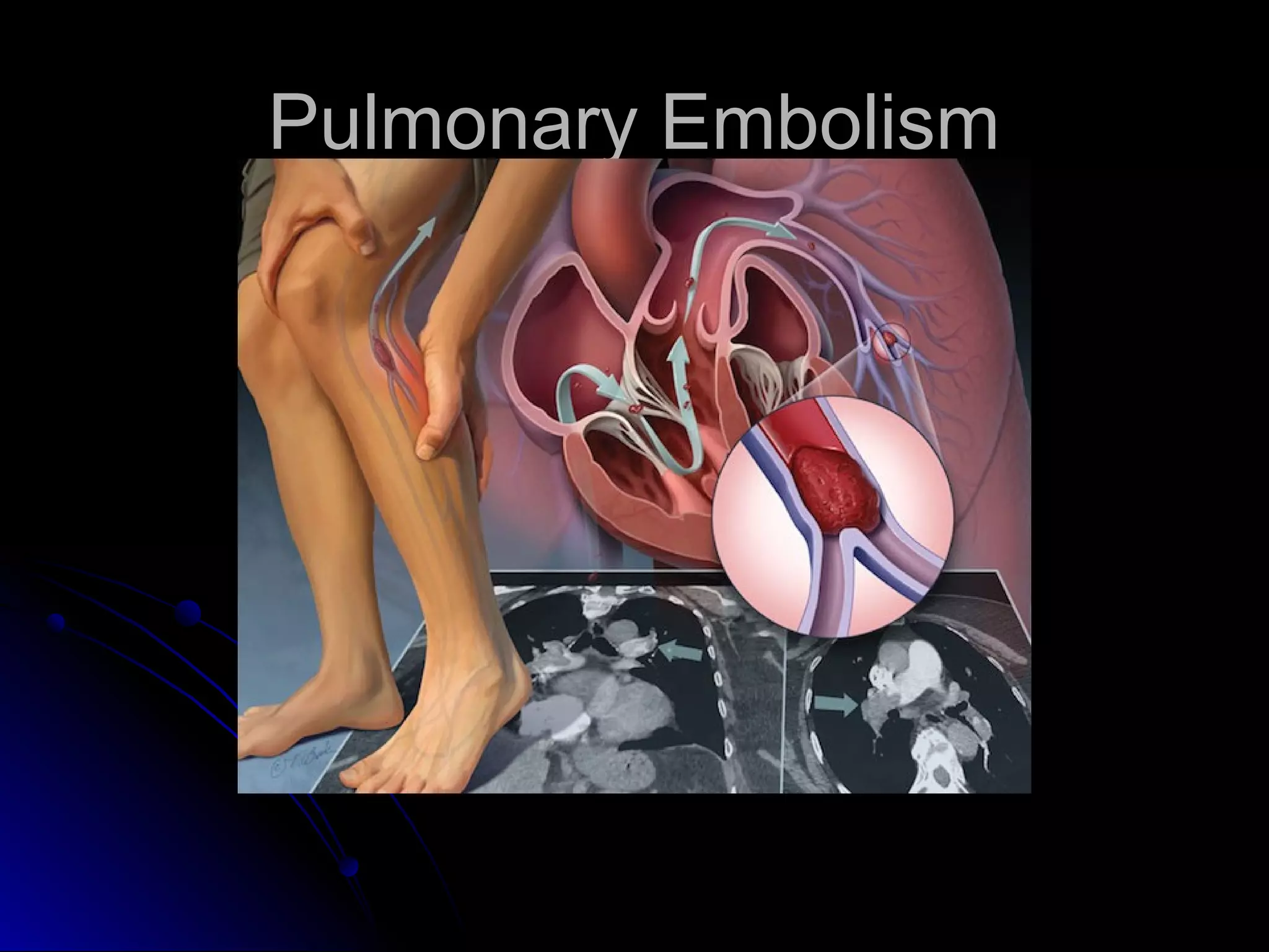
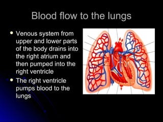
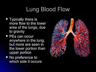
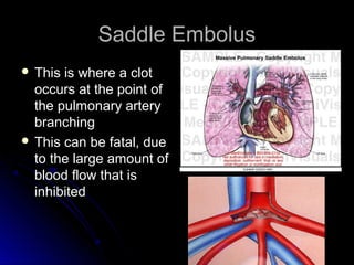
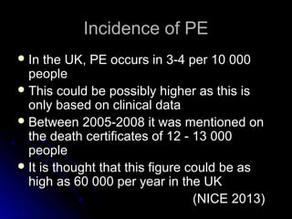
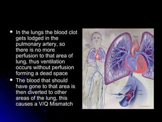
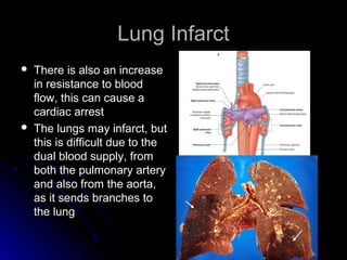
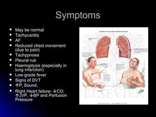
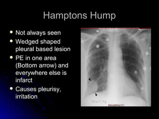
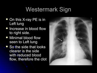
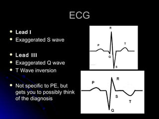
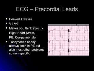
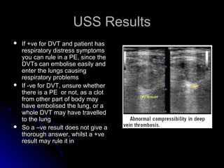
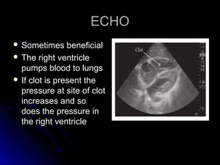
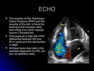
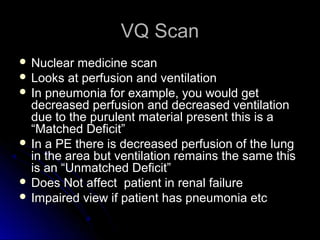
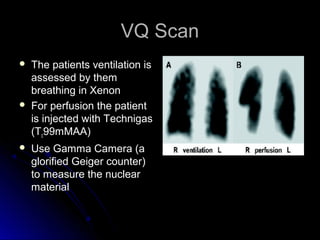
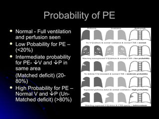
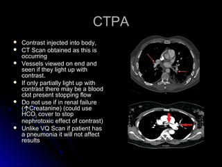
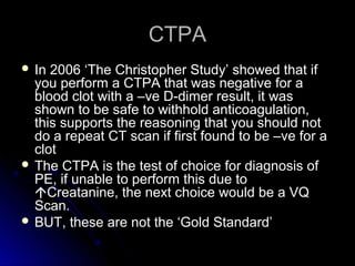
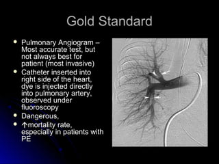
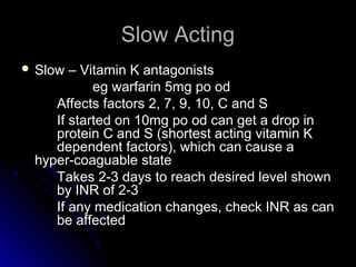
SlideShare a Scribd company logo
Pulmonary EmbolismPulmonary Embolism
Learning OutcomesLearning Outcomes
 CausesCauses
 DiagnosisDiagnosis
 Risk FactorsRisk Factors
 TreatmentTreatment
Blood flow to the lungsBlood flow to the lungs
 Venous system fromVenous system from
upper and lower partsupper and lower parts
of the body drains intoof the body drains into
the right atrium andthe right atrium and
then pumped into thethen pumped into the
right ventricleright ventricle
 The right ventricleThe right ventricle
pumps blood to thepumps blood to the
lungslungs
The Clot TravelsThe Clot Travels
 So if a clot occurs in anySo if a clot occurs in any
vein it will eventually (if itvein it will eventually (if it
breaks free) end up in thebreaks free) end up in the
right side of the heartright side of the heart
 The heart then pumps itThe heart then pumps it
into the pulmonaryinto the pulmonary
arteriesarteries
 As the pulmonary arteryAs the pulmonary artery
becomes smaller, thebecomes smaller, the
blood clot will becomeblood clot will become
caught in the lungs. Thiscaught in the lungs. This
is a Pulmonary Embolismis a Pulmonary Embolism
Lung Blood FlowLung Blood Flow
 Typically there isTypically there is
more flow to the lowermore flow to the lower
area of the lungs, duearea of the lungs, due
to gravityto gravity
 PEs can occurPEs can occur
anywhere in the lung,anywhere in the lung,
but more are seen inbut more are seen in
the lower portion thanthe lower portion than
upper portionupper portion
 No preference toNo preference to
which side it occurswhich side it occurs
Saddle EmbolusSaddle Embolus
 This is where a clotThis is where a clot
occurs at the point ofoccurs at the point of
the pulmonary arterythe pulmonary artery
branchingbranching
 This can be fatal, dueThis can be fatal, due
to the large amount ofto the large amount of
blood flow that isblood flow that is
inhibitedinhibited
Incidence of PEIncidence of PE
 In the UK, PE occurs in 3-4 per 10 000In the UK, PE occurs in 3-4 per 10 000
peoplepeople
 This could be possibly higher as this isThis could be possibly higher as this is
only based on clinical dataonly based on clinical data
 Between 2005-2008 it was mentioned onBetween 2005-2008 it was mentioned on
the death certificates of 12 - 13 000the death certificates of 12 - 13 000
peoplepeople
 It is thought that this figure could be asIt is thought that this figure could be as
high as 60 000 per year in the UKhigh as 60 000 per year in the UK
(NICE 2013)(NICE 2013)
DiagnosingDiagnosing
 A large number ofA large number of
patients arepatients are
misdiagnosed andmisdiagnosed and
only found to have aonly found to have a
PE on autopsyPE on autopsy
 We can test for themWe can test for them
but results can comebut results can come
back as negative (orback as negative (or
inconclusive)inconclusive)
DiagnosingDiagnosing
 Most PEs come fromMost PEs come from
Deep VenousDeep Venous
Thrombosis (DVT)Thrombosis (DVT)
 Most DVTs (75%) comeMost DVTs (75%) come
from the legs,from the legs,
 20% of clots embolise20% of clots embolise
with higher incidencewith higher incidence
above the knee ratherabove the knee rather
than belowthan below
 The emboli travel via theThe emboli travel via the
Inferior Vena Cava (IVC),Inferior Vena Cava (IVC),
to the heart, to the lungsto the heart, to the lungs
PEPE
 In the lungs the blood clotIn the lungs the blood clot
gets lodged in thegets lodged in the
pulmonary artery, sopulmonary artery, so
there is no morethere is no more
perfusion to that area ofperfusion to that area of
lung, thus ventilationlung, thus ventilation
occurs without perfusionoccurs without perfusion
forming a dead spaceforming a dead space
 The blood that shouldThe blood that should
have gone to that area ishave gone to that area is
then diverted to otherthen diverted to other
areas of the lung, thisareas of the lung, this
causes a V/Q Mismatchcauses a V/Q Mismatch
Lung InfarctLung Infarct
 There is also an increaseThere is also an increase
in resistance to bloodin resistance to blood
flow, this can cause aflow, this can cause a
cardiac arrestcardiac arrest
 The lungs may infarct, butThe lungs may infarct, but
this is difficult due to thethis is difficult due to the
dual blood supply, fromdual blood supply, from
both the pulmonary arteryboth the pulmonary artery
and also from the aorta,and also from the aorta,
as it sends branches toas it sends branches to
the lungthe lung
Risk FactorsRisk Factors
 Orthopaedic Procedure – THR, TKR, #sOrthopaedic Procedure – THR, TKR, #s
Lack of mobilisationLack of mobilisation
 No thrombo-prophylaxis – TEDS, Flowtrons,No thrombo-prophylaxis – TEDS, Flowtrons,
Dalteparin , WarfarinDalteparin , Warfarin
 Abdo / Pelvic surgery - especially for CaAbdo / Pelvic surgery - especially for Ca
 ObesityObesity
 Women >30yrs on oral contraceptive who smokeWomen >30yrs on oral contraceptive who smoke
 Hyper-coaguable state – Protein C & S deficiencies,Hyper-coaguable state – Protein C & S deficiencies,
Factor V LeidenFactor V Leiden
 PregnancyPregnancy
SymptomsSymptoms
 May be normalMay be normal
 TachycardiaTachycardia
 AFAF
 Reduced chest movementReduced chest movement
(due to pain)(due to pain)
 TachypnoeaTachypnoea
 Pleural rubPleural rub
 Haemoptysis (especially inHaemoptysis (especially in
lung infarction)lung infarction)
 Low grade feverLow grade fever
 Signs of DVTSigns of DVT
 PP22 Sound,Sound,
 Right Heart failure-Right Heart failure- CO,CO,
JVP,JVP, BP and PerfusionBP and Perfusion
PressurePressure
DiagnosisDiagnosis
 ABG –ABG – pH /pH /  pCOpCO22//  pOpO22 (or lower normal range)(or lower normal range)
 Respiratory AlkalosisRespiratory Alkalosis
  pOpO22 due to V/Q Mismatchdue to V/Q Mismatch
  pCOpCO22 due to tachypnoea (trying to make room for Odue to tachypnoea (trying to make room for O2)2)
 Alveolar-arterial oxygen (A-a) gradientAlveolar-arterial oxygen (A-a) gradient
 PPAAOO22 - P- PaaOO22
 Alone they are not diagnostic of pulmonary embolismAlone they are not diagnostic of pulmonary embolism
(PE), but they may be useful in excluding the diagnosis(PE), but they may be useful in excluding the diagnosis
of PE if their values fall within the normal rangeof PE if their values fall within the normal range
Alveolar-arterial oxygen (A-a)Alveolar-arterial oxygen (A-a)
gradientgradient
 PPAAOO22 - P- PaaOO22 should equal (Age (years) / 4) + 4should equal (Age (years) / 4) + 4
 PPAAOO22 = [(F= [(FiiOO22) x (760 – 47)] – (P) x (760 – 47)] – (PaaCOCO22 / 0.8)/ 0.8)
Barometric Pressure = 760mmHgBarometric Pressure = 760mmHg
Water Vapour Pressure = 47mmHgWater Vapour Pressure = 47mmHg
Respiratory Coefficient = 0.8Respiratory Coefficient = 0.8
UseUsePPaaOO22 andandPPaaCOCO22 in mmHg not KPain mmHg not KPa
Eg 40 Yr old,Eg 40 Yr old,
FFiiOO22 0.60.6
PPaaCOCO22 32.5 mmHg32.5 mmHg (4.27KPa)(4.27KPa)
PPaaOO22 65 mmHg (8.55KPa)65 mmHg (8.55KPa)
PPAAOO22 =(0.6 x 713) – 40.6 = 387.2=(0.6 x 713) – 40.6 = 387.2
PPAAOO22 - P- PaaOO22 = 387.2 – 65 = 322.2= 387.2 – 65 = 322.2
(40/4) = 10 (+4) = 14(40/4) = 10 (+4) = 14
Therefore 322.2 > 14Therefore 322.2 > 14
So if the A-a gradient > 14 it may be indicative of a PESo if the A-a gradient > 14 it may be indicative of a PE
Chest X-rayChest X-ray
 Chest X ray – most common finding with aChest X ray – most common finding with a
PE is a normal x-rayPE is a normal x-ray
 But they are useful as X-rays help to ruleBut they are useful as X-rays help to rule
out pneumonia and pneumothorax asout pneumonia and pneumothorax as
causes of dyspnoea etccauses of dyspnoea etc
 Possible signs indicating a PE are asPossible signs indicating a PE are as
followsfollows
Hamptons HumpHamptons Hump
 Not always seenNot always seen
 Wedged shapedWedged shaped
pleural based lesionpleural based lesion
 PE in one areaPE in one area
(Bottom arrow) and(Bottom arrow) and
everywhere else iseverywhere else is
infarctinfarct
 Causes pleurisy,Causes pleurisy,
irritationirritation
Westermark SignWestermark Sign
 On this X-ray PE is inOn this X-ray PE is in
Left lungLeft lung
 Increase in blood flowIncrease in blood flow
to right side.to right side.
 Minimal blood flowMinimal blood flow
seen to Left lungseen to Left lung
 So the side that looksSo the side that looks
clearer is the sideclearer is the side
with reduced bloodwith reduced blood
flow, therefore the clotflow, therefore the clot
ECGECG
 SSIIQQIIIIIITTIIIIII
 Can be seen in anyCan be seen in any
cor-pulmonalecor-pulmonale
syndrome wheresyndrome where
pulmonary arterypulmonary artery
systolic pressure issystolic pressure is
elevatedelevated
 Eg Normal PQRSTEg Normal PQRST
ECGECG
 LeadLead II
 Exaggerated S waveExaggerated S wave
 LeadLead IIIIII
 Exaggerated Q waveExaggerated Q wave
 T Wave inversionT Wave inversion
 Not specific to PE, butNot specific to PE, but
gets you to possibly thinkgets you to possibly think
of the diagnosisof the diagnosis
ECG – Precordial LeadsECG – Precordial Leads
 Peaked T wavesPeaked T waves
 V1-V4V1-V4
 Makes you think about :-Makes you think about :-
Right Heart Strain,Right Heart Strain,
PE, Cor-pulmonalePE, Cor-pulmonale
 Tachycardia nearlyTachycardia nearly
always seen in PE butalways seen in PE but
also most other problemsalso most other problems
so non-specificso non-specific
D-dimerD-dimer
 By-product of fibrinBy-product of fibrin
degradationdegradation
 As clot degraded byAs clot degraded by
enzymes, Fibrinenzymes, Fibrin
Degradation ProductsDegradation Products
(FDP) are released, one(FDP) are released, one
of these is the D-dimerof these is the D-dimer
 Little clots in body releaseLittle clots in body release
D-dimers, therefore if testD-dimers, therefore if test
+ve - do not know if it is+ve - do not know if it is
from a PE,from a PE,
 But test is so sensitiveBut test is so sensitive
that if no D-dimer ( –ve)that if no D-dimer ( –ve)
you can rule out PEyou can rule out PE
UltrasoundUltrasound
 Non-invasiveNon-invasive
 Looks at lower limbsLooks at lower limbs
 Doppler USS looks atDoppler USS looks at
vein for flow andvein for flow and
compressibilty to seecompressibilty to see
if clot presentif clot present
USS ResultsUSS Results
 If +ve for DVT and patient hasIf +ve for DVT and patient has
respiratory distress symptomsrespiratory distress symptoms
you can rule in a PE, since theyou can rule in a PE, since the
DVTs can embolise easily andDVTs can embolise easily and
enter the lungs causingenter the lungs causing
respiratory problemsrespiratory problems
 If -ve for DVT, unsure whetherIf -ve for DVT, unsure whether
there is a PE or not, as a clotthere is a PE or not, as a clot
from other part of body mayfrom other part of body may
have embolised the lung, or ahave embolised the lung, or a
whole DVT may have travelledwhole DVT may have travelled
to the lungto the lung
 So a –ve result does not give aSo a –ve result does not give a
thorough answer, whilst a +vethorough answer, whilst a +ve
result may rule it inresult may rule it in
ECHOECHO
 Sometimes beneficialSometimes beneficial
 The right ventricleThe right ventricle
pumps blood to lungspumps blood to lungs
 If clot is present theIf clot is present the
pressure at site of clotpressure at site of clot
increases and soincreases and so
does the pressure indoes the pressure in
the right ventriclethe right ventricle
ECHOECHO
 The severity of this PulmonaryThe severity of this Pulmonary
Artery Pressure (PAP) and theArtery Pressure (PAP) and the
severity of the clot, is found byseverity of the clot, is found by
looking at the tricuspid valvelooking at the tricuspid valve
and noting how much regurgeand noting how much regurge
occurs (Tricuspid jet)occurs (Tricuspid jet)
 If tricuspid jet is high then PAPIf tricuspid jet is high then PAP
differential between RA anddifferential between RA and
RV is great and the clot burdenRV is great and the clot burden
is largeis large
 All these tests may help in theAll these tests may help in the
possible diagnosis of a PE, butpossible diagnosis of a PE, but
are not definitive testsare not definitive tests
VQ ScanVQ Scan
 Nuclear medicine scanNuclear medicine scan
 Looks at perfusion and ventilationLooks at perfusion and ventilation
 In pneumonia for example, you would getIn pneumonia for example, you would get
decreased perfusion and decreased ventilationdecreased perfusion and decreased ventilation
due to the purulent material present this is adue to the purulent material present this is a
“Matched Deficit”“Matched Deficit”
 In a PE there is decreased perfusion of the lungIn a PE there is decreased perfusion of the lung
in the area but ventilation remains the same thisin the area but ventilation remains the same this
is an “Unmatched Deficit”is an “Unmatched Deficit”
 Does Not affect patient in renal failureDoes Not affect patient in renal failure
 Impaired view if patient has pneumonia etcImpaired view if patient has pneumonia etc
VQ ScanVQ Scan
 The patients ventilation isThe patients ventilation is
assessed by themassessed by them
breathing in Xenonbreathing in Xenon
 For perfusion the patientFor perfusion the patient
is injected with Technigasis injected with Technigas
(T(Tcc99mMAA)99mMAA)
 Use Gamma Camera (aUse Gamma Camera (a
glorified Geiger counter)glorified Geiger counter)
to measure the nuclearto measure the nuclear
materialmaterial
Probability of PEProbability of PE
 Normal - Full ventilationNormal - Full ventilation
and perfusion seenand perfusion seen
 Low Pobability for PE –Low Pobability for PE –
(<20%)(<20%)
 Intermediate probabilityIntermediate probability
for PE-for PE- V andV and P inP in
same areasame area
(Matched deficit) (20-(Matched deficit) (20-
80%)80%)
 High Probability for PE –High Probability for PE –
Normal V andNormal V and P (Un-P (Un-
Matched deficit) (>80%)Matched deficit) (>80%)
CTPACTPA
 Contrast injected into body,Contrast injected into body,
 CT Scan obtained as this isCT Scan obtained as this is
occurringoccurring
 Vessels viewed on end andVessels viewed on end and
seen if they light up withseen if they light up with
contrast.contrast.
 If only partially light up withIf only partially light up with
contrast there may be a bloodcontrast there may be a blood
clot present stopping flowclot present stopping flow
 Do not use if in renal failureDo not use if in renal failure
((Creatanine) (could useCreatanine) (could use
HCOHCO33 cover to stopcover to stop
nephrotoxic effect of contrast)nephrotoxic effect of contrast)
 Unlike VQ Scan if patient hasUnlike VQ Scan if patient has
a pneumonia it will not affecta pneumonia it will not affect
resultsresults
CTPACTPA
 In 2006 ‘The Christopher Study’ showed that ifIn 2006 ‘The Christopher Study’ showed that if
you perform a CTPA that was negative for ayou perform a CTPA that was negative for a
blood clot with a –ve D-dimer result, it wasblood clot with a –ve D-dimer result, it was
shown to be safe to withhold anticoagulation,shown to be safe to withhold anticoagulation,
this supports the reasoning that you should notthis supports the reasoning that you should not
do a repeat CT scan if first found to be –ve for ado a repeat CT scan if first found to be –ve for a
clotclot
 The CTPA is the test of choice for diagnosis ofThe CTPA is the test of choice for diagnosis of
PE, if unable to perform this due toPE, if unable to perform this due to
Creatanine, the next choice would be a VQCreatanine, the next choice would be a VQ
Scan.Scan.
 BUT, these are not the ‘Gold Standard’BUT, these are not the ‘Gold Standard’
Gold StandardGold Standard
 Pulmonary Angiogram –Pulmonary Angiogram –
Most accurate test, butMost accurate test, but
not always best fornot always best for
patient (most invasive)patient (most invasive)
 Catheter inserted intoCatheter inserted into
right side of the heart,right side of the heart,
dye is injected directlydye is injected directly
into pulmonary artery,into pulmonary artery,
observed underobserved under
fluoroscopyfluoroscopy
 Dangerous,Dangerous,
 mortality rate,mortality rate,
especially in patients withespecially in patients with
PEPE
TreatmentTreatment
 AnticoagulationAnticoagulation
 Eg Warfarin / HeparinEg Warfarin / Heparin
-Blood thinning-Blood thinning
-Prevent new clots-Prevent new clots
Non-thrombolyticNon-thrombolytic
treatmenttreatment
 ThrombolyticsThrombolytics
-Dissolve Clot,-Dissolve Clot,
-not solely used for-not solely used for
PE treatmentPE treatment
 DVT Treatment 3-6 monthsDVT Treatment 3-6 months
 PE Treatment 6-9 monthsPE Treatment 6-9 months
IVC FilterIVC Filter
 If anticoagulationIf anticoagulation
treatmenttreatment
contraindicatedcontraindicated IVCIVC
FilterFilter
If clot travels it getsIf clot travels it gets
caught in IVC Filtercaught in IVC Filter
IVC FilterIVC Filter
 It is possible for blood clot to be caught andIt is possible for blood clot to be caught and
develop in IVC filterdevelop in IVC filter
 Cochrane Collaboration recommends IVC forCochrane Collaboration recommends IVC for
1.1. Recurrent PE despite use of anticoagulation,Recurrent PE despite use of anticoagulation,
or in absolute contra-indication toor in absolute contra-indication to
anticoagulationanticoagulation
2.2. Proximal DVT with massive pulmonaryProximal DVT with massive pulmonary
thrombosis – next one could kill patientthrombosis – next one could kill patient
3.3. Trauma Patient – needing operationTrauma Patient – needing operation
AnticoagulationAnticoagulation
 Ideally start Fast acting and slow acting anticoagulants,Ideally start Fast acting and slow acting anticoagulants,
when slow acting anticoagulants at desired level, stopwhen slow acting anticoagulants at desired level, stop
fast actingfast acting
 Fast acting -Heparin productsFast acting -Heparin products
Heparin (IV) orHeparin (IV) or
Low Molecular Weight Heparin (S/C)Low Molecular Weight Heparin (S/C)
 Slow acting – Vitamin K AntagonistsSlow acting – Vitamin K Antagonists
WarfarinWarfarin
Fast ActingFast Acting
 Heparin productsHeparin products
IV HeparinIV Heparin
Aim for therapeutic APTTAim for therapeutic APTT
Can be startedCan be started
immediatelyimmediately
Quick AnticoagulationQuick Anticoagulation
actionaction
If bleeding occurs canIf bleeding occurs can
stop it and reducedstop it and reduced
effects occur within feweffects occur within few
hours as it has a shorthours as it has a short
half life (45-60min)half life (45-60min)
Heparin InducedHeparin Induced
ThrombocytopaeniaThrombocytopaenia
Can cause highest incidence of HITCan cause highest incidence of HIT
-Usually occurs 5-14 days after starting-Usually occurs 5-14 days after starting HeparinHeparin
(even if discontinued) or sooner if previously had(even if discontinued) or sooner if previously had
heparinheparin
-Immune response to heparin-Immune response to heparin
-Antibodies combine with heparin and platelets-Antibodies combine with heparin and platelets
causing platelet activation of microparticles whichcausing platelet activation of microparticles which
initiate the formation of blood clots; the plateletinitiate the formation of blood clots; the platelet
count falls as a result, So,count falls as a result, So,
-Platelet count drops by >50%-Platelet count drops by >50%
-Get hyper-coaguable state, get more clotting-Get hyper-coaguable state, get more clotting
even though platelet count loweven though platelet count low
-If this occurs change from heparin products-If this occurs change from heparin products
Fast ActingFast Acting
 Low Molecular Weight Heparin (SC)Low Molecular Weight Heparin (SC)
Eg clexane, dalteparin, fondaparinuxEg clexane, dalteparin, fondaparinux
Give twice per day dependent on Creatanine Clearance (CrCl)Give twice per day dependent on Creatanine Clearance (CrCl)
(20-29ml/min)(20-29ml/min)
Or if CrCl ≥30ml/min give once a dayOr if CrCl ≥30ml/min give once a day
Problem is if given, anticoagulation is present for next 12 orProblem is if given, anticoagulation is present for next 12 or
24hrs (as half life is four times as long as heparin, about 4 hrs)24hrs (as half life is four times as long as heparin, about 4 hrs)
Incidence of HIT is lower than IV HeparinIncidence of HIT is lower than IV Heparin
Easy and quick to administerEasy and quick to administer
 Also available is rivaroxaban, a fast acting oral medication, TheAlso available is rivaroxaban, a fast acting oral medication, The
effects lasts 8 to 12 hours, but factor Xa activity does not return toeffects lasts 8 to 12 hours, but factor Xa activity does not return to
normal within 24 hours so once-daily dosing is possible.normal within 24 hours so once-daily dosing is possible.
To work out CrClTo work out CrCl
 Takes into accountTakes into account
Patients Age, Gender,Patients Age, Gender,
Ideal Weight (Kg) SerumIdeal Weight (Kg) Serum
Creatanine (μmol/L) fromCreatanine (μmol/L) from
last 24hrslast 24hrs
 Use the equationUse the equation
((140-Age) x (ideal weight) /((140-Age) x (ideal weight) /
Cr) x1.23(male) = CrClCr) x1.23(male) = CrCl
((140-Age) x (ideal weight) /((140-Age) x (ideal weight) /
Cr) x1.04(female) = CrClCr) x1.04(female) = CrCl
Use STH treatment ofUse STH treatment of
Thromboembolic DiseaseThromboembolic Disease
Dalteparin PrescriptionDalteparin Prescription
ChartChart
ExamplesExamples
Eg Age 67 yrsEg Age 67 yrs
Gender - MaleGender - Male
Ideal Weight 85KgIdeal Weight 85Kg
Cr 73 μmol/LCr 73 μmol/L
Eg (140-67 = 73)Eg (140-67 = 73)  73 x 85 = 620573 x 85 = 6205  6205/ 73 =856205/ 73 =85 85 x 1.23 = 104.ml/min85 x 1.23 = 104.ml/min
So, 18 000 iu Dalteparin once per day as CrCl > 30ml/minSo, 18 000 iu Dalteparin once per day as CrCl > 30ml/min
Age 85 yrsAge 85 yrs
Gender – FemaleGender – Female
Ideal Weight 60KgIdeal Weight 60Kg
Cr – 160 μmol/LCr – 160 μmol/L
(140-85 = 55)(140-85 = 55)  55 x 60 = 330055 x 60 = 3300  3300 / 160 = 20.63300 / 160 = 20.6  20.6 x 1.04 =20.6 x 1.04 =
21.24ml/min21.24ml/min
So, 5000iu Dalteparin am and 5000iu Dalteparin pm as CrCl 20-29ml/minSo, 5000iu Dalteparin am and 5000iu Dalteparin pm as CrCl 20-29ml/min
If patient weighed more than 100Kg split dose if CrCl ≥30ml/minIf patient weighed more than 100Kg split dose if CrCl ≥30ml/min
Slow ActingSlow Acting
 Slow – Vitamin K antagonistsSlow – Vitamin K antagonists
eg warfarin 5mg po odeg warfarin 5mg po od
Affects factors 2, 7, 9, 10, C and SAffects factors 2, 7, 9, 10, C and S
If started on 10mg po od can get a drop inIf started on 10mg po od can get a drop in
protein C and S (shortest acting vitamin Kprotein C and S (shortest acting vitamin K
dependent factors), which can cause adependent factors), which can cause a
hyper-coaguable statehyper-coaguable state
Takes 2-3 days to reach desired level shownTakes 2-3 days to reach desired level shown
by INR of 2-3by INR of 2-3
If any medication changes, check INR as canIf any medication changes, check INR as can
be affectedbe affected
PreventionPrevention
 Bilateral Lower ExtremityBilateral Lower Extremity
Sequential CompressionSequential Compression
DevicesDevices
TEDs and FlowtronsTEDs and Flowtrons
When the legs areWhen the legs are
squeezed the veins release asqueezed the veins release a
factor which thins the bloodfactor which thins the blood
stopping clot formation,stopping clot formation,
the rhythmic motion copiesthe rhythmic motion copies
that of leg movementthat of leg movement
 ThromboprophylaxisThromboprophylaxis
DalteparinDalteparin
Genetic Blood TestsGenetic Blood Tests
 25-50% of patients with VTE have an inherited disorder25-50% of patients with VTE have an inherited disorder
 There are genetic causes of metabolism which may be tested forThere are genetic causes of metabolism which may be tested for
- Factor V Leiden – causes increased clotting as variant cannot beFactor V Leiden – causes increased clotting as variant cannot be
inactivated by Factor Protein C (5% of popinactivated by Factor Protein C (5% of popnn
and 20% of pts withand 20% of pts with
thrombus)thrombus)
- Factor Protein C Deficiency – results in normal cleaving of FactorFactor Protein C Deficiency – results in normal cleaving of Factor
Va and Factor VIIIaVa and Factor VIIIa
- 20210 (prothrombin) Mutation – 2-3 times risk of clot formation20210 (prothrombin) Mutation – 2-3 times risk of clot formation
- MTHMFR affects regulation of homocysteineMTHMFR affects regulation of homocysteine
- Lupus anticoagulant- prothrombotic agent which can causeLupus anticoagulant- prothrombotic agent which can cause
inappropriate clottinginappropriate clotting
- Anti phospholipid antibody – confused autoimmune responseAnti phospholipid antibody – confused autoimmune response
If any of these are positive and patient has a clot then may needIf any of these are positive and patient has a clot then may need
treatment for longertreatment for longer
So, RememberSo, Remember

More Related Content

What's hot

Pulmonary embolism
Pulmonary embolismPulmonary embolism
Pulmonary embolism
Farrukh Masood
 
Pulmonary embolism
Pulmonary embolismPulmonary embolism
Pulmonary embolism
Amir Mahmoud
 
Pulmonary embolism,overview
Pulmonary embolism,overviewPulmonary embolism,overview
Pulmonary embolism,overview
Eman Mahmoud
 
Pulmonary Artery Anatomy and Pulmonary Embolism
Pulmonary Artery Anatomy and Pulmonary EmbolismPulmonary Artery Anatomy and Pulmonary Embolism
Pulmonary Artery Anatomy and Pulmonary Embolism
Gamal Agmy
 
Aortic anurysm
Aortic anurysmAortic anurysm
Aortic anurysm
Monika Devi NR
 
Cardiomegaly
CardiomegalyCardiomegaly
Cardiomegaly
SheelamannilJohn
 
Pulmonary thromboembolism
Pulmonary thromboembolismPulmonary thromboembolism
Pulmonary thromboembolism
Bajanagaraju
 
Pulmonary embolism ( PE)
Pulmonary embolism ( PE)Pulmonary embolism ( PE)
Pulmonary embolism ( PE)
Mohamed Elbhnasawy
 
Pneumothorax
PneumothoraxPneumothorax
Pneumothorax
Waqas Ahmed Khan
 
Pneumothorax sagar gandhi
Pneumothorax sagar gandhiPneumothorax sagar gandhi
Pneumothorax sagar gandhi
Sagar Gandhi
 
Right Ventricular Failure
Right Ventricular FailureRight Ventricular Failure
Right Ventricular Failure
Spectrum Health System
 
Pulmonary embolism Nursing mx..(ESTHER)
Pulmonary embolism Nursing mx..(ESTHER)Pulmonary embolism Nursing mx..(ESTHER)
Pulmonary embolism Nursing mx..(ESTHER)
Esther Mary Mathew
 
Imaging of Aortic Dissection
Imaging of Aortic DissectionImaging of Aortic Dissection
Imaging of Aortic Dissection
Sakher Alkhaderi
 
Pulmonary embolism
Pulmonary embolism Pulmonary embolism
Pulmonary embolism
RahulGupta1687
 
Transesophageal echocardiography
Transesophageal echocardiographyTransesophageal echocardiography
Transesophageal echocardiography
Amit Gulati
 
Aortic regurgitation
Aortic regurgitationAortic regurgitation
Aortic regurgitation
Vitrag Shah
 
Constrictive pericarditis
Constrictive pericarditis Constrictive pericarditis
Constrictive pericarditis
Ramachandra Barik
 
Ultrasonography Fundamentals In Critical Care: Lung Ultrasound, Pleural Ultra...
Ultrasonography Fundamentals In Critical Care: Lung Ultrasound, Pleural Ultra...Ultrasonography Fundamentals In Critical Care: Lung Ultrasound, Pleural Ultra...
Ultrasonography Fundamentals In Critical Care: Lung Ultrasound, Pleural Ultra...
Bassel Ericsoussi, MD
 
Hemodyanmic features of Constrictive pericarditis and Restrictive cardiomyopathy
Hemodyanmic features of Constrictive pericarditis and Restrictive cardiomyopathyHemodyanmic features of Constrictive pericarditis and Restrictive cardiomyopathy
Hemodyanmic features of Constrictive pericarditis and Restrictive cardiomyopathy
Himanshu Rana
 
Pulmonary Embolism- Diagnosis by Dr.Tinku Joseph
Pulmonary Embolism- Diagnosis  by Dr.Tinku JosephPulmonary Embolism- Diagnosis  by Dr.Tinku Joseph
Pulmonary Embolism- Diagnosis by Dr.Tinku Joseph
Dr.Tinku Joseph
 

What's hot (20)

Pulmonary embolism
Pulmonary embolismPulmonary embolism
Pulmonary embolism
 
Pulmonary embolism
Pulmonary embolismPulmonary embolism
Pulmonary embolism
 
Pulmonary embolism,overview
Pulmonary embolism,overviewPulmonary embolism,overview
Pulmonary embolism,overview
 
Pulmonary Artery Anatomy and Pulmonary Embolism
Pulmonary Artery Anatomy and Pulmonary EmbolismPulmonary Artery Anatomy and Pulmonary Embolism
Pulmonary Artery Anatomy and Pulmonary Embolism
 
Aortic anurysm
Aortic anurysmAortic anurysm
Aortic anurysm
 
Cardiomegaly
CardiomegalyCardiomegaly
Cardiomegaly
 
Pulmonary thromboembolism
Pulmonary thromboembolismPulmonary thromboembolism
Pulmonary thromboembolism
 
Pulmonary embolism ( PE)
Pulmonary embolism ( PE)Pulmonary embolism ( PE)
Pulmonary embolism ( PE)
 
Pneumothorax
PneumothoraxPneumothorax
Pneumothorax
 
Pneumothorax sagar gandhi
Pneumothorax sagar gandhiPneumothorax sagar gandhi
Pneumothorax sagar gandhi
 
Right Ventricular Failure
Right Ventricular FailureRight Ventricular Failure
Right Ventricular Failure
 
Pulmonary embolism Nursing mx..(ESTHER)
Pulmonary embolism Nursing mx..(ESTHER)Pulmonary embolism Nursing mx..(ESTHER)
Pulmonary embolism Nursing mx..(ESTHER)
 
Imaging of Aortic Dissection
Imaging of Aortic DissectionImaging of Aortic Dissection
Imaging of Aortic Dissection
 
Pulmonary embolism
Pulmonary embolism Pulmonary embolism
Pulmonary embolism
 
Transesophageal echocardiography
Transesophageal echocardiographyTransesophageal echocardiography
Transesophageal echocardiography
 
Aortic regurgitation
Aortic regurgitationAortic regurgitation
Aortic regurgitation
 
Constrictive pericarditis
Constrictive pericarditis Constrictive pericarditis
Constrictive pericarditis
 
Ultrasonography Fundamentals In Critical Care: Lung Ultrasound, Pleural Ultra...
Ultrasonography Fundamentals In Critical Care: Lung Ultrasound, Pleural Ultra...Ultrasonography Fundamentals In Critical Care: Lung Ultrasound, Pleural Ultra...
Ultrasonography Fundamentals In Critical Care: Lung Ultrasound, Pleural Ultra...
 
Hemodyanmic features of Constrictive pericarditis and Restrictive cardiomyopathy
Hemodyanmic features of Constrictive pericarditis and Restrictive cardiomyopathyHemodyanmic features of Constrictive pericarditis and Restrictive cardiomyopathy
Hemodyanmic features of Constrictive pericarditis and Restrictive cardiomyopathy
 
Pulmonary Embolism- Diagnosis by Dr.Tinku Joseph
Pulmonary Embolism- Diagnosis  by Dr.Tinku JosephPulmonary Embolism- Diagnosis  by Dr.Tinku Joseph
Pulmonary Embolism- Diagnosis by Dr.Tinku Joseph
 

Viewers also liked

Pulmonary embolism 2
Pulmonary embolism 2Pulmonary embolism 2
Pulmonary embolism 2
samirelansary
 
Pulmonary embolism
Pulmonary embolismPulmonary embolism
Pulmonary embolism
salaheldin abusin
 
Pulmonary embolism ppt
Pulmonary embolism pptPulmonary embolism ppt
Pulmonary embolism ppt
resmigs
 
Pulmonary embolism
Pulmonary embolism Pulmonary embolism
Pulmonary embolism
Pulmonary embolismPulmonary embolism
Pulmonary embolism
TeleClinEd
 
GEMC: Acute Pulmonary Emergencies: Pulmonary Embolism, Pulmonary Edema and Pa...
GEMC: Acute Pulmonary Emergencies: Pulmonary Embolism, Pulmonary Edema and Pa...GEMC: Acute Pulmonary Emergencies: Pulmonary Embolism, Pulmonary Edema and Pa...
GEMC: Acute Pulmonary Emergencies: Pulmonary Embolism, Pulmonary Edema and Pa...
Open.Michigan
 
GEMC: The Emergency Department Management of Pulmonary Embolism
GEMC: The Emergency Department Management of Pulmonary EmbolismGEMC: The Emergency Department Management of Pulmonary Embolism
GEMC: The Emergency Department Management of Pulmonary Embolism
Open.Michigan
 
Pulmonary Embolism
Pulmonary EmbolismPulmonary Embolism
Pulmonary Embolism
meducationdotnet
 
Pulmonary Embolism
Pulmonary EmbolismPulmonary Embolism
Pulmonary Embolism
drmcbansal
 
Imaging of Pulmonary Embolism
Imaging of Pulmonary Embolism  Imaging of Pulmonary Embolism
Imaging of Pulmonary Embolism
Gamal Agmy
 
Pe final
Pe finalPe final
Pe final
Frank Meissner
 
Gerber Pulmonary Embolism
Gerber Pulmonary EmbolismGerber Pulmonary Embolism
Gerber Pulmonary Embolism
MedicineAndHealthCancer
 
Figure 3 sign fleischner sign-fissure sign-headcheese sign-hilar overlay sign...
Figure 3 sign fleischner sign-fissure sign-headcheese sign-hilar overlay sign...Figure 3 sign fleischner sign-fissure sign-headcheese sign-hilar overlay sign...
Figure 3 sign fleischner sign-fissure sign-headcheese sign-hilar overlay sign...
Minstry of health ,Ibn alnafis hoapital, Damascus
 
Acute Pulmonary Embolism
Acute Pulmonary EmbolismAcute Pulmonary Embolism
Acute Pulmonary Embolism
Sariu Ali
 
CTA OF PULMONARY EMBOLISM
CTA OF PULMONARY EMBOLISMCTA OF PULMONARY EMBOLISM
CTA OF PULMONARY EMBOLISM
Xiu Srithammasit
 
Blood
BloodBlood
Blood
cuithbert
 
Deep Venous Thrombosis Wells Criteria
Deep Venous Thrombosis Wells CriteriaDeep Venous Thrombosis Wells Criteria
Deep Venous Thrombosis Wells Criteria
DJ CrissCross
 
Thrombolytics for Pulmonary Embolism
Thrombolytics for Pulmonary EmbolismThrombolytics for Pulmonary Embolism
Thrombolytics for Pulmonary Embolism
chrispartyka
 
Thrombotic and nonthrombotic pulmonary embolism
Thrombotic and nonthrombotic pulmonary embolismThrombotic and nonthrombotic pulmonary embolism
Thrombotic and nonthrombotic pulmonary embolism
Gamal Agmy
 

Viewers also liked (20)

Pulmonary embolism 2
Pulmonary embolism 2Pulmonary embolism 2
Pulmonary embolism 2
 
Pulmonary embolism
Pulmonary embolismPulmonary embolism
Pulmonary embolism
 
Pulmonary embolism ppt
Pulmonary embolism pptPulmonary embolism ppt
Pulmonary embolism ppt
 
Pulmonary embolism
Pulmonary embolism Pulmonary embolism
Pulmonary embolism
 
Pulmonary embolism
Pulmonary embolismPulmonary embolism
Pulmonary embolism
 
GEMC: Acute Pulmonary Emergencies: Pulmonary Embolism, Pulmonary Edema and Pa...
GEMC: Acute Pulmonary Emergencies: Pulmonary Embolism, Pulmonary Edema and Pa...GEMC: Acute Pulmonary Emergencies: Pulmonary Embolism, Pulmonary Edema and Pa...
GEMC: Acute Pulmonary Emergencies: Pulmonary Embolism, Pulmonary Edema and Pa...
 
GEMC: The Emergency Department Management of Pulmonary Embolism
GEMC: The Emergency Department Management of Pulmonary EmbolismGEMC: The Emergency Department Management of Pulmonary Embolism
GEMC: The Emergency Department Management of Pulmonary Embolism
 
Pulmonary Embolism
Pulmonary EmbolismPulmonary Embolism
Pulmonary Embolism
 
Pulmonary Embolism
Pulmonary EmbolismPulmonary Embolism
Pulmonary Embolism
 
Imaging of Pulmonary Embolism
Imaging of Pulmonary Embolism  Imaging of Pulmonary Embolism
Imaging of Pulmonary Embolism
 
Pe final
Pe finalPe final
Pe final
 
Gerber Pulmonary Embolism
Gerber Pulmonary EmbolismGerber Pulmonary Embolism
Gerber Pulmonary Embolism
 
Figure 3 sign fleischner sign-fissure sign-headcheese sign-hilar overlay sign...
Figure 3 sign fleischner sign-fissure sign-headcheese sign-hilar overlay sign...Figure 3 sign fleischner sign-fissure sign-headcheese sign-hilar overlay sign...
Figure 3 sign fleischner sign-fissure sign-headcheese sign-hilar overlay sign...
 
Pulmonary embolism
Pulmonary embolismPulmonary embolism
Pulmonary embolism
 
Acute Pulmonary Embolism
Acute Pulmonary EmbolismAcute Pulmonary Embolism
Acute Pulmonary Embolism
 
CTA OF PULMONARY EMBOLISM
CTA OF PULMONARY EMBOLISMCTA OF PULMONARY EMBOLISM
CTA OF PULMONARY EMBOLISM
 
Blood
BloodBlood
Blood
 
Deep Venous Thrombosis Wells Criteria
Deep Venous Thrombosis Wells CriteriaDeep Venous Thrombosis Wells Criteria
Deep Venous Thrombosis Wells Criteria
 
Thrombolytics for Pulmonary Embolism
Thrombolytics for Pulmonary EmbolismThrombolytics for Pulmonary Embolism
Thrombolytics for Pulmonary Embolism
 
Thrombotic and nonthrombotic pulmonary embolism
Thrombotic and nonthrombotic pulmonary embolismThrombotic and nonthrombotic pulmonary embolism
Thrombotic and nonthrombotic pulmonary embolism
 

Similar to Pulmonary Embolism - (PE)

Acute Cardiogenic Pulmonary Oedema - (ACPO)
Acute Cardiogenic Pulmonary Oedema - (ACPO)Acute Cardiogenic Pulmonary Oedema - (ACPO)
Acute Cardiogenic Pulmonary Oedema - (ACPO)
steveclaydon1970
 
Pulmonary embolism dr.bayazid
Pulmonary embolism dr.bayazidPulmonary embolism dr.bayazid
Pulmonary embolism dr.bayazid
DrMuhammad Bayazid
 
Hemodynamics
HemodynamicsHemodynamics
Hemodynamics
Sherry Knowles
 
Hemodynamics
HemodynamicsHemodynamics
Hemodynamics
Sherry Knowles
 
Extrapleural Pneumonectomy
Extrapleural PneumonectomyExtrapleural Pneumonectomy
Extrapleural Pneumonectomy
Navy Savchenko
 
Advanced Hemodynamics
Advanced HemodynamicsAdvanced Hemodynamics
Advanced Hemodynamics
Sherry Knowles
 
Finaale pulmonary stenosis
Finaale pulmonary stenosisFinaale pulmonary stenosis
Finaale pulmonary stenosis
Fuad Farooq
 
Clinical features & Diagnosis of Pulmonary Vascular Diseases
Clinical features & Diagnosis of Pulmonary Vascular DiseasesClinical features & Diagnosis of Pulmonary Vascular Diseases
Clinical features & Diagnosis of Pulmonary Vascular Diseases
Bala Murugan
 
Abnormal drainage of the pulmonary veins jf.pptx
Abnormal drainage of the pulmonary veins jf.pptxAbnormal drainage of the pulmonary veins jf.pptx
Abnormal drainage of the pulmonary veins jf.pptx
Jackfrimpong
 
Diagnosis & Classification of Pulmonary Hypertension
Diagnosis & Classification of Pulmonary HypertensionDiagnosis & Classification of Pulmonary Hypertension
Diagnosis & Classification of Pulmonary Hypertension
mediwaves
 
Pulmonary embolism investigations
Pulmonary embolism investigationsPulmonary embolism investigations
Pulmonary embolism investigations
Fatima Awadh
 
Clinical Examination of CVS
Clinical Examination of CVSClinical Examination of CVS
Clinical Examination of CVS
Prajwal Rk
 
CYANOTIC CONGENITAL HEART DISEASES WITH DECREASED BLOOD FLOW
CYANOTIC CONGENITAL HEART DISEASES WITH DECREASED BLOOD FLOWCYANOTIC CONGENITAL HEART DISEASES WITH DECREASED BLOOD FLOW
CYANOTIC CONGENITAL HEART DISEASES WITH DECREASED BLOOD FLOW
badrik19
 
Nir Hus Absite review q6
Nir Hus Absite review q6Nir Hus Absite review q6
Nir Hus Absite review q6
Nir Hus MD, PhD, FACS
 
Cvs examination
Cvs examination Cvs examination
Cvs examination
Kishor Thapa
 
Cyanotic Heart Disease
Cyanotic Heart DiseaseCyanotic Heart Disease
Cyanotic Heart Disease
Dang Thanh Tuan
 
Cyanotic heart disease
Cyanotic heart diseaseCyanotic heart disease
Cyanotic heart disease
SREEJITH HARIHARAN
 
Patent Ductus Arteriosus (PDA) Echocardiographic Assessment: Anatomy, Flow & ...
Patent Ductus Arteriosus (PDA) Echocardiographic Assessment: Anatomy, Flow & ...Patent Ductus Arteriosus (PDA) Echocardiographic Assessment: Anatomy, Flow & ...
Patent Ductus Arteriosus (PDA) Echocardiographic Assessment: Anatomy, Flow & ...
ABD EL-SALAM AL-ETHAWI
 
Functional echocardiography ppt
Functional echocardiography   pptFunctional echocardiography   ppt
Functional echocardiography ppt
rajasthan govt
 
Ebsteins anomaly.pptx
Ebsteins anomaly.pptxEbsteins anomaly.pptx
Ebsteins anomaly.pptx
HolaHumble
 

Similar to Pulmonary Embolism - (PE) (20)

Acute Cardiogenic Pulmonary Oedema - (ACPO)
Acute Cardiogenic Pulmonary Oedema - (ACPO)Acute Cardiogenic Pulmonary Oedema - (ACPO)
Acute Cardiogenic Pulmonary Oedema - (ACPO)
 
Pulmonary embolism dr.bayazid
Pulmonary embolism dr.bayazidPulmonary embolism dr.bayazid
Pulmonary embolism dr.bayazid
 
Hemodynamics
HemodynamicsHemodynamics
Hemodynamics
 
Hemodynamics
HemodynamicsHemodynamics
Hemodynamics
 
Extrapleural Pneumonectomy
Extrapleural PneumonectomyExtrapleural Pneumonectomy
Extrapleural Pneumonectomy
 
Advanced Hemodynamics
Advanced HemodynamicsAdvanced Hemodynamics
Advanced Hemodynamics
 
Finaale pulmonary stenosis
Finaale pulmonary stenosisFinaale pulmonary stenosis
Finaale pulmonary stenosis
 
Clinical features & Diagnosis of Pulmonary Vascular Diseases
Clinical features & Diagnosis of Pulmonary Vascular DiseasesClinical features & Diagnosis of Pulmonary Vascular Diseases
Clinical features & Diagnosis of Pulmonary Vascular Diseases
 
Abnormal drainage of the pulmonary veins jf.pptx
Abnormal drainage of the pulmonary veins jf.pptxAbnormal drainage of the pulmonary veins jf.pptx
Abnormal drainage of the pulmonary veins jf.pptx
 
Diagnosis & Classification of Pulmonary Hypertension
Diagnosis & Classification of Pulmonary HypertensionDiagnosis & Classification of Pulmonary Hypertension
Diagnosis & Classification of Pulmonary Hypertension
 
Pulmonary embolism investigations
Pulmonary embolism investigationsPulmonary embolism investigations
Pulmonary embolism investigations
 
Clinical Examination of CVS
Clinical Examination of CVSClinical Examination of CVS
Clinical Examination of CVS
 
CYANOTIC CONGENITAL HEART DISEASES WITH DECREASED BLOOD FLOW
CYANOTIC CONGENITAL HEART DISEASES WITH DECREASED BLOOD FLOWCYANOTIC CONGENITAL HEART DISEASES WITH DECREASED BLOOD FLOW
CYANOTIC CONGENITAL HEART DISEASES WITH DECREASED BLOOD FLOW
 
Nir Hus Absite review q6
Nir Hus Absite review q6Nir Hus Absite review q6
Nir Hus Absite review q6
 
Cvs examination
Cvs examination Cvs examination
Cvs examination
 
Cyanotic Heart Disease
Cyanotic Heart DiseaseCyanotic Heart Disease
Cyanotic Heart Disease
 
Cyanotic heart disease
Cyanotic heart diseaseCyanotic heart disease
Cyanotic heart disease
 
Patent Ductus Arteriosus (PDA) Echocardiographic Assessment: Anatomy, Flow & ...
Patent Ductus Arteriosus (PDA) Echocardiographic Assessment: Anatomy, Flow & ...Patent Ductus Arteriosus (PDA) Echocardiographic Assessment: Anatomy, Flow & ...
Patent Ductus Arteriosus (PDA) Echocardiographic Assessment: Anatomy, Flow & ...
 
Functional echocardiography ppt
Functional echocardiography   pptFunctional echocardiography   ppt
Functional echocardiography ppt
 
Ebsteins anomaly.pptx
Ebsteins anomaly.pptxEbsteins anomaly.pptx
Ebsteins anomaly.pptx
 

Recently uploaded

Cardiac Assessment for B.sc Nursing Student.pdf
Cardiac Assessment for B.sc Nursing Student.pdfCardiac Assessment for B.sc Nursing Student.pdf
Cardiac Assessment for B.sc Nursing Student.pdf
shivalingatalekar1
 
CHEMOTHERAPY_RDP_CHAPTER 3_ANTIFUNGAL AGENT.pdf
CHEMOTHERAPY_RDP_CHAPTER 3_ANTIFUNGAL AGENT.pdfCHEMOTHERAPY_RDP_CHAPTER 3_ANTIFUNGAL AGENT.pdf
CHEMOTHERAPY_RDP_CHAPTER 3_ANTIFUNGAL AGENT.pdf
rishi2789
 
Cell Therapy Expansion and Challenges in Autoimmune Disease
Cell Therapy Expansion and Challenges in Autoimmune DiseaseCell Therapy Expansion and Challenges in Autoimmune Disease
Cell Therapy Expansion and Challenges in Autoimmune Disease
Health Advances
 
Part II - Body Grief: Losing parts of ourselves and our identity before, duri...
Part II - Body Grief: Losing parts of ourselves and our identity before, duri...Part II - Body Grief: Losing parts of ourselves and our identity before, duri...
Part II - Body Grief: Losing parts of ourselves and our identity before, duri...
bkling
 
Basavarajeeyam - Ayurvedic heritage book of Andhra pradesh
Basavarajeeyam - Ayurvedic heritage book of Andhra pradeshBasavarajeeyam - Ayurvedic heritage book of Andhra pradesh
Basavarajeeyam - Ayurvedic heritage book of Andhra pradesh
Dr. Madduru Muni Haritha
 
CHEMOTHERAPY_RDP_CHAPTER 6_Anti Malarial Drugs.pdf
CHEMOTHERAPY_RDP_CHAPTER 6_Anti Malarial Drugs.pdfCHEMOTHERAPY_RDP_CHAPTER 6_Anti Malarial Drugs.pdf
CHEMOTHERAPY_RDP_CHAPTER 6_Anti Malarial Drugs.pdf
rishi2789
 
ABDOMINAL TRAUMA in pediatrics part one.
ABDOMINAL TRAUMA in pediatrics part one.ABDOMINAL TRAUMA in pediatrics part one.
ABDOMINAL TRAUMA in pediatrics part one.
drhasanrajab
 
Dehradun #ℂall #gIRLS Oyo Hotel 8107221448 #ℂall #gIRL in Dehradun
Dehradun #ℂall #gIRLS Oyo Hotel 8107221448 #ℂall #gIRL in DehradunDehradun #ℂall #gIRLS Oyo Hotel 8107221448 #ℂall #gIRL in Dehradun
Dehradun #ℂall #gIRLS Oyo Hotel 8107221448 #ℂall #gIRL in Dehradun
chandankumarsmartiso
 
How STIs Influence the Development of Pelvic Inflammatory Disease.pptx
How STIs Influence the Development of Pelvic Inflammatory Disease.pptxHow STIs Influence the Development of Pelvic Inflammatory Disease.pptx
How STIs Influence the Development of Pelvic Inflammatory Disease.pptx
FFragrant
 
Efficacy of Avartana Sneha in Ayurveda
Efficacy of Avartana Sneha in AyurvedaEfficacy of Avartana Sneha in Ayurveda
Efficacy of Avartana Sneha in Ayurveda
Dr. Jyothirmai Paindla
 
share - Lions, tigers, AI and health misinformation, oh my!.pptx
share - Lions, tigers, AI and health misinformation, oh my!.pptxshare - Lions, tigers, AI and health misinformation, oh my!.pptx
share - Lions, tigers, AI and health misinformation, oh my!.pptx
Tina Purnat
 
Journal Article Review on Rasamanikya
Journal Article Review on RasamanikyaJournal Article Review on Rasamanikya
Journal Article Review on Rasamanikya
Dr. Jyothirmai Paindla
 
THERAPEUTIC ANTISENSE MOLECULES .pptx
THERAPEUTIC ANTISENSE MOLECULES    .pptxTHERAPEUTIC ANTISENSE MOLECULES    .pptx
THERAPEUTIC ANTISENSE MOLECULES .pptx
70KRISHPATEL
 
Top 10 Best Ayurvedic Kidney Stone Syrups in India
Top 10 Best Ayurvedic Kidney Stone Syrups in IndiaTop 10 Best Ayurvedic Kidney Stone Syrups in India
Top 10 Best Ayurvedic Kidney Stone Syrups in India
Swastik Ayurveda
 
TEST BANK For Community Health Nursing A Canadian Perspective, 5th Edition by...
TEST BANK For Community Health Nursing A Canadian Perspective, 5th Edition by...TEST BANK For Community Health Nursing A Canadian Perspective, 5th Edition by...
TEST BANK For Community Health Nursing A Canadian Perspective, 5th Edition by...
Donc Test
 
Hemodialysis: Chapter 4, Dialysate Circuit - Dr.Gawad
Hemodialysis: Chapter 4, Dialysate Circuit - Dr.GawadHemodialysis: Chapter 4, Dialysate Circuit - Dr.Gawad
Hemodialysis: Chapter 4, Dialysate Circuit - Dr.Gawad
NephroTube - Dr.Gawad
 
OCT Training Course for clinical practice Part 1
OCT Training Course for clinical practice Part 1OCT Training Course for clinical practice Part 1
OCT Training Course for clinical practice Part 1
KafrELShiekh University
 
Osteoporosis - Definition , Evaluation and Management .pdf
Osteoporosis - Definition , Evaluation and Management .pdfOsteoporosis - Definition , Evaluation and Management .pdf
Osteoporosis - Definition , Evaluation and Management .pdf
Jim Jacob Roy
 
The Best Ayurvedic Antacid Tablets in India
The Best Ayurvedic Antacid Tablets in IndiaThe Best Ayurvedic Antacid Tablets in India
The Best Ayurvedic Antacid Tablets in India
Swastik Ayurveda
 
Best Ayurvedic medicine for Gas and Indigestion
Best Ayurvedic medicine for Gas and IndigestionBest Ayurvedic medicine for Gas and Indigestion
Best Ayurvedic medicine for Gas and Indigestion
Swastik Ayurveda
 

Recently uploaded (20)

Cardiac Assessment for B.sc Nursing Student.pdf
Cardiac Assessment for B.sc Nursing Student.pdfCardiac Assessment for B.sc Nursing Student.pdf
Cardiac Assessment for B.sc Nursing Student.pdf
 
CHEMOTHERAPY_RDP_CHAPTER 3_ANTIFUNGAL AGENT.pdf
CHEMOTHERAPY_RDP_CHAPTER 3_ANTIFUNGAL AGENT.pdfCHEMOTHERAPY_RDP_CHAPTER 3_ANTIFUNGAL AGENT.pdf
CHEMOTHERAPY_RDP_CHAPTER 3_ANTIFUNGAL AGENT.pdf
 
Cell Therapy Expansion and Challenges in Autoimmune Disease
Cell Therapy Expansion and Challenges in Autoimmune DiseaseCell Therapy Expansion and Challenges in Autoimmune Disease
Cell Therapy Expansion and Challenges in Autoimmune Disease
 
Part II - Body Grief: Losing parts of ourselves and our identity before, duri...
Part II - Body Grief: Losing parts of ourselves and our identity before, duri...Part II - Body Grief: Losing parts of ourselves and our identity before, duri...
Part II - Body Grief: Losing parts of ourselves and our identity before, duri...
 
Basavarajeeyam - Ayurvedic heritage book of Andhra pradesh
Basavarajeeyam - Ayurvedic heritage book of Andhra pradeshBasavarajeeyam - Ayurvedic heritage book of Andhra pradesh
Basavarajeeyam - Ayurvedic heritage book of Andhra pradesh
 
CHEMOTHERAPY_RDP_CHAPTER 6_Anti Malarial Drugs.pdf
CHEMOTHERAPY_RDP_CHAPTER 6_Anti Malarial Drugs.pdfCHEMOTHERAPY_RDP_CHAPTER 6_Anti Malarial Drugs.pdf
CHEMOTHERAPY_RDP_CHAPTER 6_Anti Malarial Drugs.pdf
 
ABDOMINAL TRAUMA in pediatrics part one.
ABDOMINAL TRAUMA in pediatrics part one.ABDOMINAL TRAUMA in pediatrics part one.
ABDOMINAL TRAUMA in pediatrics part one.
 
Dehradun #ℂall #gIRLS Oyo Hotel 8107221448 #ℂall #gIRL in Dehradun
Dehradun #ℂall #gIRLS Oyo Hotel 8107221448 #ℂall #gIRL in DehradunDehradun #ℂall #gIRLS Oyo Hotel 8107221448 #ℂall #gIRL in Dehradun
Dehradun #ℂall #gIRLS Oyo Hotel 8107221448 #ℂall #gIRL in Dehradun
 
How STIs Influence the Development of Pelvic Inflammatory Disease.pptx
How STIs Influence the Development of Pelvic Inflammatory Disease.pptxHow STIs Influence the Development of Pelvic Inflammatory Disease.pptx
How STIs Influence the Development of Pelvic Inflammatory Disease.pptx
 
Efficacy of Avartana Sneha in Ayurveda
Efficacy of Avartana Sneha in AyurvedaEfficacy of Avartana Sneha in Ayurveda
Efficacy of Avartana Sneha in Ayurveda
 
share - Lions, tigers, AI and health misinformation, oh my!.pptx
share - Lions, tigers, AI and health misinformation, oh my!.pptxshare - Lions, tigers, AI and health misinformation, oh my!.pptx
share - Lions, tigers, AI and health misinformation, oh my!.pptx
 
Journal Article Review on Rasamanikya
Journal Article Review on RasamanikyaJournal Article Review on Rasamanikya
Journal Article Review on Rasamanikya
 
THERAPEUTIC ANTISENSE MOLECULES .pptx
THERAPEUTIC ANTISENSE MOLECULES    .pptxTHERAPEUTIC ANTISENSE MOLECULES    .pptx
THERAPEUTIC ANTISENSE MOLECULES .pptx
 
Top 10 Best Ayurvedic Kidney Stone Syrups in India
Top 10 Best Ayurvedic Kidney Stone Syrups in IndiaTop 10 Best Ayurvedic Kidney Stone Syrups in India
Top 10 Best Ayurvedic Kidney Stone Syrups in India
 
TEST BANK For Community Health Nursing A Canadian Perspective, 5th Edition by...
TEST BANK For Community Health Nursing A Canadian Perspective, 5th Edition by...TEST BANK For Community Health Nursing A Canadian Perspective, 5th Edition by...
TEST BANK For Community Health Nursing A Canadian Perspective, 5th Edition by...
 
Hemodialysis: Chapter 4, Dialysate Circuit - Dr.Gawad
Hemodialysis: Chapter 4, Dialysate Circuit - Dr.GawadHemodialysis: Chapter 4, Dialysate Circuit - Dr.Gawad
Hemodialysis: Chapter 4, Dialysate Circuit - Dr.Gawad
 
OCT Training Course for clinical practice Part 1
OCT Training Course for clinical practice Part 1OCT Training Course for clinical practice Part 1
OCT Training Course for clinical practice Part 1
 
Osteoporosis - Definition , Evaluation and Management .pdf
Osteoporosis - Definition , Evaluation and Management .pdfOsteoporosis - Definition , Evaluation and Management .pdf
Osteoporosis - Definition , Evaluation and Management .pdf
 
The Best Ayurvedic Antacid Tablets in India
The Best Ayurvedic Antacid Tablets in IndiaThe Best Ayurvedic Antacid Tablets in India
The Best Ayurvedic Antacid Tablets in India
 
Best Ayurvedic medicine for Gas and Indigestion
Best Ayurvedic medicine for Gas and IndigestionBest Ayurvedic medicine for Gas and Indigestion
Best Ayurvedic medicine for Gas and Indigestion
 

Pulmonary Embolism - (PE)

  • 2. Learning OutcomesLearning Outcomes  CausesCauses  DiagnosisDiagnosis  Risk FactorsRisk Factors  TreatmentTreatment
  • 3. Blood flow to the lungsBlood flow to the lungs  Venous system fromVenous system from upper and lower partsupper and lower parts of the body drains intoof the body drains into the right atrium andthe right atrium and then pumped into thethen pumped into the right ventricleright ventricle  The right ventricleThe right ventricle pumps blood to thepumps blood to the lungslungs
  • 4. The Clot TravelsThe Clot Travels  So if a clot occurs in anySo if a clot occurs in any vein it will eventually (if itvein it will eventually (if it breaks free) end up in thebreaks free) end up in the right side of the heartright side of the heart  The heart then pumps itThe heart then pumps it into the pulmonaryinto the pulmonary arteriesarteries  As the pulmonary arteryAs the pulmonary artery becomes smaller, thebecomes smaller, the blood clot will becomeblood clot will become caught in the lungs. Thiscaught in the lungs. This is a Pulmonary Embolismis a Pulmonary Embolism
  • 5. Lung Blood FlowLung Blood Flow  Typically there isTypically there is more flow to the lowermore flow to the lower area of the lungs, duearea of the lungs, due to gravityto gravity  PEs can occurPEs can occur anywhere in the lung,anywhere in the lung, but more are seen inbut more are seen in the lower portion thanthe lower portion than upper portionupper portion  No preference toNo preference to which side it occurswhich side it occurs
  • 6. Saddle EmbolusSaddle Embolus  This is where a clotThis is where a clot occurs at the point ofoccurs at the point of the pulmonary arterythe pulmonary artery branchingbranching  This can be fatal, dueThis can be fatal, due to the large amount ofto the large amount of blood flow that isblood flow that is inhibitedinhibited
  • 7. Incidence of PEIncidence of PE  In the UK, PE occurs in 3-4 per 10 000In the UK, PE occurs in 3-4 per 10 000 peoplepeople  This could be possibly higher as this isThis could be possibly higher as this is only based on clinical dataonly based on clinical data  Between 2005-2008 it was mentioned onBetween 2005-2008 it was mentioned on the death certificates of 12 - 13 000the death certificates of 12 - 13 000 peoplepeople  It is thought that this figure could be asIt is thought that this figure could be as high as 60 000 per year in the UKhigh as 60 000 per year in the UK (NICE 2013)(NICE 2013)
  • 8. DiagnosingDiagnosing  A large number ofA large number of patients arepatients are misdiagnosed andmisdiagnosed and only found to have aonly found to have a PE on autopsyPE on autopsy  We can test for themWe can test for them but results can comebut results can come back as negative (orback as negative (or inconclusive)inconclusive)
  • 9. DiagnosingDiagnosing  Most PEs come fromMost PEs come from Deep VenousDeep Venous Thrombosis (DVT)Thrombosis (DVT)  Most DVTs (75%) comeMost DVTs (75%) come from the legs,from the legs,  20% of clots embolise20% of clots embolise with higher incidencewith higher incidence above the knee ratherabove the knee rather than belowthan below  The emboli travel via theThe emboli travel via the Inferior Vena Cava (IVC),Inferior Vena Cava (IVC), to the heart, to the lungsto the heart, to the lungs PEPE
  • 10.  In the lungs the blood clotIn the lungs the blood clot gets lodged in thegets lodged in the pulmonary artery, sopulmonary artery, so there is no morethere is no more perfusion to that area ofperfusion to that area of lung, thus ventilationlung, thus ventilation occurs without perfusionoccurs without perfusion forming a dead spaceforming a dead space  The blood that shouldThe blood that should have gone to that area ishave gone to that area is then diverted to otherthen diverted to other areas of the lung, thisareas of the lung, this causes a V/Q Mismatchcauses a V/Q Mismatch
  • 11. Lung InfarctLung Infarct  There is also an increaseThere is also an increase in resistance to bloodin resistance to blood flow, this can cause aflow, this can cause a cardiac arrestcardiac arrest  The lungs may infarct, butThe lungs may infarct, but this is difficult due to thethis is difficult due to the dual blood supply, fromdual blood supply, from both the pulmonary arteryboth the pulmonary artery and also from the aorta,and also from the aorta, as it sends branches toas it sends branches to the lungthe lung
  • 12. Risk FactorsRisk Factors  Orthopaedic Procedure – THR, TKR, #sOrthopaedic Procedure – THR, TKR, #s Lack of mobilisationLack of mobilisation  No thrombo-prophylaxis – TEDS, Flowtrons,No thrombo-prophylaxis – TEDS, Flowtrons, Dalteparin , WarfarinDalteparin , Warfarin  Abdo / Pelvic surgery - especially for CaAbdo / Pelvic surgery - especially for Ca  ObesityObesity  Women >30yrs on oral contraceptive who smokeWomen >30yrs on oral contraceptive who smoke  Hyper-coaguable state – Protein C & S deficiencies,Hyper-coaguable state – Protein C & S deficiencies, Factor V LeidenFactor V Leiden  PregnancyPregnancy
  • 13. SymptomsSymptoms  May be normalMay be normal  TachycardiaTachycardia  AFAF  Reduced chest movementReduced chest movement (due to pain)(due to pain)  TachypnoeaTachypnoea  Pleural rubPleural rub  Haemoptysis (especially inHaemoptysis (especially in lung infarction)lung infarction)  Low grade feverLow grade fever  Signs of DVTSigns of DVT  PP22 Sound,Sound,  Right Heart failure-Right Heart failure- CO,CO, JVP,JVP, BP and PerfusionBP and Perfusion PressurePressure
  • 14. DiagnosisDiagnosis  ABG –ABG – pH /pH /  pCOpCO22//  pOpO22 (or lower normal range)(or lower normal range)  Respiratory AlkalosisRespiratory Alkalosis   pOpO22 due to V/Q Mismatchdue to V/Q Mismatch   pCOpCO22 due to tachypnoea (trying to make room for Odue to tachypnoea (trying to make room for O2)2)  Alveolar-arterial oxygen (A-a) gradientAlveolar-arterial oxygen (A-a) gradient  PPAAOO22 - P- PaaOO22  Alone they are not diagnostic of pulmonary embolismAlone they are not diagnostic of pulmonary embolism (PE), but they may be useful in excluding the diagnosis(PE), but they may be useful in excluding the diagnosis of PE if their values fall within the normal rangeof PE if their values fall within the normal range
  • 15. Alveolar-arterial oxygen (A-a)Alveolar-arterial oxygen (A-a) gradientgradient  PPAAOO22 - P- PaaOO22 should equal (Age (years) / 4) + 4should equal (Age (years) / 4) + 4  PPAAOO22 = [(F= [(FiiOO22) x (760 – 47)] – (P) x (760 – 47)] – (PaaCOCO22 / 0.8)/ 0.8) Barometric Pressure = 760mmHgBarometric Pressure = 760mmHg Water Vapour Pressure = 47mmHgWater Vapour Pressure = 47mmHg Respiratory Coefficient = 0.8Respiratory Coefficient = 0.8 UseUsePPaaOO22 andandPPaaCOCO22 in mmHg not KPain mmHg not KPa Eg 40 Yr old,Eg 40 Yr old, FFiiOO22 0.60.6 PPaaCOCO22 32.5 mmHg32.5 mmHg (4.27KPa)(4.27KPa) PPaaOO22 65 mmHg (8.55KPa)65 mmHg (8.55KPa) PPAAOO22 =(0.6 x 713) – 40.6 = 387.2=(0.6 x 713) – 40.6 = 387.2 PPAAOO22 - P- PaaOO22 = 387.2 – 65 = 322.2= 387.2 – 65 = 322.2 (40/4) = 10 (+4) = 14(40/4) = 10 (+4) = 14 Therefore 322.2 > 14Therefore 322.2 > 14 So if the A-a gradient > 14 it may be indicative of a PESo if the A-a gradient > 14 it may be indicative of a PE
  • 16. Chest X-rayChest X-ray  Chest X ray – most common finding with aChest X ray – most common finding with a PE is a normal x-rayPE is a normal x-ray  But they are useful as X-rays help to ruleBut they are useful as X-rays help to rule out pneumonia and pneumothorax asout pneumonia and pneumothorax as causes of dyspnoea etccauses of dyspnoea etc  Possible signs indicating a PE are asPossible signs indicating a PE are as followsfollows
  • 17. Hamptons HumpHamptons Hump  Not always seenNot always seen  Wedged shapedWedged shaped pleural based lesionpleural based lesion  PE in one areaPE in one area (Bottom arrow) and(Bottom arrow) and everywhere else iseverywhere else is infarctinfarct  Causes pleurisy,Causes pleurisy, irritationirritation
  • 18. Westermark SignWestermark Sign  On this X-ray PE is inOn this X-ray PE is in Left lungLeft lung  Increase in blood flowIncrease in blood flow to right side.to right side.  Minimal blood flowMinimal blood flow seen to Left lungseen to Left lung  So the side that looksSo the side that looks clearer is the sideclearer is the side with reduced bloodwith reduced blood flow, therefore the clotflow, therefore the clot
  • 19. ECGECG  SSIIQQIIIIIITTIIIIII  Can be seen in anyCan be seen in any cor-pulmonalecor-pulmonale syndrome wheresyndrome where pulmonary arterypulmonary artery systolic pressure issystolic pressure is elevatedelevated  Eg Normal PQRSTEg Normal PQRST
  • 20. ECGECG  LeadLead II  Exaggerated S waveExaggerated S wave  LeadLead IIIIII  Exaggerated Q waveExaggerated Q wave  T Wave inversionT Wave inversion  Not specific to PE, butNot specific to PE, but gets you to possibly thinkgets you to possibly think of the diagnosisof the diagnosis
  • 21. ECG – Precordial LeadsECG – Precordial Leads  Peaked T wavesPeaked T waves  V1-V4V1-V4  Makes you think about :-Makes you think about :- Right Heart Strain,Right Heart Strain, PE, Cor-pulmonalePE, Cor-pulmonale  Tachycardia nearlyTachycardia nearly always seen in PE butalways seen in PE but also most other problemsalso most other problems so non-specificso non-specific
  • 22. D-dimerD-dimer  By-product of fibrinBy-product of fibrin degradationdegradation  As clot degraded byAs clot degraded by enzymes, Fibrinenzymes, Fibrin Degradation ProductsDegradation Products (FDP) are released, one(FDP) are released, one of these is the D-dimerof these is the D-dimer  Little clots in body releaseLittle clots in body release D-dimers, therefore if testD-dimers, therefore if test +ve - do not know if it is+ve - do not know if it is from a PE,from a PE,  But test is so sensitiveBut test is so sensitive that if no D-dimer ( –ve)that if no D-dimer ( –ve) you can rule out PEyou can rule out PE
  • 23. UltrasoundUltrasound  Non-invasiveNon-invasive  Looks at lower limbsLooks at lower limbs  Doppler USS looks atDoppler USS looks at vein for flow andvein for flow and compressibilty to seecompressibilty to see if clot presentif clot present
  • 24. USS ResultsUSS Results  If +ve for DVT and patient hasIf +ve for DVT and patient has respiratory distress symptomsrespiratory distress symptoms you can rule in a PE, since theyou can rule in a PE, since the DVTs can embolise easily andDVTs can embolise easily and enter the lungs causingenter the lungs causing respiratory problemsrespiratory problems  If -ve for DVT, unsure whetherIf -ve for DVT, unsure whether there is a PE or not, as a clotthere is a PE or not, as a clot from other part of body mayfrom other part of body may have embolised the lung, or ahave embolised the lung, or a whole DVT may have travelledwhole DVT may have travelled to the lungto the lung  So a –ve result does not give aSo a –ve result does not give a thorough answer, whilst a +vethorough answer, whilst a +ve result may rule it inresult may rule it in
  • 25. ECHOECHO  Sometimes beneficialSometimes beneficial  The right ventricleThe right ventricle pumps blood to lungspumps blood to lungs  If clot is present theIf clot is present the pressure at site of clotpressure at site of clot increases and soincreases and so does the pressure indoes the pressure in the right ventriclethe right ventricle
  • 26. ECHOECHO  The severity of this PulmonaryThe severity of this Pulmonary Artery Pressure (PAP) and theArtery Pressure (PAP) and the severity of the clot, is found byseverity of the clot, is found by looking at the tricuspid valvelooking at the tricuspid valve and noting how much regurgeand noting how much regurge occurs (Tricuspid jet)occurs (Tricuspid jet)  If tricuspid jet is high then PAPIf tricuspid jet is high then PAP differential between RA anddifferential between RA and RV is great and the clot burdenRV is great and the clot burden is largeis large  All these tests may help in theAll these tests may help in the possible diagnosis of a PE, butpossible diagnosis of a PE, but are not definitive testsare not definitive tests
  • 27. VQ ScanVQ Scan  Nuclear medicine scanNuclear medicine scan  Looks at perfusion and ventilationLooks at perfusion and ventilation  In pneumonia for example, you would getIn pneumonia for example, you would get decreased perfusion and decreased ventilationdecreased perfusion and decreased ventilation due to the purulent material present this is adue to the purulent material present this is a “Matched Deficit”“Matched Deficit”  In a PE there is decreased perfusion of the lungIn a PE there is decreased perfusion of the lung in the area but ventilation remains the same thisin the area but ventilation remains the same this is an “Unmatched Deficit”is an “Unmatched Deficit”  Does Not affect patient in renal failureDoes Not affect patient in renal failure  Impaired view if patient has pneumonia etcImpaired view if patient has pneumonia etc
  • 28. VQ ScanVQ Scan  The patients ventilation isThe patients ventilation is assessed by themassessed by them breathing in Xenonbreathing in Xenon  For perfusion the patientFor perfusion the patient is injected with Technigasis injected with Technigas (T(Tcc99mMAA)99mMAA)  Use Gamma Camera (aUse Gamma Camera (a glorified Geiger counter)glorified Geiger counter) to measure the nuclearto measure the nuclear materialmaterial
  • 29. Probability of PEProbability of PE  Normal - Full ventilationNormal - Full ventilation and perfusion seenand perfusion seen  Low Pobability for PE –Low Pobability for PE – (<20%)(<20%)  Intermediate probabilityIntermediate probability for PE-for PE- V andV and P inP in same areasame area (Matched deficit) (20-(Matched deficit) (20- 80%)80%)  High Probability for PE –High Probability for PE – Normal V andNormal V and P (Un-P (Un- Matched deficit) (>80%)Matched deficit) (>80%)
  • 30. CTPACTPA  Contrast injected into body,Contrast injected into body,  CT Scan obtained as this isCT Scan obtained as this is occurringoccurring  Vessels viewed on end andVessels viewed on end and seen if they light up withseen if they light up with contrast.contrast.  If only partially light up withIf only partially light up with contrast there may be a bloodcontrast there may be a blood clot present stopping flowclot present stopping flow  Do not use if in renal failureDo not use if in renal failure ((Creatanine) (could useCreatanine) (could use HCOHCO33 cover to stopcover to stop nephrotoxic effect of contrast)nephrotoxic effect of contrast)  Unlike VQ Scan if patient hasUnlike VQ Scan if patient has a pneumonia it will not affecta pneumonia it will not affect resultsresults
  • 31. CTPACTPA  In 2006 ‘The Christopher Study’ showed that ifIn 2006 ‘The Christopher Study’ showed that if you perform a CTPA that was negative for ayou perform a CTPA that was negative for a blood clot with a –ve D-dimer result, it wasblood clot with a –ve D-dimer result, it was shown to be safe to withhold anticoagulation,shown to be safe to withhold anticoagulation, this supports the reasoning that you should notthis supports the reasoning that you should not do a repeat CT scan if first found to be –ve for ado a repeat CT scan if first found to be –ve for a clotclot  The CTPA is the test of choice for diagnosis ofThe CTPA is the test of choice for diagnosis of PE, if unable to perform this due toPE, if unable to perform this due to Creatanine, the next choice would be a VQCreatanine, the next choice would be a VQ Scan.Scan.  BUT, these are not the ‘Gold Standard’BUT, these are not the ‘Gold Standard’
  • 32. Gold StandardGold Standard  Pulmonary Angiogram –Pulmonary Angiogram – Most accurate test, butMost accurate test, but not always best fornot always best for patient (most invasive)patient (most invasive)  Catheter inserted intoCatheter inserted into right side of the heart,right side of the heart, dye is injected directlydye is injected directly into pulmonary artery,into pulmonary artery, observed underobserved under fluoroscopyfluoroscopy  Dangerous,Dangerous,  mortality rate,mortality rate, especially in patients withespecially in patients with PEPE
  • 33. TreatmentTreatment  AnticoagulationAnticoagulation  Eg Warfarin / HeparinEg Warfarin / Heparin -Blood thinning-Blood thinning -Prevent new clots-Prevent new clots Non-thrombolyticNon-thrombolytic treatmenttreatment  ThrombolyticsThrombolytics -Dissolve Clot,-Dissolve Clot, -not solely used for-not solely used for PE treatmentPE treatment  DVT Treatment 3-6 monthsDVT Treatment 3-6 months  PE Treatment 6-9 monthsPE Treatment 6-9 months
  • 34. IVC FilterIVC Filter  If anticoagulationIf anticoagulation treatmenttreatment contraindicatedcontraindicated IVCIVC FilterFilter If clot travels it getsIf clot travels it gets caught in IVC Filtercaught in IVC Filter
  • 35. IVC FilterIVC Filter  It is possible for blood clot to be caught andIt is possible for blood clot to be caught and develop in IVC filterdevelop in IVC filter  Cochrane Collaboration recommends IVC forCochrane Collaboration recommends IVC for 1.1. Recurrent PE despite use of anticoagulation,Recurrent PE despite use of anticoagulation, or in absolute contra-indication toor in absolute contra-indication to anticoagulationanticoagulation 2.2. Proximal DVT with massive pulmonaryProximal DVT with massive pulmonary thrombosis – next one could kill patientthrombosis – next one could kill patient 3.3. Trauma Patient – needing operationTrauma Patient – needing operation
  • 36. AnticoagulationAnticoagulation  Ideally start Fast acting and slow acting anticoagulants,Ideally start Fast acting and slow acting anticoagulants, when slow acting anticoagulants at desired level, stopwhen slow acting anticoagulants at desired level, stop fast actingfast acting  Fast acting -Heparin productsFast acting -Heparin products Heparin (IV) orHeparin (IV) or Low Molecular Weight Heparin (S/C)Low Molecular Weight Heparin (S/C)  Slow acting – Vitamin K AntagonistsSlow acting – Vitamin K Antagonists WarfarinWarfarin
  • 37. Fast ActingFast Acting  Heparin productsHeparin products IV HeparinIV Heparin Aim for therapeutic APTTAim for therapeutic APTT Can be startedCan be started immediatelyimmediately Quick AnticoagulationQuick Anticoagulation actionaction If bleeding occurs canIf bleeding occurs can stop it and reducedstop it and reduced effects occur within feweffects occur within few hours as it has a shorthours as it has a short half life (45-60min)half life (45-60min)
  • 38. Heparin InducedHeparin Induced ThrombocytopaeniaThrombocytopaenia Can cause highest incidence of HITCan cause highest incidence of HIT -Usually occurs 5-14 days after starting-Usually occurs 5-14 days after starting HeparinHeparin (even if discontinued) or sooner if previously had(even if discontinued) or sooner if previously had heparinheparin -Immune response to heparin-Immune response to heparin -Antibodies combine with heparin and platelets-Antibodies combine with heparin and platelets causing platelet activation of microparticles whichcausing platelet activation of microparticles which initiate the formation of blood clots; the plateletinitiate the formation of blood clots; the platelet count falls as a result, So,count falls as a result, So, -Platelet count drops by >50%-Platelet count drops by >50% -Get hyper-coaguable state, get more clotting-Get hyper-coaguable state, get more clotting even though platelet count loweven though platelet count low -If this occurs change from heparin products-If this occurs change from heparin products
  • 39. Fast ActingFast Acting  Low Molecular Weight Heparin (SC)Low Molecular Weight Heparin (SC) Eg clexane, dalteparin, fondaparinuxEg clexane, dalteparin, fondaparinux Give twice per day dependent on Creatanine Clearance (CrCl)Give twice per day dependent on Creatanine Clearance (CrCl) (20-29ml/min)(20-29ml/min) Or if CrCl ≥30ml/min give once a dayOr if CrCl ≥30ml/min give once a day Problem is if given, anticoagulation is present for next 12 orProblem is if given, anticoagulation is present for next 12 or 24hrs (as half life is four times as long as heparin, about 4 hrs)24hrs (as half life is four times as long as heparin, about 4 hrs) Incidence of HIT is lower than IV HeparinIncidence of HIT is lower than IV Heparin Easy and quick to administerEasy and quick to administer  Also available is rivaroxaban, a fast acting oral medication, TheAlso available is rivaroxaban, a fast acting oral medication, The effects lasts 8 to 12 hours, but factor Xa activity does not return toeffects lasts 8 to 12 hours, but factor Xa activity does not return to normal within 24 hours so once-daily dosing is possible.normal within 24 hours so once-daily dosing is possible.
  • 40. To work out CrClTo work out CrCl  Takes into accountTakes into account Patients Age, Gender,Patients Age, Gender, Ideal Weight (Kg) SerumIdeal Weight (Kg) Serum Creatanine (μmol/L) fromCreatanine (μmol/L) from last 24hrslast 24hrs  Use the equationUse the equation ((140-Age) x (ideal weight) /((140-Age) x (ideal weight) / Cr) x1.23(male) = CrClCr) x1.23(male) = CrCl ((140-Age) x (ideal weight) /((140-Age) x (ideal weight) / Cr) x1.04(female) = CrClCr) x1.04(female) = CrCl Use STH treatment ofUse STH treatment of Thromboembolic DiseaseThromboembolic Disease Dalteparin PrescriptionDalteparin Prescription ChartChart
  • 41. ExamplesExamples Eg Age 67 yrsEg Age 67 yrs Gender - MaleGender - Male Ideal Weight 85KgIdeal Weight 85Kg Cr 73 μmol/LCr 73 μmol/L Eg (140-67 = 73)Eg (140-67 = 73)  73 x 85 = 620573 x 85 = 6205  6205/ 73 =856205/ 73 =85 85 x 1.23 = 104.ml/min85 x 1.23 = 104.ml/min So, 18 000 iu Dalteparin once per day as CrCl > 30ml/minSo, 18 000 iu Dalteparin once per day as CrCl > 30ml/min Age 85 yrsAge 85 yrs Gender – FemaleGender – Female Ideal Weight 60KgIdeal Weight 60Kg Cr – 160 μmol/LCr – 160 μmol/L (140-85 = 55)(140-85 = 55)  55 x 60 = 330055 x 60 = 3300  3300 / 160 = 20.63300 / 160 = 20.6  20.6 x 1.04 =20.6 x 1.04 = 21.24ml/min21.24ml/min So, 5000iu Dalteparin am and 5000iu Dalteparin pm as CrCl 20-29ml/minSo, 5000iu Dalteparin am and 5000iu Dalteparin pm as CrCl 20-29ml/min If patient weighed more than 100Kg split dose if CrCl ≥30ml/minIf patient weighed more than 100Kg split dose if CrCl ≥30ml/min
  • 42. Slow ActingSlow Acting  Slow – Vitamin K antagonistsSlow – Vitamin K antagonists eg warfarin 5mg po odeg warfarin 5mg po od Affects factors 2, 7, 9, 10, C and SAffects factors 2, 7, 9, 10, C and S If started on 10mg po od can get a drop inIf started on 10mg po od can get a drop in protein C and S (shortest acting vitamin Kprotein C and S (shortest acting vitamin K dependent factors), which can cause adependent factors), which can cause a hyper-coaguable statehyper-coaguable state Takes 2-3 days to reach desired level shownTakes 2-3 days to reach desired level shown by INR of 2-3by INR of 2-3 If any medication changes, check INR as canIf any medication changes, check INR as can be affectedbe affected
  • 43. PreventionPrevention  Bilateral Lower ExtremityBilateral Lower Extremity Sequential CompressionSequential Compression DevicesDevices TEDs and FlowtronsTEDs and Flowtrons When the legs areWhen the legs are squeezed the veins release asqueezed the veins release a factor which thins the bloodfactor which thins the blood stopping clot formation,stopping clot formation, the rhythmic motion copiesthe rhythmic motion copies that of leg movementthat of leg movement  ThromboprophylaxisThromboprophylaxis DalteparinDalteparin
  • 44. Genetic Blood TestsGenetic Blood Tests  25-50% of patients with VTE have an inherited disorder25-50% of patients with VTE have an inherited disorder  There are genetic causes of metabolism which may be tested forThere are genetic causes of metabolism which may be tested for - Factor V Leiden – causes increased clotting as variant cannot beFactor V Leiden – causes increased clotting as variant cannot be inactivated by Factor Protein C (5% of popinactivated by Factor Protein C (5% of popnn and 20% of pts withand 20% of pts with thrombus)thrombus) - Factor Protein C Deficiency – results in normal cleaving of FactorFactor Protein C Deficiency – results in normal cleaving of Factor Va and Factor VIIIaVa and Factor VIIIa - 20210 (prothrombin) Mutation – 2-3 times risk of clot formation20210 (prothrombin) Mutation – 2-3 times risk of clot formation - MTHMFR affects regulation of homocysteineMTHMFR affects regulation of homocysteine - Lupus anticoagulant- prothrombotic agent which can causeLupus anticoagulant- prothrombotic agent which can cause inappropriate clottinginappropriate clotting - Anti phospholipid antibody – confused autoimmune responseAnti phospholipid antibody – confused autoimmune response If any of these are positive and patient has a clot then may needIf any of these are positive and patient has a clot then may need treatment for longertreatment for longer