The document discusses the pulmonary artery catheter, including its indications, contraindications, preparation, technique, interpretation of physiological values and waveforms, and complications. The pulmonary artery catheter can be used diagnostically to differentiate causes of shock, types of pulmonary edema, and detect intracardiac shunts. It can also be used therapeutically to help manage high-risk surgery patients, sepsis, heart failure, and guide pharmacologic therapy. Placement involves inserting the catheter into the internal jugular or subclavian vein and advancing it into the pulmonary artery using pressure readings and waveforms as guides. Measurements obtained include pressures, cardiac output, oxygen saturation, and derived values like vascular resistances. Complications include bleeding, arr
This document provides information about central venous cannulation and central venous pressure (CVP) measurement. It discusses the indications, contraindications, sites, techniques, and complications of central venous cannulation. The internal jugular vein and subclavian vein are described as common sites. Ultrasound guidance can help with visualization and decreases complications rates. CVP is measured by connecting a manometer to the central line and observing the fluid level, with normal ranges being from 3-8 cmH2O. Peripherally inserted central catheters are also discussed as an alternative approach.
The document discusses various artificial airways used in respiratory therapy, including oropharyngeal airways, nasopharyngeal airways, endotracheal tubes, and tracheostomy tubes. Oropharyngeal airways are used to maintain the airway in unconscious patients and protect endotracheal tubes from being bitten. Nasopharyngeal airways are used for airway maintenance when oral airway placement is difficult. Endotracheal tubes are inserted into the trachea to provide a clear airway and facilitate mechanical ventilation. Tracheostomy tubes are inserted through an opening in the neck to provide a direct airway to the trachea.
This document provides information on central venous catheterization, including indications, contraindications, complications, techniques, and tips. It discusses the Seldinger technique for placement and locations for catheter insertion, including the internal jugular, subclavian, and femoral veins. Precautions are outlined for each approach. Ultrasound guidance is becoming standard to visualize the vein and compress it during insertion.
This document provides information about cardiopulmonary bypass (CPB), including its goals, components, and processes. It discusses how CPB circuits divert blood flow away from the heart and lungs using a pump and oxygenator, allowing for surgery on a bloodless field. Key components that are described include the pump, oxygenator, heat exchanger, cannulas, and filters used. The document outlines the steps of priming, anticoagulation, cannulation, initiation and maintenance of bypass, as well as weaning and termination from bypass. Potential complications are also briefly mentioned.
This document discusses pulmonary artery pressure monitoring using a pulmonary artery catheter. It describes how Swan-Ganz catheters are inserted into the pulmonary artery to measure pressures. The document outlines the normal values of pressures in the heart and lungs. It also discusses how the catheter is used to monitor cardiac output through thermodilution and continuous cardiac output methods. Potential complications of the procedure are mentioned.
This document discusses options for lung isolation during surgery, including double lumen tubes (DLTs) and bronchial blockers. It provides details on:
1) The history and development of DLTs from the 1950s onward, including specific DLT designs like the Carlens tube.
2) Guidance on proper DLT placement using bronchoscopy to position the endobronchial cuff below the carina in the left or right bronchus.
3) Both advantages and disadvantages of DLTs and bronchial blockers for lung isolation are outlined. Positioning DLTs requires bronchoscopy while blockers can be placed through a standard endotracheal tube but dislodge
This document discusses various modes of mechanical ventilation. It begins by describing the basic components and functions of a ventilator. The document then explains the key parameters that ventilators can control including tidal volume, frequency, pressure, and time settings. Several common ventilation modes are described including controlled mandatory ventilation (CMV), assist-control ventilation, intermittent mandatory ventilation (IMV), and synchronized intermittent mandatory ventilation (SIMV). Each mode is defined by how the ventilator delivers breaths in terms of being time-triggered or patient-triggered and how breaths are cycled. The advantages and disadvantages of different modes are also briefly discussed.
This document discusses the criteria and process for extubating a patient from mechanical ventilation. It begins by defining extubation as the removal of the endotracheal tube, which should only be done once a patient no longer requires ventilation and can protect their airway. Specific criteria are provided for assessing a patient's readiness, including hemodynamic stability, adequate oxygenation, normal blood gases, resolution of underlying conditions, and intact neurological function. Potential complications are also reviewed. The process of extubation involves preparing equipment, explaining to the patient, continuously monitoring their condition, and carefully removing the tube while suctioning and assessing airway patency.
This document provides an overview of arterial blood pressure monitoring. It discusses the history and development of non-invasive blood pressure measurement techniques. It then focuses on the components, principles, and technical aspects of invasive arterial blood pressure monitoring using an intra-arterial catheter connected to a transducer system. Key points covered include the components of the measuring system, optimizing the system's natural frequency and damping, and the importance of zeroing and leveling the transducer.
Hemodynamic monitoring involves measuring pressures, flows, and oxygen levels within the cardiovascular system both invasively and non-invasively. Invasive monitoring includes measuring systemic and pulmonary arterial pressures, central venous pressure, pulmonary artery wedge pressure, and venous and arterial oxygen saturations. Non-invasive monitoring includes pulse oximetry and arterial blood gas analysis. Invasive arterial pressure monitoring uses an arterial catheter and transducer to continuously measure blood pressure and is used to guide treatment for conditions like shock, respiratory failure, or during procedures. Pulmonary artery catheters can be used to measure pressures and oxygen saturations in the heart and lungs to guide fluid management and treatment of conditions like acute respiratory distress syndrome or heart failure. Comp
This document provides information on arterial line insertion and monitoring. It discusses indications for arterial lines, equipment needed, insertion techniques, complications, and troubleshooting. The radial artery is typically used as it has a low complication rate and is superficial, allowing for easy compression if needed. Continuous monitoring of arterial waveforms is important to ensure accurate blood pressure readings and detect any issues. Troubleshooting involves assessing the waveform, equipment, and catheter placement to address potential problems like dampening or resonance in the tracing.
Non Invasive and Invasive Blood pressure monitoring RRTRanjith Thampi
This document discusses non-invasive and invasive blood pressure monitoring. Non-invasive methods include auscultation, oscillometry, plethysmography, and tonometry. Invasive arterial monitoring requires arterial catheterization, usually in the radial, femoral, axillary, or brachial arteries. It provides accurate continuous readings and is used when frequent measurements are needed. Factors like waveforms, technical maintenance like patency, leveling, and zeroing affect accuracy. Invasive monitoring carries risks but provides benefits for critically ill patients that require close blood pressure monitoring.
1. Functional residual capacity (FRC) is the amount of air in the lungs after a normal expiration and is dependent on factors like sex, age, height, and weight. FRC increases with age and decreases with weight.
2. Positive end-expiratory pressure (PEEP) maintains a positive pressure during expiration to keep alveoli inflated, which increases functional residual capacity and improves oxygenation. PEEP is indicated for refractory hypoxemia, intrapulmonary shunts, and decreased FRC and lung compliance.
3. Complications of PEEP include decreased venous return, decreased cardiac output, barotrauma, increased intracranial pressure, and altered renal function
Mechanical ventilation is a therapeutic method that uses physical devices to assist or replace spontaneous breathing. There are two main types: negative pressure ventilation which applies pressure lower than atmospheric to the chest, and positive pressure ventilation which applies pressure higher than atmospheric to the lungs. Positive pressure ventilation is more commonly used today. It is important to carefully monitor patients on mechanical ventilation to optimize ventilation and prevent lung injury, through monitoring pressures, volumes, oxygen levels and CO2 levels. The goals are to provide adequate gas exchange while applying the lowest possible pressures and volumes to the lungs.
This document discusses central venous pressure (CVP), including indications for CVP monitoring, measurement, waveform interpretation, and techniques for central venous cannulation. It notes that CVP can be used to assess intravascular volume status, right ventricular function, and is indicated for major procedures involving fluid shifts. The internal jugular vein and subclavian vein are common access sites, and ultrasound guidance can help with cannulation. Potential complications include arterial puncture, pneumothorax, and infection.
This document discusses the use of capnography, or the monitoring of end-tidal carbon dioxide levels (EtCO2). It begins by stating that capnography is the most reliable method to confirm proper endotracheal tube placement. It then covers the physiology of respiration and how factors like increased/decreased cardiac output, bronchospasm, or hypo/hyperventilation can affect EtCO2 levels. Normal EtCO2 ranges from 35-45 mmHg. The document outlines the four main applications of capnography: assessing asthma severity, monitoring head injuries, during cardiac arrest, and tube confirmation. It provides examples of normal and abnormal waveforms and discusses how capnography can be used to guide treatment and evaluate
This document discusses hemodynamic monitoring in pediatrics. It begins with definitions of hemodynamics and hemodynamic monitoring. Both invasive and noninvasive monitoring methods are described, including arterial blood pressure monitoring, central venous pressure monitoring, and pulmonary artery catheterization. Complications of invasive methods are outlined. Normal hemodynamic parameters for children are provided. The document emphasizes the importance of hemodynamic monitoring in critically ill children to guide treatment and optimize tissue perfusion and oxygen delivery.
This document provides an overview of capnography including:
1) The objectives of describing ventilation, perfusion, and their relationship as assessed by capnography.
2) A description of the normal capnogram waveform and factors that can cause abnormal waveforms related to airway, breathing, and circulation problems.
3) Clinical applications of capnography including confirming endotracheal tube placement, assessing ventilation status, and predicting outcomes of cardiac arrest resuscitation.
In critical care medicine the invasive life saving techniques are often employed and when all goes well such interventions will be withdrawn to all for normal physiology to resume. Identifying this point for safe withdrawal for the resumption of normal respiratory function is of utmost importance.
Central venous catheterization and venous cut down techniques were presented. Central venous catheterization involves placing lines into large neck, chest, or groin veins and should only be done aseptically in operating rooms or high dependency units. It has indications for monitoring, infusing irritant drugs, pacing, dialysis, and emergencies. Sites include the subclavian, internal jugular, and femoral veins, each with advantages and disadvantages. Ultrasound guidance is becoming standard. Complications include infections, arterial puncture, and pneumothorax. Venous cut down is an open surgical technique to access veins and remains useful when other methods fail or are unavailable.
Extubation is the removal of an artificial airway such as an endotracheal tube. For successful extubation, a patient must be able to breathe spontaneously and maintain an open airway. Factors like underlying medical conditions, respiratory status, and readiness for extubation are considered. After extubation, patients are monitored for complications like stridor, reintubation may be needed in some cases. Corticosteroids and other treatments can help treat post-extubation stridor.
Comprehensive presentation on intra arterial blood pressure with a good insight into the the basic physics and brief look into the risks and complications.
One Lung Ventilation (OLV) is a technique that isolates ventilation to one lung during surgery using double lumen tubes (DLTs) or bronchial blockers. DLTs allow control of ventilation to each lung and switching between single and dual lung ventilation. Placement is confirmed with fiberoptic bronchoscopy. OLV reduces the risk of cross contamination during certain procedures. Preoperative pulmonary function tests assess risk, with an FEV1 <40% or DLCO <40% indicating high risk. During OLV, hypoxic pulmonary vasoconstriction and gravity divert blood flow away from the non-ventilated lung to reduce shunting. Anesthesia aims to maintain cardiovascular stability and minimize inhibition of hypo
Pulmonary artery catheterisation, Cardiac surgeries, Non cardiac surgeries, LVEDD and PA pressure relationship, Technique and complications of PA placement
The pulmonary artery catheter, also known as a Swan-Ganz catheter, provides diagnostic information about critically ill patients by measuring pressures and oxygen saturation in the pulmonary artery, right atrium and right ventricle. It gives continuously monitored values like pulmonary artery pressure, central venous pressure and mixed venous oxygen saturation. Intermittently monitored values include pulmonary capillary wedge pressure and cardiac output. Derived calculations include systemic and pulmonary vascular resistances and cardiac index. The catheter is positioned from the subclavian vein through the right side of the heart into the pulmonary artery.
Pulmonary artery catheterization is a technique used to monitor cardiac pressures and output. The catheter is inserted into the internal jugular, subclavian, or femoral veins and threaded into the pulmonary artery. It has four lumens to measure pressures in the right atrium, right ventricle, pulmonary artery, and by wedging in a pulmonary capillary. Indications include major surgery, heart disease, or hemodynamic instability. Complications include arrhythmias, thromboembolism, and pulmonary artery rupture. Abnormal waveforms provide information on conditions like mitral regurgitation. Controversy exists on outcomes, with some studies finding increased risk while others found no effect or benefit in selected patients.
This document discusses rupture of the pulmonary artery caused by placement of a Swan Ganz catheter, which occurs in approximately 0.4% of cases. Risk factors include age over 65, anticoagulation therapy, pulmonary artery hypertension, and chronic steroid use. Symptoms may include hemoptysis, which can range from massive to mild, and may have an early or delayed onset. Diagnosis can be made through CT angiography or angiography. Treatment options include watchful waiting, transcatheter coil embolization, thrombin injection, or emergency surgery like ligation of the pulmonary artery or lobectomy in cases of massive bleeding.
The Swan-Ganz catheter, also known as a pulmonary artery catheter, is a specialized catheter used to monitor a patient's hemodynamics. It is inserted into the internal jugular or subclavian vein and threaded through the heart into the pulmonary artery. This allows direct measurement of pressures in the right atrium, right ventricle, pulmonary artery, and indirect measurement of left-sided pressures. The catheter is useful for diagnosis and management of conditions affecting heart function or pulmonary circulation. However, randomized controlled trials found no improvement in outcomes with its use and increased risks, so the catheter's benefits must be weighed against risks for each individual patient.
Acute Decompensated Heart Failure : What is New ?drucsamal
Prof. U. C. SAMAL is an expert in cardiology who has held leadership positions in several cardiological societies. The document discusses the management of acute decompensated heart failure and summarizes recent changes to guidelines. It provides an overview of pharmacological interventions for acute heart failure such as diuretics, vasodilators, and inotropes. Non-invasive ventilation and risk stratification scores are also mentioned. The document emphasizes the importance of both short-term stabilization and long-term management through multi-disciplinary programs to prevent readmissions.
This document summarizes different types of shock, including their definitions, classifications, pathophysiology, diagnostic approaches, and management strategies. The main types discussed are hypovolemic, cardiogenic, extracardiac obstructive, and distributive shock. Diagnosis involves clinical evaluation, laboratory tests, imaging, and hemodynamic monitoring using a pulmonary artery catheter. Treatment goals are hemodynamic support and reversal of the underlying cause of shock.
Advanced haemodynamic monitoring involves closely monitoring parameters of the circulatory system such as preload, contractility, and afterload. This summary provides an overview of some key aspects of advanced haemodynamic monitoring discussed in the document:
Central venous pressure (CVP) monitoring is commonly used but CVP is an indirect measure influenced by many factors and does not always accurately reflect cardiac preload. Cardiac output can be measured using techniques such as thermodilution which involves injecting cold saline through a pulmonary artery catheter. Pulmonary artery catheters allow measurement of pulmonary pressures and cardiac output but require an invasive procedure and have some limitations. Advanced monitoring provides more detailed information than basic monitoring but also has greater risks and limitations
The document discusses pharmacological treatment for hypertension. It recommends using thiazide-type diuretics as first-line treatment for most hypertensives, in combination with other drug classes where multiple drugs are required. Clinical trial data indicates that lowering blood pressure with antihypertensive drugs effectively reduces cardiovascular outcomes. Outcome benefits have been seen particularly with regimens based on ACE inhibitors, ARBs, CCBs, and diuretics like chlorthalidone. RAS agents have also been shown to be beneficial in patients with conditions like CAD, diabetes, or CKD. Tight blood pressure control through pharmacological treatment is fundamental for preventing cardiovascular disease.
Andreas Vesalius in 1555 suggested opening the trachea and inserting a tube to allow the lung to reinflate and strengthen the heart, representing one of the earliest descriptions of mechanical ventilation.
Dr. Nikhil Yadav's document discusses various modes of mechanical ventilation including controlled modes like volume control and pressure control ventilation, assisted modes like assist-control and synchronized intermittent mandatory ventilation, and spontaneous breathing modes like pressure support ventilation and proportional assist ventilation. The summary provides a high-level overview of the key topics and historical context covered in the document.
The EEG records electrical activity in the brain from the scalp using electrodes placed according to the 10-20 system. There are different types of brain waves seen on EEG including alpha, beta, theta, and delta waves which vary in frequency and amplitude. Factors like age, consciousness, medications, and stimuli can influence the brain waves observed on EEG. Hans Berger first recorded human EEG waves in 1929, establishing EEG as a tool for examining brain function.
This document discusses arterial blood gas analysis, including the physiology of oxygenation and factors that influence hemoglobin's affinity for oxygen. It provides reference ranges for blood gas values and guidelines for interpreting results. Techniques for obtaining blood samples and potential complications are outlined. Blood gas analyzers and quality assurance procedures are also reviewed.
This document summarizes arterial blood gas (ABG) analysis, including indications, sample collection techniques, measurement interpretations, and compensation mechanisms. It describes how ABG tests are used to assess respiratory and acid-base status by measuring pH, pCO2, pO2, HCO3, and other values. The radial artery is most commonly used for puncture due to low risk of complications. Interpretation of ABG values indicates acid-base and oxygenation status, and whether the lungs or kidneys are compensating. Normal ABG ranges are also provided for reference.
This document summarizes blood gas analysis and acid-base balance. It describes how pH is maintained between 7.36-7.44 through bicarbonate and phosphate buffer systems. Respiratory and metabolic acidosis and alkalosis are explained in relation to changes in CO2 and bicarbonate levels. Key factors in analyzing acid-base disturbances including anion gap, predicted respiratory pH, and metabolic components are outlined. Different types of acid-base disorders and their diagnoses are also summarized.
Este documento describe el catéter Swan Ganz, incluyendo sus características, funciones, vías de inserción, resultados normales y cuidados de enfermería. El catéter Swan Ganz se usa para medir presiones cardíacas y pulmonares y calcular el gasto cardiaco de manera invasiva. Requiere cuidados especiales antes, durante y después de su inserción para prevenir complicaciones.
Interpretation of the Arterial Blood Gas analysisVishal Golay
The document discusses the basics of acid-base balance, the role of kidneys in homeostasis, and a step-wise approach to diagnosing acid-base disorders from arterial blood gas results including evaluating pH, PCO2, HCO3, and other electrolytes and looking at changes from normal values. It also covers proper sampling techniques for arterial blood gases and interpreting various values calculated from the measured results.
This document summarizes different methods for measuring cardiac output, including clinical assessment, minimally invasive techniques, and invasive pulmonary artery catheterization. Clinical assessment involves evaluating end organ perfusion rather than direct cardiac output measurements. Minimally invasive techniques discussed include thoracic bioimpedance and esophageal Doppler. Invasive pulmonary artery catheterization provides direct cardiac output measurements via thermodilution but carries risks of complications. The document evaluates the advantages, limitations, and evidence for various cardiac output monitoring methods.
Interventional Radiology in General Surgery.pptxPRAGATISHUKLA40
This document provides an overview of interventional radiology procedures in general surgery. It discusses:
1. The history of interventional radiology, including pioneers like Seldinger who developed catheterization techniques.
2. Methods for arterial and venous vascular access, such as the Seldinger technique for arteries and identifying veins via Doppler.
3. Common tools used like guidewires, catheters, stents, and filters.
4. Clinical applications for procedures like thrombolysis, embolization, and stent placement to treat conditions like DVTs, aneurysms, and arterial blockages.
Cardiac catheterization at a glance (including instruments, view, dye)Md Rahman
1. Cardiac catheterization involves inserting flexible tubes into the heart chambers under fluoroscopic guidance for diagnostic or therapeutic purposes. It was first performed in animals in 1844 and in humans in 1929.
2. Diagnostic catheterization provides hemodynamic and anatomical data to evaluate congenital heart defects. Therapeutic uses include closing defects, valvuloplasty, angioplasty, and pacemaker insertion. Prognostic catheterization assesses outcomes after procedures.
3. Equipment includes catheters, guidewires, sheaths, and fluoroscopy. Vascular access is typically through the femoral vessels. Precautions are taken to prevent complications like infection, bleeding, or arrhythmia.
This document discusses various types of venous access in children, including peripheral IVs, peripherally inserted central catheters (PICCs), midlines, central venous catheters (CVCs), tunneled catheters, and implanted ports. It provides details on insertion sites such as the forearm, hand, femoral, subclavian, and internal jugular veins. Complications of placement like infiltration, extravasation, and infection are also reviewed. The document emphasizes using the safest and most distal sites first when placing peripheral IVs and discusses techniques and landmarks for central line placement.
This document provides information on procedures in the pediatric intensive care unit (PICU) with a focus on central venous line insertion and intercostal drainage tube insertion. It discusses indications, contraindications, equipment, positioning, approaches and complications for central line placement via the internal jugular vein and subclavian vein. It also covers tunneled central venous catheters. For intercostal drainage, it reviews indications, contraindications, tube size, drainage systems, positioning, site selection and the basic procedure steps. Complications associated with both central lines and chest tubes are also summarized.
Heart transplantation involves surgically removing a failing heart and replacing it with a donor heart. Some key points about heart transplantation include:
- It was first successfully performed in humans in 1967 and involved overcoming major immunological barriers to transplantation.
- Candidate selection considers the severity of heart failure symptoms and prognosis without transplantation against risks of the procedure and long-term outcomes.
- With improvements in immunosuppression and care, 1-year survival rates now exceed 85% although long-term survival remains limited, with a median of 10-15 years.
- Over 4,000 heart transplants are performed annually worldwide, representing the standard of care for carefully selected patients with end-stage heart failure.
There are many interventional cardiac procedure those need a trans septal puncture of the interatrial septum. This presentation clearly elaborates everything you need to know about the TSP.
Technique of peripheral angiogram and complicationMai Parachy
The document discusses techniques for peripheral angiograms and potential complications. It covers operating room preparation including equipment such as needles, guide wires, sheaths, and catheters. Access site selection is discussed including the common femoral, popliteal, tibial, brachial, subclavian, and radial arteries. The angiogram procedure is outlined including artery puncture, sheath placement, guidewire insertion, catheter selection, contrast injection, and closure techniques such as manual compression or closure devices. Complications from the procedure are also mentioned.
This document discusses tunneled central vein catheters that are often used as temporary access for hemodialysis. It can describe where catheters are typically placed, including the right internal jugular vein, left internal jugular vein, and femoral vein. Complications of tunneled catheters discussed include malfunction, infection, and central vein stenosis. The goal of recognizing complications early is to prevent issues like inadequate dialysis or loss of the vascular site. Photographs in the document aim to help identify common catheter problems.
This document provides an overview and introduction to an atlas about dialysis vascular access. It discusses how dialysis depends on vascular access but it is often overlooked. The atlas aims to improve understanding of access anatomy and problems through images. It is intended for physicians, nurses and technicians involved in dialysis access care. The author thanks the groups and individuals who assisted with the project and hopes it leads to improved access care quality.
This document discusses central line placement, including:
1) It provides a brief history of central line development and discusses their increasing common use today.
2) Major uses of central lines include access for volume infusion, determination of cardiovascular function, and nutrition.
3) Potential complications include mechanical issues like arterial puncture or cannulation in 5-19% of cases, and infectious complications in 5-26% of cases.
Dr. Yashveer Singh discusses the history and techniques of shunt surgery. Key points include:
- Shunt surgery diverts cerebrospinal fluid (CSF) from the brain ventricles to another site to treat hydrocephalus. It has evolved from early 20th century drainage techniques to modern shunt systems using silicone catheters and programmable valves.
- Careful planning is required to determine the optimal insertion sites for ventricular and distal catheters based on patient history and anatomy. Meticulous surgical technique and attention to details can reduce complications like infection and blockage.
- The goals of shunt surgery are to achieve normal intracranial pressure and neurological function while
Central venous catheters (CVCs) are commonly used in critically ill children for medication administration, monitoring, and other purposes. The document discusses CVC insertion techniques and sites, including the internal jugular, subclavian, and femoral veins. Proper insertion involves strict sterility, ultrasound guidance when possible, local anesthesia, and confirming proper placement to avoid complications like bleeding, infection, and accidental arterial puncture.
The document describes the RUSH (Rapid Ultrasound in SHock) exam, a 3-step shock ultrasound protocol to rapidly evaluate patients in shock. Step 1 is evaluation of the pump (heart) using focused echocardiography to check for pericardial effusions, assess left ventricular contractility, and compare right and left ventricular sizes. Step 2 evaluates the tank (intravascular volume status) by examining the inferior vena cava size and respiratory changes, jugular veins, lungs, and abdomen. Step 3 evaluates the pipes (arteries and veins) for aneurysms, dissections, and deep vein thromboses. The goal is a quick bedside assessment of the cause of shock to guide initial
This document discusses vascular access options for hemodialysis in children, including peritoneal dialysis, arteriovenous fistulas, synthetic grafts, and central venous catheters. Peritoneal dialysis is generally preferred for neonates and small children due to its simplicity compared to hemodialysis. Arteriovenous fistulas are the best long-term option but have a slow maturation process and risk of failure. Synthetic grafts have increased infection risks and expected lifespan of only 3-5 years. Central venous catheters are used for acute cases but carry risks of infection and thrombosis. Patient size, vascular anatomy, and dialysis needs must all be considered to determine the most appropriate access.
The document provides an overview of cisternography, a radiological procedure to identify the location of cerebrospinal fluid (CSF) leakage. It discusses the anatomy of CSF cisterns in the brain, indications for cisternography including traumatic and non-traumatic CSF leaks, contraindications, and the procedure which involves a lumbar puncture to inject contrast followed by imaging. Patient preparation, consent, and post-procedure care are also outlined.
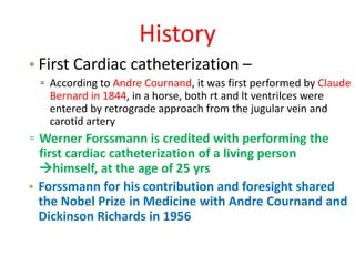
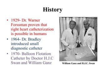
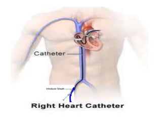
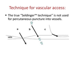
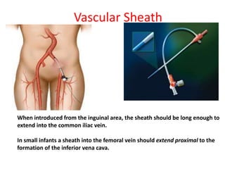
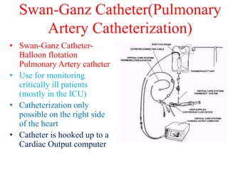
The document discusses the history and technique of transseptal puncture (TSP). It describes how TSP provides direct access to the left atrium and has become a routine skill for electrophysiologists performing procedures like atrial fibrillation ablation. The technique involves using a Brockenbrough needle and Mullins sheath inserted via the femoral or jugular vein to puncture the interatrial septum, usually at the fossa ovalis. Landmarks, equipment, steps of the procedure, challenges, and complications are reviewed in detail. The summary emphasizes the importance and increasing use of TSP as well as reviews key aspects of the technique and potential complications.
Bedside Ultrasound in Neurosurgery Part 1/3Liew Boon Seng
This document provides an overview of ultrasound training in neurosurgery. It introduces various applications of ultrasound in neurosurgery such as fetal neurosonograms, cranial ultrasonography of newborns, assessing ventricular shunt patency, spinal ultrasound in infants, and transcranial insonation of blood vessels. It also summarizes techniques for different types of Doppler imaging and discusses pathologies that can be detected using ultrasound like intraventricular hemorrhage.
Recent advances in interventional pediatric cardiology include improving existing techniques such as atrial septal defect closure and balloon valvuloplasty, developing new methods like percutaneous pulmonary valve replacement, and exploring hybrid procedures combining surgery and catheterization. New devices now allow closure of defects that were previously not suitable for catheter procedures. Stenting has expanded the options for treating lesions like coarctation of the aorta. Overall, interventional techniques are helping manage more complex congenital heart disease with less invasive approaches.
Cebu city in the Philippines was visited in February 2013. The document appears to be a travel log or diary from a trip to Cebu city in the Philippines in February 2013, as it lists the location and date but does not provide any other details about the trip.
The document describes a visit to Cebu City in the Philippines in February 2013. It mentions Cebu City and the email address drtoufiq19711@yahoo.com, but does not provide any other details about the visit or activities.
This document provides information about an echo evaluation for undergraduates and postgraduate fellows conducted by Dr. Md. Toufiqur Rahman, an associate professor of cardiology. It lists Dr. Rahman's academic credentials and positions at various hospitals and healthcare centers. It concludes by thanking all participants and providing Dr. Rahman's email for contact.
This document provides information about an ECG evaluation for undergraduates and post graduates fellows led by Dr. Md. Toufiqur Rahman, an associate professor of cardiology. It lists Dr. Rahman's academic qualifications and positions at various institutions including the National Institute of Cardiovascular Diseases. It concludes by thanking participants and providing Dr. Rahman's email for contact.
This document provides information about an echo evaluation training session for undergraduates and post graduates led by Dr. Md. Toufiqur Rahman. It lists his academic credentials and positions at various institutions including as an Associate Professor of Cardiology at the National Institute of Cardiovascular Diseases. The document concludes by thanking all participants and providing Dr. Rahman's email for contact.
Dr. Md. Toufiqur Rahman is an associate professor of cardiology at the National Institute of Cardiovascular Diseases in Dhaka, Bangladesh. He holds numerous medical certifications and acts as a consultant at several hospitals and clinics in Dhaka, including Medinova, Apollo Hospital, and STS Life Care Centre. His contact email is provided.
Cardio-cerebral resuscitation is a new approach for treating patients experiencing primary cardiac arrest that emphasizes minimizing interruptions to chest compressions, passive ventilation, and early epinephrine administration. It has been shown to significantly increase survival rates compared to traditional CPR, which focused more on ventilations and allowed for interruptions in chest compressions. Cardiac arrest is a major public health problem, and solutions require accurately measuring the effectiveness of local resuscitation systems of care through data collection.
The document outlines parameters for reporting on the left ventricle based on echocardiography findings. It describes assessing and reporting on cavity size, wall thickness, ventricular mass, shape, systolic and diastolic function, thrombus, mass, and ventricular septal defects. Measurement of ejection fraction and fractional shortening are outlined for evaluating systolic function, and diastolic filling patterns are described for assessing diastolic function. The document provides detailed guidelines for documenting the presence, size, location and characteristics of any abnormalities found.
This document provides guidelines for reporting echocardiography exams. It recommends that reports contain three sections: 1) Demographic and identifying information about the patient and exam, 2) A detailed echocardiographic and Doppler evaluation of cardiac structures and measurements, and 3) A summary. The demographic section should include patient name, age, gender, indications for the test, and physician information. The evaluation section describes assessing structures like the ventricles, valves, and vessels and providing quantitative measurements when possible. Measurements of structures like ventricular size and function, valvular stenosis or regurgitation, and cardiac shunts are recommended.
This document discusses techniques for coronary angiography including cannulating coronary arteries and grafts, angiographic views, and interpreting angiograms. Key points include different techniques for cannulating the left and right coronary arteries as well as grafts like saphenous veins and internal mammary arteries. Common angiographic views are described for visualizing different coronary segments. The document also covers quantitatively and visually assessing coronary narrowings and diagnosing coronary spasm.
This document discusses procedures for achieving hemostasis after vascular access procedures. It describes applying gentle pressure to the puncture site when removing catheters and sheaths. Firm pressure should then be applied for 15-30 minutes to achieve manual hemostasis. Adhesive bandages can then be used to cover the wound. Vascular closure devices can also help achieve hemostasis more quickly and allow for earlier ambulation. Protamine may be given to reverse heparin effects before sheath removal if needed. Precautions are discussed for patients receiving insulin. Radial access hemostasis involves using a pressurized bracelet over the puncture site.
Beta-blockers are a class of drugs that are used to manage various cardiac conditions by blocking the effects of epinephrine and other stress hormones on beta receptors. They were first developed in the 1950s and revolutionized cardiology. Beta-blockers are indicated for conditions like hypertension, arrhythmias, heart attack, and glaucoma. While they provide important benefits, they can also cause adverse effects like fatigue, dizziness, and bronchospasm. Different beta-blockers have varying levels of selectivity for beta-1 versus beta-2 receptors and some have additional alpha-blocking properties. Guidelines provide recommendations on the appropriate use of specific beta-blockers for different cardiac indications.
This study assessed the severity of mitral stenosis before and after percutaneous transvenous mitral commissurotomy (PTMC) using the mitral leaflet separation (MLS) index. 275 patients undergoing PTMC were evaluated clinically and via echocardiography before and after the procedure. Before PTMC, a good correlation was observed between mitral valve area (MVA) measurements and MLS index. Post-PTMC, MVA increased significantly while MLS index also increased and continued to correlate well with MVA. The study concludes that MLS index can be used as a complementary method to assess mitral stenosis severity both before and after PTMC.
Local Advanced Lung Cancer: Artificial Intelligence, Synergetics, Complex Sys...Oleg Kshivets
Overall life span (LS) was 1671.7±1721.6 days and cumulative 5YS reached 62.4%, 10 years – 50.4%, 20 years – 44.6%. 94 LCP lived more than 5 years without cancer (LS=2958.6±1723.6 days), 22 – more than 10 years (LS=5571±1841.8 days). 67 LCP died because of LC (LS=471.9±344 days). AT significantly improved 5YS (68% vs. 53.7%) (P=0.028 by log-rank test). Cox modeling displayed that 5YS of LCP significantly depended on: N0-N12, T3-4, blood cell circuit, cell ratio factors (ratio between cancer cells-CC and blood cells subpopulations), LC cell dynamics, recalcification time, heparin tolerance, prothrombin index, protein, AT, procedure type (P=0.000-0.031). Neural networks, genetic algorithm selection and bootstrap simulation revealed relationships between 5YS and N0-12 (rank=1), thrombocytes/CC (rank=2), segmented neutrophils/CC (3), eosinophils/CC (4), erythrocytes/CC (5), healthy cells/CC (6), lymphocytes/CC (7), stick neutrophils/CC (8), leucocytes/CC (9), monocytes/CC (10). Correct prediction of 5YS was 100% by neural networks computing (error=0.000; area under ROC curve=1.0).
These lecture slides, by Dr Sidra Arshad, offer a quick overview of the physiological basis of a normal electrocardiogram.
Learning objectives:
1. Define an electrocardiogram (ECG) and electrocardiography
2. Describe how dipoles generated by the heart produce the waveforms of the ECG
3. Describe the components of a normal electrocardiogram of a typical bipolar lead (limb II)
4. Differentiate between intervals and segments
5. Enlist some common indications for obtaining an ECG
6. Describe the flow of current around the heart during the cardiac cycle
7. Discuss the placement and polarity of the leads of electrocardiograph
8. Describe the normal electrocardiograms recorded from the limb leads and explain the physiological basis of the different records that are obtained
9. Define mean electrical vector (axis) of the heart and give the normal range
10. Define the mean QRS vector
11. Describe the axes of leads (hexagonal reference system)
12. Comprehend the vectorial analysis of the normal ECG
13. Determine the mean electrical axis of the ventricular QRS and appreciate the mean axis deviation
14. Explain the concepts of current of injury, J point, and their significance
Study Resources:
1. Chapter 11, Guyton and Hall Textbook of Medical Physiology, 14th edition
2. Chapter 9, Human Physiology - From Cells to Systems, Lauralee Sherwood, 9th edition
3. Chapter 29, Ganong’s Review of Medical Physiology, 26th edition
4. Electrocardiogram, StatPearls - https://www.ncbi.nlm.nih.gov/books/NBK549803/
5. ECG in Medical Practice by ABM Abdullah, 4th edition
6. Chapter 3, Cardiology Explained, https://www.ncbi.nlm.nih.gov/books/NBK2214/
7. ECG Basics, http://www.nataliescasebook.com/tag/e-c-g-basics
NVBDCP.pptx Nation vector borne disease control programSapna Thakur
NVBDCP was launched in 2003-2004 . Vector-Borne Disease: Disease that results from an infection transmitted to humans and other animals by blood-feeding arthropods, such as mosquitoes, ticks, and fleas. Examples of vector-borne diseases include Dengue fever, West Nile Virus, Lyme disease, and malaria.
ABDOMINAL TRAUMA in pediatrics part one.drhasanrajab
Abdominal trauma in pediatrics refers to injuries or damage to the abdominal organs in children. It can occur due to various causes such as falls, motor vehicle accidents, sports-related injuries, and physical abuse. Children are more vulnerable to abdominal trauma due to their unique anatomical and physiological characteristics. Signs and symptoms include abdominal pain, tenderness, distension, vomiting, and signs of shock. Diagnosis involves physical examination, imaging studies, and laboratory tests. Management depends on the severity and may involve conservative treatment or surgical intervention. Prevention is crucial in reducing the incidence of abdominal trauma in children.
Basavarajeeyam is a Sreshta Sangraha grantha (Compiled book ), written by Neelkanta kotturu Basavaraja Virachita. It contains 25 Prakaranas, First 24 Chapters related to Rogas& 25th to Rasadravyas.
Recomendações da OMS sobre cuidados maternos e neonatais para uma experiência pós-natal positiva.
Em consonância com os ODS – Objetivos do Desenvolvimento Sustentável e a Estratégia Global para a Saúde das Mulheres, Crianças e Adolescentes, e aplicando uma abordagem baseada nos direitos humanos, os esforços de cuidados pós-natais devem expandir-se para além da cobertura e da simples sobrevivência, de modo a incluir cuidados de qualidade.
Estas diretrizes visam melhorar a qualidade dos cuidados pós-natais essenciais e de rotina prestados às mulheres e aos recém-nascidos, com o objetivo final de melhorar a saúde e o bem-estar materno e neonatal.
Uma “experiência pós-natal positiva” é um resultado importante para todas as mulheres que dão à luz e para os seus recém-nascidos, estabelecendo as bases para a melhoria da saúde e do bem-estar a curto e longo prazo. Uma experiência pós-natal positiva é definida como aquela em que as mulheres, pessoas que gestam, os recém-nascidos, os casais, os pais, os cuidadores e as famílias recebem informação consistente, garantia e apoio de profissionais de saúde motivados; e onde um sistema de saúde flexível e com recursos reconheça as necessidades das mulheres e dos bebês e respeite o seu contexto cultural.
Estas diretrizes consolidadas apresentam algumas recomendações novas e já bem fundamentadas sobre cuidados pós-natais de rotina para mulheres e neonatos que recebem cuidados no pós-parto em unidades de saúde ou na comunidade, independentemente dos recursos disponíveis.
É fornecido um conjunto abrangente de recomendações para cuidados durante o período puerperal, com ênfase nos cuidados essenciais que todas as mulheres e recém-nascidos devem receber, e com a devida atenção à qualidade dos cuidados; isto é, a entrega e a experiência do cuidado recebido. Estas diretrizes atualizam e ampliam as recomendações da OMS de 2014 sobre cuidados pós-natais da mãe e do recém-nascido e complementam as atuais diretrizes da OMS sobre a gestão de complicações pós-natais.
O estabelecimento da amamentação e o manejo das principais intercorrências é contemplada.
Recomendamos muito.
Vamos discutir essas recomendações no nosso curso de pós-graduação em Aleitamento no Instituto Ciclos.
Esta publicação só está disponível em inglês até o momento.
Prof. Marcus Renato de Carvalho
www.agostodourado.com
Novas diretrizes da OMS para os cuidados perinatais de mais qualidade
Pulmonary artery catheterization
1. Pulmonary Artery Catheterization
Dr. Md.Toufiqur Rahman
MBBS, FCPS, MD, FACC, FESC, FRCPE, FSCAI,
FAPSC, FAPSIC, FAHA, FCCP, FRCPG
Associate Professor of Cardiology
National Institute of Cardiovascular Diseases(NICVD),
Sher-e-Bangla Nagar, Dhaka-1207
Consultant, Medinova, Malibagh branch
Honorary Consultant, Apollo Hospitals, Dhaka and
STS Life Care Centre, Dhanmondi
drtoufiq19711@yahoo.com
CRT 2014
Washington
DC, USA
2.
3. History
• First Cardiac catheterization –
▫ According to Andre Cournand, it was first performed by Claude
Bernard in 1844, in a horse, both rt and lt ventrilces were
entered by retrograde approach from the jugular vein and
carotid artery
▫ Werner Forssmann is credited with performing the
first cardiac catheterization of a living person
himself, at the age of 25 yrs
• Forssmann for his contribution and foresight shared
the Nobel Prize in Medicine with Andre Cournand and
Dickinson Richards in 1956
4. History
• 1929- Dr. Warner
Forssman proven that
right heart catheterization
is possible in humans
• 1964- Dr. Bradley
introduced small
diagnostic catheter
• 1970- Balloon Flotation
Catheter by Doctor H.J.C
Swan and William Ganz
5.
6. Technique for vascular access:
The true “Seldinger™ technique” is not used
for percutaneous puncture into vessels.
7. Vascular Sheath
Percutaneous introduction and then the use of an
indwelling vascular sheath in vessels is the standard
technique for catheterization of pediatric and congenital
heart patients.
Ideal sheath should have:
1. Dilator
1. long, fine and smoothly tapered tip.
2. inner lumen of the dilator tip should tightly fit over the guide wire
3. tip of the dilator should have a smooth, fine transitional taper onto
the surface of the wire.
2. female Lure™ lock connecting hub at the proximal end
3. back-bleed valve
4. Lateral tube / flush port
8. Vascular Sheath
When introduced from the inguinal area, the sheath should be long enough to
extend into the common iliac vein.
In small infants a sheath into the femoral vein should extend proximal to the
formation of the inferior vena cava.
9. Vascular Sheath
Ideal short sheath (7.5 cm long) for venous site –
5 Fr for an infant or child (<15– 20 Kg) and
7 Fr for a larger child or adult
Extra long sheaths (45 to 90 cm ) are used to –
1. guide catheters directly and repeatedly to an area
within the heart itself (biopsies, blade catheters),
2. for trans septal procedures,
3. to deliver special devices within the heart or great
vessels (stents, occlusion devices), and
4. for the withdrawal of foreign bodies from the vascular
system.
10. Swan-Ganz Catheter(Pulmonary
Artery Catheterization)
• Swan-Ganz Catheter-
Balloon flotation
Pulmonary Artery catheter
• Use for monitoring
critically ill patients
(mostly in the ICU)
• Catheterization only
possible on the right side
of the heart
• Catheter is hooked up to a
Cardiac Output computer