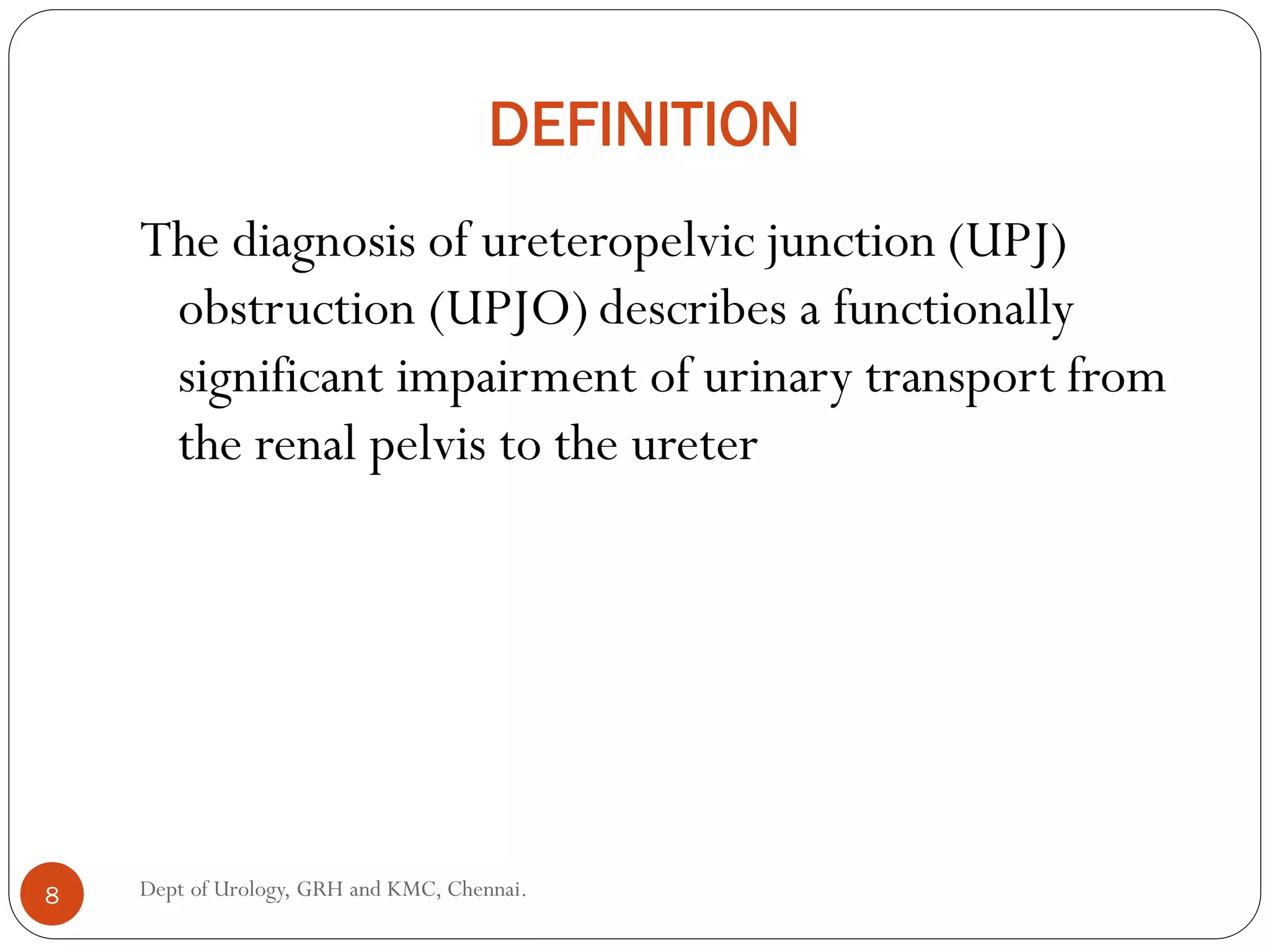
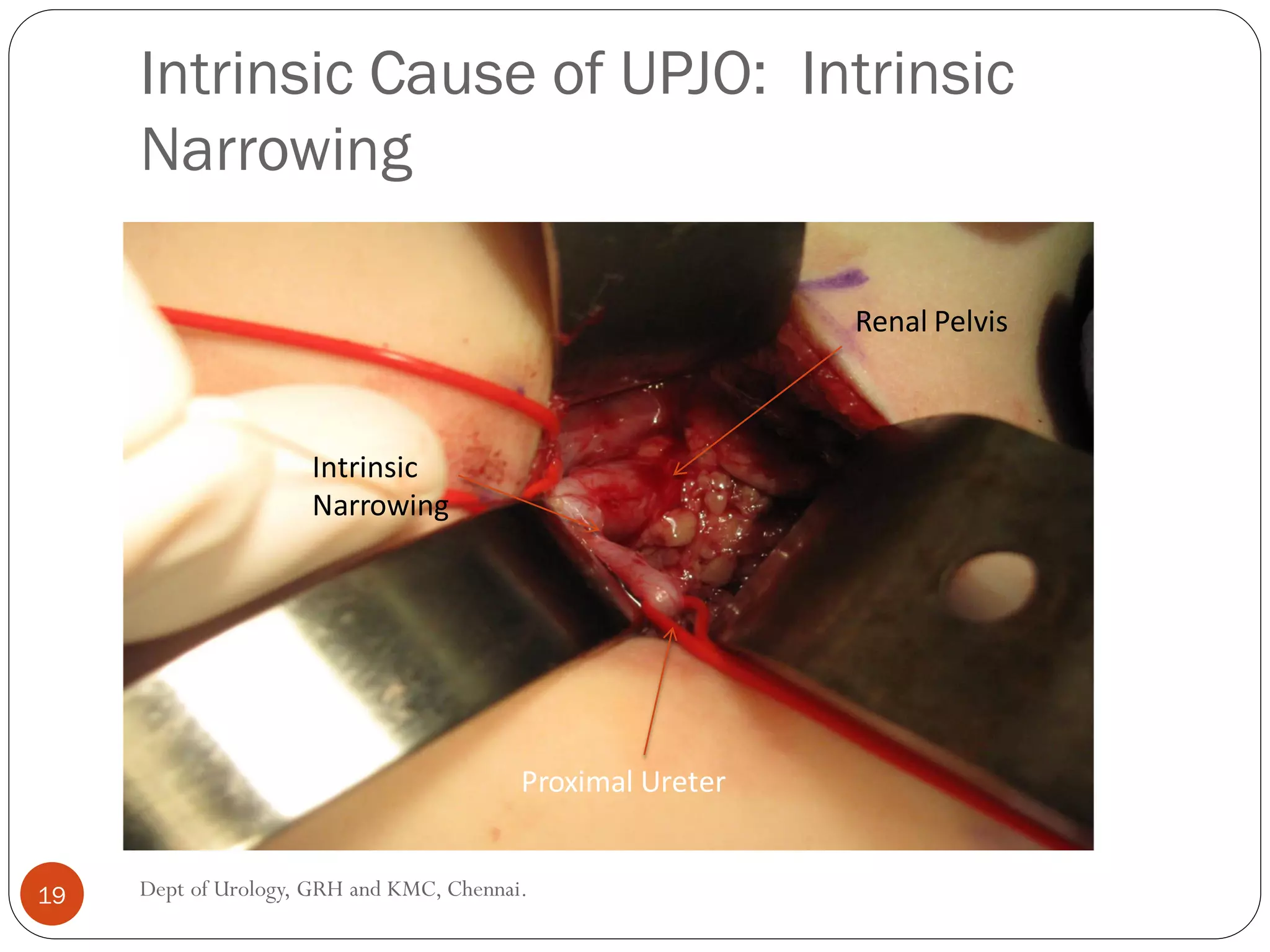
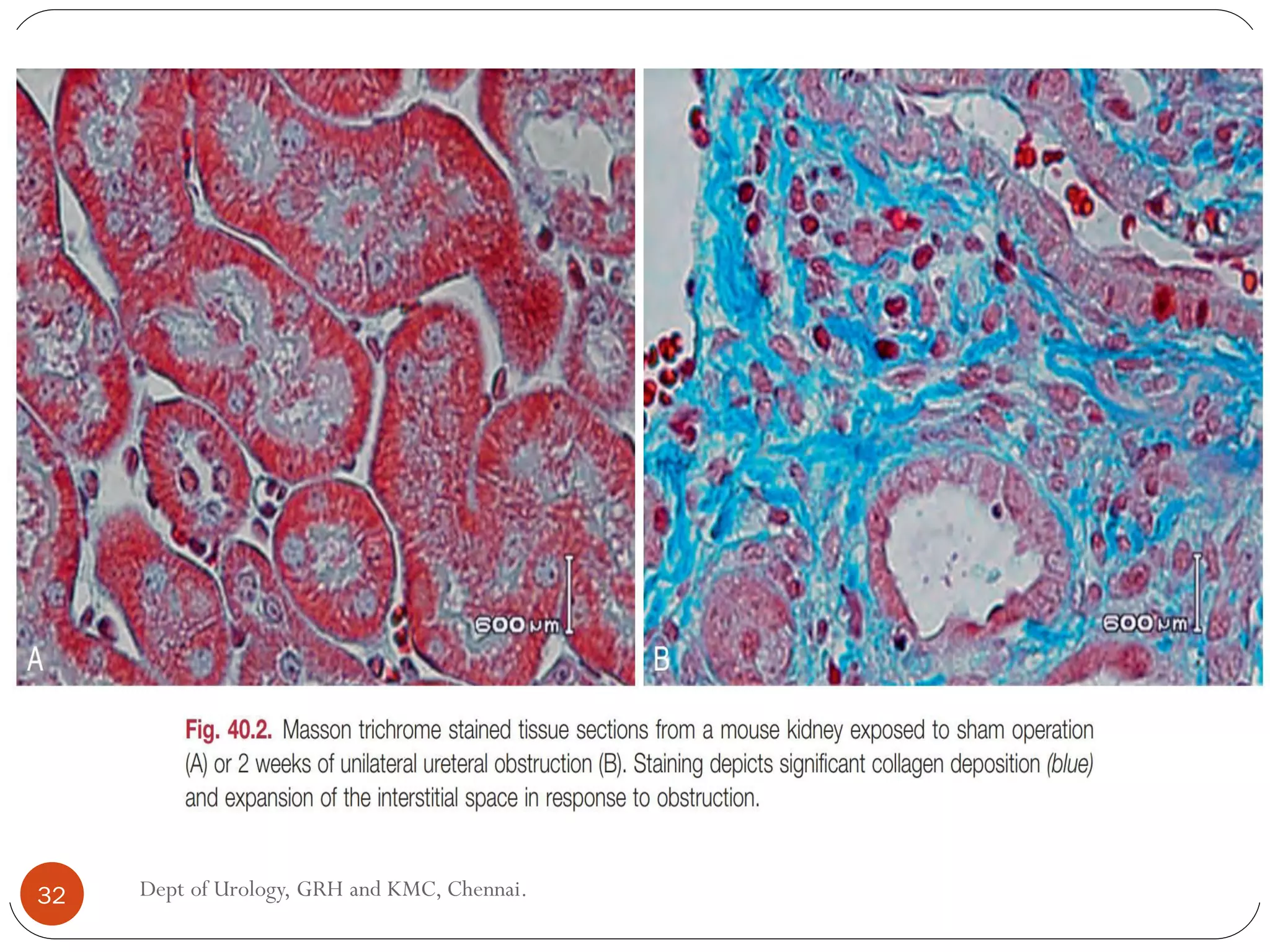
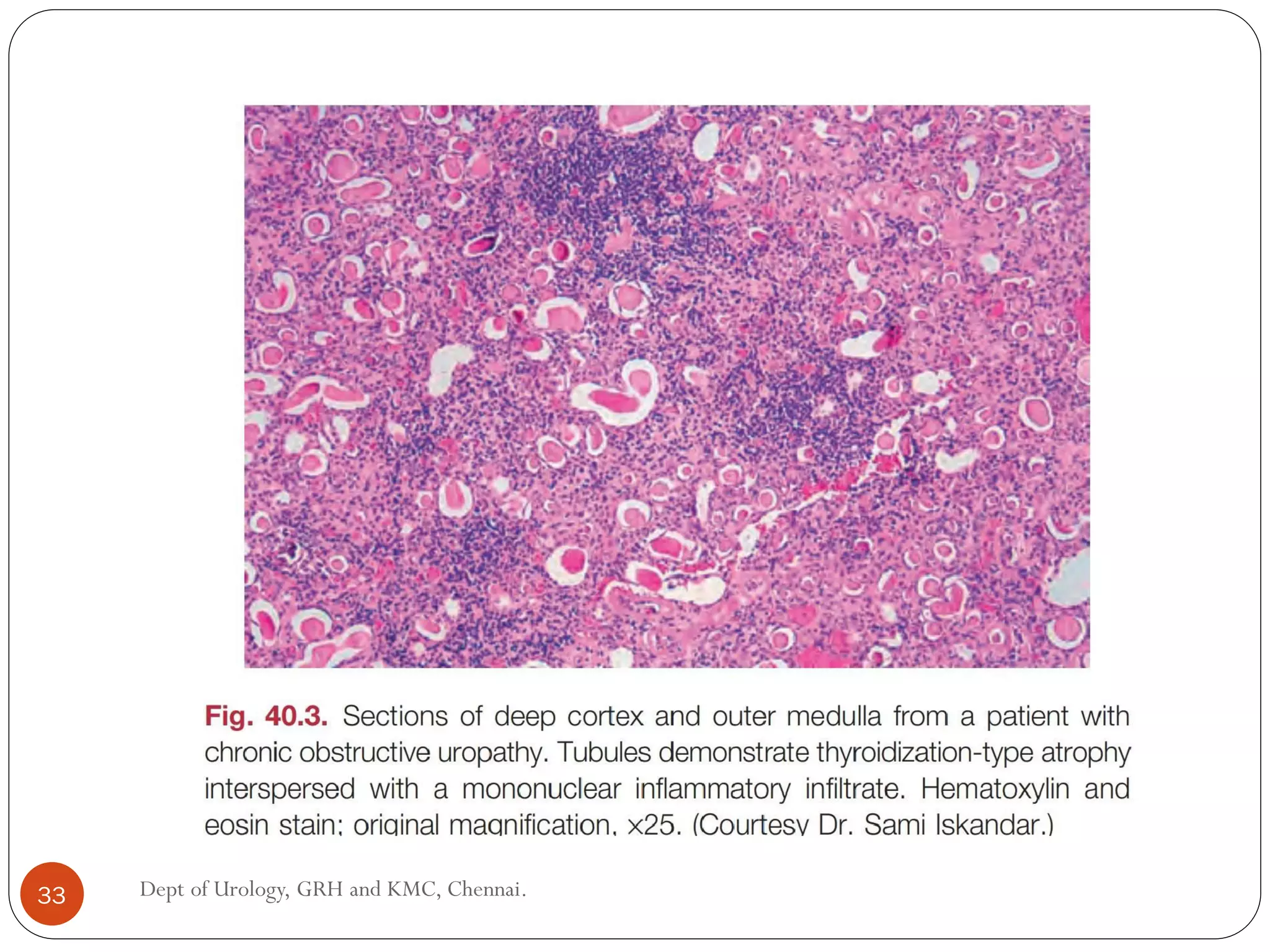
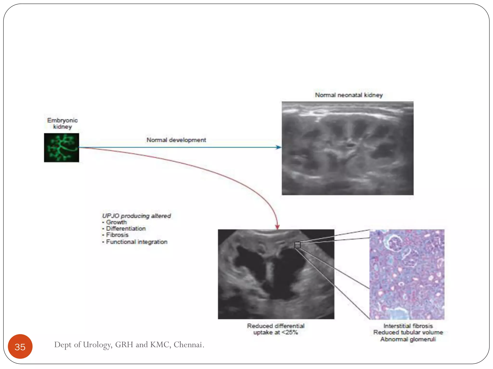
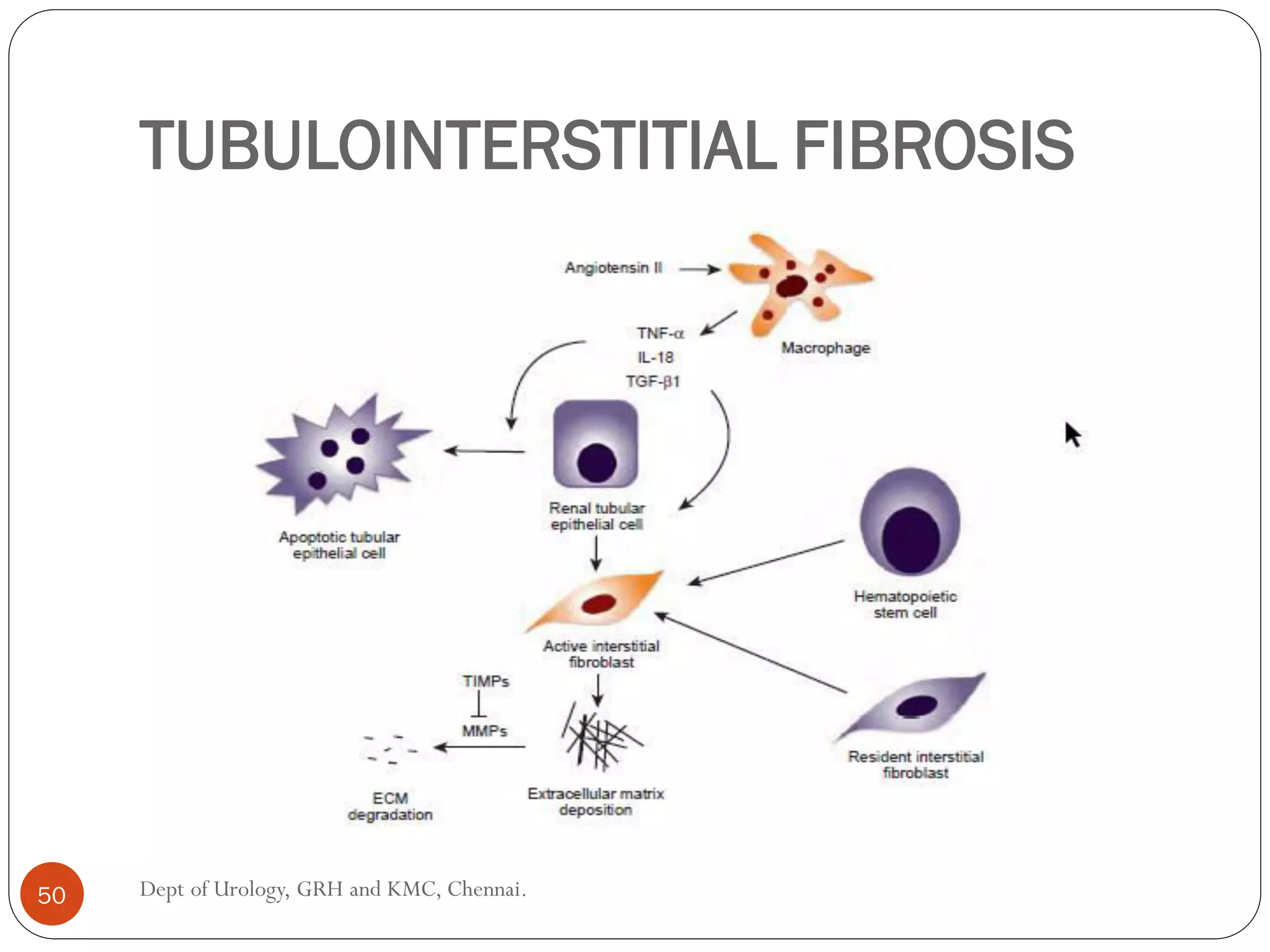
Bangalore Call Girls Nelamangala Number 7001035870 Meetin With Bangalore Esc...
Pediatric urology:Pujo- etiopathogenesis and presentation
1. Dept of Urology
Govt Royapettah Hospital and Kilpauk Medical College
Chennai
ETIOPATHOGENESIS AND PRESENTATION
OF CONGENITAL PELVI-URETERIC
JUNCTION OBSTRUCTION(PUJO)
1
2. Moderators:
Professors:
Prof. Dr. G. Sivasankar, M.S., M.Ch.,
Prof. Dr.A. Senthilvel, M.S., M.Ch.,
Asst Professors:
Dr. J. Sivabalan, M.S., M.Ch.,
Dr. R. Bhargavi, M.S., M.Ch.,
Dr. S. Raju, M.S., M.Ch.,
Dr. K. Muthurathinam, M.S., M.Ch.,
Dr. D.Tamilselvan, M.S., M.Ch.,
Dr. K. Senthilkumar, M.S., M.Ch.
Dept of Urology, GRH and KMC, Chennai.
2
3. HISTORY
PUJ obstruction was first described by Dietl in
1864.
The fibrotic change at the PUJ was demonstrated
by AllenTD in 1970.
3 Dept of Urology, GRH and KMC, Chennai.
4. ANATOMY OF PUJ
Area where renal pelvis funnels down in to upper
ureter
No clear demarcation-endoscopically- mucosal
rossettes(long folds)
Length in adult-7mm
At the level of L2- L side and slightly lower on R
4 Dept of Urology, GRH and KMC, Chennai.
5. RELATIONS
Ant- hep flexure on right and splenic flexure on L
Post- psoas major
Lat- lower pole of resp kidney
Med- right- 2nd duodenum,left- 4th duodenum,DJ
5 Dept of Urology, GRH and KMC, Chennai.
8. DEFINITION
The diagnosis of ureteropelvic junction (UPJ)
obstruction (UPJO) describes a functionally
significant impairment of urinary transport from
the renal pelvis to the ureter
8 Dept of Urology, GRH and KMC, Chennai.
9. EPIDEMIOLOGY
Pelvi-ureteric junction (PUJ) obstruction is the
commonest cause of pediatric hydronephrosis
occurring in 1 in 1000-2000 live births.
Accounts for 64% of infants with unresolved post-
natal hydronephrosis
Males > Females (2:1 in newborns)
Left >Right (2:1)
10% bilateral Synchronous or asynchronous
May be inheritable
9 Dept of Urology, GRH and KMC, Chennai.
10. Among children, hydrone-phrosis appears to be
somewhat more prevalent in boys, and the
majority of cases occur in subjects younger than 1
year.
Indeed, obstructive nephrouropathies constitute
the single largest entity leading to renal
insufficiency in male children younger than 1 year
of age (Benfield, 2003;Seikaly et al., 2003;
USRDS, 2015).
10 Dept of Urology, GRH and KMC, Chennai.
11. ETIOLOGY
The precise cause of UPJ obstruction remains
elusive despite investigation along a number of
lines:
? embryologic
? anatomic
whether this is a result of developmental
arrest ; or of incomplete recanalization of
the ureter is not yet known
11 Dept of Urology, GRH and KMC, Chennai.
12. ETIOLOGY
1)PRIMARY
congenital
2) SECONDARY
Acquired
extrinsic compression (crossing
vessel)
Stone disease
Post op stricture (adhesions)
Post inflammatory (TB)
TCC upper tract
VUR (secondary pujo)
Fibroepithelial polyps
12 Dept of Urology, GRH and KMC, Chennai.
13. CONGENITAL PUJO:ETIOPATHOGENESIS
Intrinsic (M/C)
Aperistaltic segment (M/C)
kinks or valves produced by infoldings of the
ureteral mucosa and musculature
Decresed density of cajal cell
true ureteral stricture (less frequent)
(Abnormalities of ureteral musculature -
electron microscopy -excessive collagen
deposition at the site of the stricture )
13 Dept of Urology, GRH and KMC, Chennai.
14. cytokine production in the urothelium has also
been proposed to exacerbate UPJO
studies have implicated transforming growth
factor-!, epidermal growth factor expression,
nitric oxide, and neuropeptideY in UPJ stenosis
14 Dept of Urology, GRH and KMC, Chennai.
15. The microphotograph is taken through the ureteropelvic
junction.
There is marked attenuation of smooth muscles and smooth
muscle in disarray and hypertrophy surrounding the urothelial
lining
15 Dept of Urology, GRH and KMC, Chennai.
17. Lower Pole Vessel
UPJ Anterior
High Insertion Kinking
Crossing Vessels
17 Dept of Urology, GRH and KMC, Chennai.
18. INTRINSIC:APRISTALTIC SEGMENT
spiral musculature normally present has been
replaced by abnormal longitudinal muscle
bundles or fibrous tissue
This results in failure to develop a normal
peristaltic wave for propagation of urine from the
renal pelvis to the ureter
18 Dept of Urology, GRH and KMC, Chennai.
19. Intrinsic Cause of UPJO: Intrinsic
Narrowing
Renal Pelvis
Proximal Ureter
Intrinsic
Narrowing
19 Dept of Urology, GRH and KMC, Chennai.
20. KINKS AND VALVES
Intrinsic obstruction at the UPJ may also result from
kinks or valves produced by infoldings of the ureteral
mucosa and musculature (Maizels and Stephens, 1980).
In these patients the obstruction may actually be at the
level of the proximal ureter.
This phenomenon appears to result from retention or
exaggeration of congenital folds normally found in the
ureter of developing fetuses.
20 Dept of Urology, GRH and KMC, Chennai.
21. Ostling’s folds
Present in 92% newborns
Result from differential
growth of ureter vs body
Usually resolves in
childhood
Persistent fetal convolution
TRANSVERSEFOLDS IN
PROXIMALURETER
21 Dept of Urology, GRH and KMC, Chennai.
22. In some of these patients the defects are bridged by ureteral adventitia.
Grossly,this can manifest as external bands or adhesions that appear to be
causing the obstruction.
In fact, Johnston et al. (1977) reported that lysis of external adhesions can
at times reestablish flow without pyeloplasty.
In the majority of patients, these bands or adhesions are likely to be a
secondary phenomenon associated with intrinsic obstruction, thus
operative pyeloplasty would usually be most effective.
The presence of these kinks, valves, bands, or adhesions may also produce
angulation of the ureter at the lower margin of the renal pelvis in such a
manner that as the pelvis dilates anteriorly and inferiorly, the ureteral
insertion is carried further proximally
22 Dept of Urology, GRH and KMC, Chennai.
23. . In these patients the most dependent portion of the
pelvis is inadequately drained, and the apparent high
insertion of the ureteral ostium is actually a secondary
phenomenon (Kelalis, 1976).
In at least some patients, however, the high insertion is
likely the primary obstructing lesion because this
phenomenon is found more frequently in the presence of
renal ectopia or fusion anomalies (Das and Amar, 1984;
Zincke et al., 1974).
23 Dept of Urology, GRH and KMC, Chennai.
24. ROLE OF ABERRANT VESSELS IN
CAUSE OF PUJO
Controversy persists regarding the potential
role
Significant crossing vessels have been noted in up to 63%
of patients with UPJO but in as little as 20% of
individuals with normal kidneys
Although these lower pole vessels have often been
referred to as aberrant, these segmental vessels, which
may be branches from the main renal artery or arise
directly from the aorta, are usually normal variants
24 Dept of Urology, GRH and KMC, Chennai.
25. Historically, it has been believed that the associated
vessel alone does not cause the primary obstruction
(Hanna, 1978).
In fact, the true cause is an intrinsic lesion at the UPJ or
proximal ureter that causes dilation and ballooning of the
renal pelvis over the polar or aberrant vessel.
Recent studies using three-dimensional (3D)
multidetector row CT demonstrated that the precise
location of crossing vessels did not correspond to the
obstructive transition point in patients with UPJO
25 Dept of Urology, GRH and KMC, Chennai.
29. PATHOLOGICAL CHANGES OF
OBSTRUCTION
1.Pathological Changes in the Mature Kidney
The gross pathological changes that occur in the mature
kidney in response to obstruction have been well described in
animal models and parallel findings in humans.
After 42 hours of obstruction, there is dilation of the
collecting system and blunting of the papillary tips associated
with an increased weight of the kidney.
29 Dept of Urology, GRH and KMC, Chennai.
30. Collecting system dilation and renal weight further increase
and the parenchyma becomes edematous after 7 days of
obstruction.
Further collecting system dilation develops after 12 days of
obstruction, but after 21 to 28 days the cortex and medullary
tissue in the obstructed kidney become diffusely thinned.
After 6 weeks, the obstructed kidney is enlarged with a
cystic appearance,but lower weight, as compared with the
normal contralateral kidney
30 Dept of Urology, GRH and KMC, Chennai.
31. The histologic derangements associated with early
obstruction are localized primarily to the
tubulointerstitial compartment of the kidney and
include
massive tubular dilation,
progressive tubulointerstitial fibrosis,
inflammatory cell infiltration, and
apoptotic renal tubular cell death.
long-standing obstruction ultimately results in glo-
merulosclerosis most likely as a result of chronic inflammation
and/or hyperfiltration injury
31 Dept of Urology, GRH and KMC, Chennai.
36. PATHOLOGICAL CHANGES IN THE
DEVELOPING KIDNEY
General Observations
Histologic examination of obstructed renal tissues has shown
disorganization of structure with primitive forms of renal
tissues, pathologically defined as dysplasia .
Structural alterations
fibrosis
increased interstitial tissues,
abnormal tubules and glomeruli
layered organization of the kidney with the cortical and
medullary areas with inner and outer stripes is often
distorted or absent
36 Dept of Urology, GRH and KMC, Chennai.
37. summarized as producing alterations in the
1. regulation of growth,
2. tissue differentiation,
3. extracellular matrix, and fibrosis, and
4. in altering the functional integration of the kidney.
The latter is largely a result of the first three major factors and
refers to the mechanisms producing vascular, neural, and
humoral homeostasis and in the regulation of inflammatory
cascades.
37 Dept of Urology, GRH and KMC, Chennai.
38. 1.GROWTH
obstructed kidney may demonstrate impaired or accelerated
growth
small, obstructed kidney is not atrophic as may be seen in the
adult, but it is hypoplastic.The growth it should have
experienced never occurred
generalized impairment of all parts of the kidney, with
reduced numbers of nephrons as well as smaller nephrons
38 Dept of Urology, GRH and KMC, Chennai.
39. Reduced renal mass can be associated with
hypertension
reduced filtration function, and
loss of tubular mass will affect
electrolyte and acid-base homeostasis,
as well as water balance.
39 Dept of Urology, GRH and KMC, Chennai.
40. The factors that predict altered growth
severity of
obstruction
timing of the
obstructive
effect
40 Dept of Urology, GRH and KMC, Chennai.
41. a)GROWTH REGULATION OF THE
KIDNEY
extremely complex and dynamic through development
Obstructive conditions have been shown to alter expression
of growth-regulatory genes as well as the presence of the
proteins coded by these genes
EGF has been shown to reduce some of the growth effects of
obstruction when administered exogenously
41 Dept of Urology, GRH and KMC, Chennai.
44. b)APOPTOSIS REGULATION
The early fetal kidney is extremely active in terms of new
cell formation as well as turnover
This permits remodeling during development, as well as
providing a control system over unregulated growth
apoptotic activity is regulated by
cytokines
mechanical factors
mediators may be measurable in the urine or blood, and they may
permit therapeutic manipulation
44 Dept of Urology, GRH and KMC, Chennai.
45. 2.DIFFERENTIATION
Differentiation is the process of cells attaining specific
functional traits to permit specialized functions and
organization into tissues and is the basis for the many
functions of the kidney
Abnormal epithelial-to-mesenchymal transformation (EMT)
is one alteration in differentiation that does occur in the adult
and can be reversible
A variety of mediators of EMT have been described,
includingTGF-', plasminogen and leukocyte, as well as
inhibitors, such as hepatocyte growth factor (HGF)
45 Dept of Urology, GRH and KMC, Chennai.
46. Induction Process
Renal differentiation begins with induction and from then on
is exquisitely sensitive to various outside effects that may
disrupt the normal sequence of cellular changes that results
in a kidney.
46 Dept of Urology, GRH and KMC, Chennai.
49. FIBROSIS
•A universal characteristic of obstructive nephropathy
appears to be renal fibrosis
• It is seen as infiltration of the interstitium with abnormal
amounts of ECM, including collagens, fibronectin, and other
connective tissue proteins.
•Their presence disrupts the normal interconnections between
cells that permit functional integration of the renal tissues.
.
49 Dept of Urology, GRH and KMC, Chennai.
51. • Modulation of renal fibrosis may be a significant potential target for
managing obstructive nephropathy,
• but the delicate balance of these factors needs to be understood to a
greater degree than at present
• Nitric oxide has also been shown to regulate the development of
obstructive fibrosis postnatally and may play a similar role prenatally
• Increased nitric oxide generation reduces the degree of interstitial
fibrosis
51 Dept of Urology, GRH and KMC, Chennai.
52. CLINICAL FEATURE
UPJO, although most often a congenital problem, Can
present clinically at any time of life.
Asymptomatic
HistoricallyThe most common presentation in neonates
and infants was the findiing of a palpable flank mass
However, the use of maternal prenatal
ultrasonography has led to a dramatic increase
in the number of asymptomatic newborns being
diagnosed with hydronephrosis, many of whom
are subsequently found to have UPJO
52 Dept of Urology, GRH and KMC, Chennai.
53. During evaluation of azotemia, which may result from
bilateral obstruction in a functionally or anatomically
solitary kidney.
UPJ obstruction may also be incidentally found during
studies performed to evaluate unrelated anomalies such
as congenital heart disease
53 Dept of Urology, GRH and KMC, Chennai.
54. In older children or adults, intermittent abdominal or
flank pain, at times associated with nausea or vomiting,
Hematuria, either spontaneous or associated with
otherwise relatively minor trauma,
Laboratory findings of microhematuria, pyuria, or frank
urinary tract infection may also bring an otherwise
asymptomatic patient to the urologist
Hypertension
54 Dept of Urology, GRH and KMC, Chennai.
55. ASSOCIATED ANOMALIES
Contralateral UPJO: M.C.: 10-40%.
VUR : low grade: 40%.
Renal dysplasia and multicystic dysplastic kidneys.
Duplicated system (usually lower moiety)
Horseshoe / ectopic kidney.
Unilateral agenesis: 5%
VATER syndrome: 21% pts have UPJO
55 Dept of Urology, GRH and KMC, Chennai.