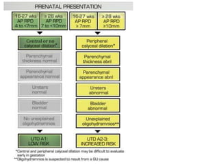
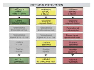
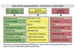
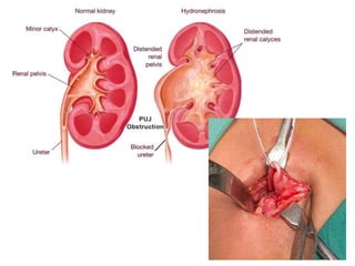
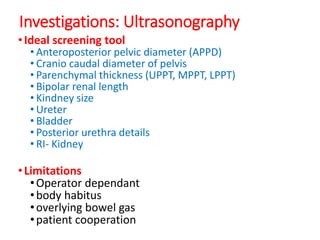
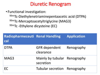
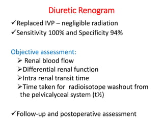
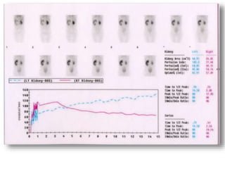
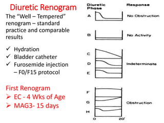
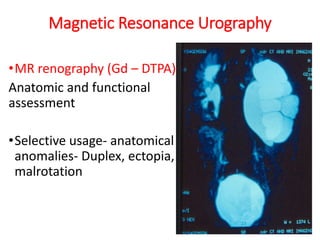
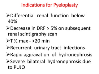
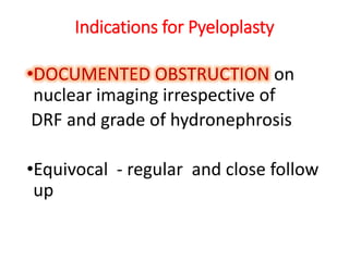
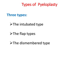
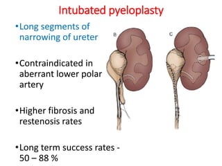
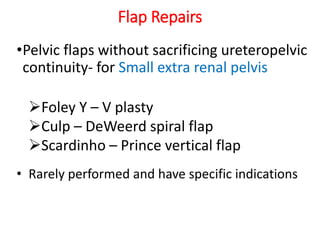
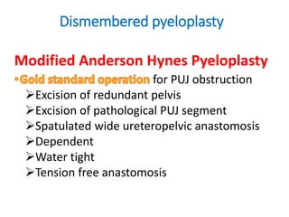
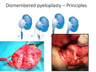
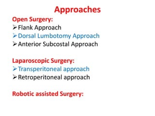
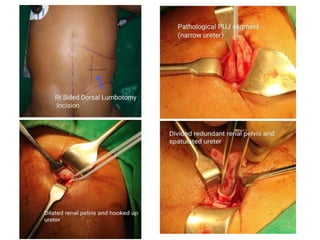
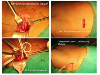
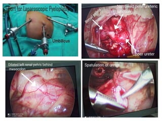
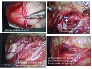
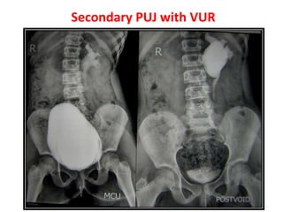
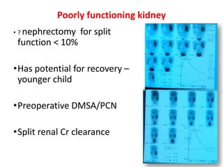
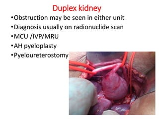
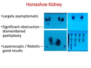
Pelvi-Ureteric Junction
Obstruction
Dr. Arjun A. Pawar
MBBS, MS,
M. Ch. Pediatric Surgery,
DNB Pediatric Surgery,
FMAS,
FIAGES.
Divine Pediatric Surgery Centre. i.
a. w.
Hydronephrosis- Aseptic dilatation of
pelvicalyceal system
SFU
UTD – Urinary tract dilatation
Causes of Antenatal Hydronephrosis
Introduction
Incidence - 1 in 1250 live births
M:F=2:1
PUJO- Adynamic segment
High grade vs Low grade
Intrinsic Vs Extrinsic
Etiology
Primary PUJO:
Intrinsic obstruction – Scarring of ureteric
valves
Ureteral hypoplasia- Inhibit the natural
peristaltic emptying
Abnormal or high insertion of the ureter
Crossing lower-pole renal vessel(s)
Renal ectopy
Renal Hypermobility
Secondary PUJO: Renal stone disease,
Failed repair,
Pathophysiology
Adynamic segment in upper ureter,
Obstructing lesions(Valves, Polyps)
Folds(Persistent Ostlings folds).
Complete obstruction- Rapid deterioration
of function
Partial obstruction- Gradual deterioration
of function
Clinical Presentation
Increasing prenatal
detection- 80%
Asymtomatic PUJO in infants
Abdominal lump
Abdominal pain
Urinary tract infection
Dietl’s crisis
Hematuria, Hypertension
Association with other
anomalies- Anorectal,
syndromes-MRKH
Evaluation
•Blood Ix: CBC, KFT, Urine, HIV, HbsAg,
•USG
•MCU
•IVP
•EC Scan/ MAG3 scan +- DMSA scan
•CT KUB scan
•MR Urography
Investigations: Ultrasonography
•Ideal screening tool
• Anteroposterior pelvic diameter (APPD)
• Cranio caudal diameter of pelvis
• Parenchymal thickness (UPPT, MPPT, LPPT)
• Bipolar renal length
• Kindney size
• Ureter
• Bladder
• Posterior urethra details
• RI- Kidney
•Limitations
•Operator dependant
•body habitus
•overlying bowel gas
•patient cooperation
USG
•The first postnatal USG 48 to 72
hrs after birth
•RI > 0.75 - obstruction requiring
surgery
•Sensitivity ~ 90 – 95%,
•Specificity ~ 85%
Diuretic Renogram
•Functional investigation:
99mTc-Diethylenetriaminepentaacetic acid (DTPA)
99mTc-Mercaptoacetyltriglycine (MAG3)
99mTc -Ethylene dicysteine (EC)
Radiopharmeceuti
cal
Renal Handling Application
DTPA GFR dependent
clearance
Renography
MAG3 Mainly by tubular
secretion
Renography
EC Tubular secretion Renography
Diuretic Renogram
Replaced IVP – negligible radiation
Sensitivity 100% and Specificity 94%
Objective assessment:
 Renal blood flow
Differential renal function
Intra renal transit time
Time taken for radioisotope washout from
the pelvicalyceal system (t½)
Follow-up and postoperative assessment
Diuretic Renogram
The “Well – Tempered”
renogram – standard
practice and comparable
results
 Hydration
 Bladder catheter
 Furosemide injection
– F0/F15 protocol
First Renogram
 EC - 4 Wks of Age
 MAG3- 15 days
Magnetic Resonance Urography
•MR renography (Gd – DTPA)
Anatomic and functional
assessment
•Selective usage- anatomical
anomalies- Duplex, ectopia,
malrotation
Indications for Pyeloplasty
Differential renal function below
40%
Decrease in DRF > 5% on subsequent
renal scintigraphy scan
T ½ max - >20 min
Recurrent urinary tract infections
Rapid aggravation of hydronephrosis
Severe bilateral hydronephrosis due
to PUJO
Indications for Pyeloplasty
•DOCUMENTED OBSTRUCTION on
nuclear imaging irrespective of
DRF and grade of hydronephrosis
•Equivocal - regular and close follow
up
Types of Pyeloplasty
Three types:
The intubated type
The flap types
The dismembered type
Intubated pyeloplasty
•Long segments of
narrowing of ureter
•Contraindicated in
aberrant lower polar
artery
•Higher fibrosis and
restenosis rates
•Long term success rates -
50 – 88 %
Flap Repairs
•Pelvic flaps without sacrificing ureteropelvic
continuity- for Small extra renal pelvis
Foley Y – V plasty
Culp – DeWeerd spiral flap
Scardinho – Prince vertical flap
• Rarely performed and have specific indications
Dismembered pyeloplasty
Modified Anderson Hynes Pyeloplasty
for PUJ obstruction
Excision of redundant pelvis
Excision of pathological PUJ segment
Spatulated wide ureteropelvic anastomosis
Dependent
Water tight
Tension free anastomosis
Dismembered pyeloplasty – Principles
Approaches
Open Surgery:
Flank Approach
Dorsal Lumbotomy Approach
Anterior Subcostal Approach
Laparoscopic Surgery:
Transperitoneal approach
Retroperitoneal approach
Robotic assisted Surgery:
Complications and Outcome
Bleeding and infection
Prolonged urinary extravasation
Perirenal Urinoma
Urosepsis
D-J stent migration
Delayed opening of the anastomosis
Anastomotic stricture
SPECIAL SITUATIONS
PUJ and polar vessels
•Often a surprise
•Rarely associated
with intrinsic PUJ
obstruction
•Pyelopyelostomy
•A-H Pyeloplasty
PUJ and VUJ obstruction
•The saline flush test for distal patency
•Missed VUJ obstruction – large low pelvis
•DJ stent / nephrostomy till VUJ is addressed
(Ureteric reimplantation )
PUJ obstruction and VUR
•10% PUJO – concurrent reflux
•Routine MCU
Bilateral HDN
Ureteric dilatation on preop USG
• Dilated ureter at operation
• Pyeloplasty - post op MCU / DMSA
• Manage VUR on merits
Secondary PUJ with VUR
Poorly functioning kidney
• ? nephrectomy for split
function < 10%
•Has potential for recovery –
younger child
•Preoperative DMSA/PCN
•Split renal Cr clearance
Duplex kidney
•Obstruction may be seen in either unit
•Diagnosis usually on radionuclide scan
•MCU /IVP/MRU
•AH pyeloplasty
•Pyeloureterostomy
Horseshoe Kidney
•Largely asymptomatic
•Significant obstruction –
dismembered
pyeloplasty
•Laparoscopic / Robotic –
good results
Pelvi-ureteric junction obstruction

Pelvi-ureteric junction obstruction

  • 1.
    Pelvi-Ureteric Junction Obstruction Dr. ArjunA. Pawar MBBS, MS, M. Ch. Pediatric Surgery, DNB Pediatric Surgery, FMAS, FIAGES. Divine Pediatric Surgery Centre. i. a. w.
  • 2.
    Hydronephrosis- Aseptic dilatationof pelvicalyceal system SFU
  • 3.
    UTD – Urinarytract dilatation
  • 5.
    Causes of AntenatalHydronephrosis
  • 9.
    Introduction Incidence - 1in 1250 live births M:F=2:1 PUJO- Adynamic segment High grade vs Low grade Intrinsic Vs Extrinsic
  • 11.
    Etiology Primary PUJO: Intrinsic obstruction– Scarring of ureteric valves Ureteral hypoplasia- Inhibit the natural peristaltic emptying Abnormal or high insertion of the ureter Crossing lower-pole renal vessel(s) Renal ectopy Renal Hypermobility Secondary PUJO: Renal stone disease, Failed repair,
  • 12.
    Pathophysiology Adynamic segment inupper ureter, Obstructing lesions(Valves, Polyps) Folds(Persistent Ostlings folds). Complete obstruction- Rapid deterioration of function Partial obstruction- Gradual deterioration of function
  • 13.
    Clinical Presentation Increasing prenatal detection-80% Asymtomatic PUJO in infants Abdominal lump Abdominal pain Urinary tract infection Dietl’s crisis Hematuria, Hypertension Association with other anomalies- Anorectal, syndromes-MRKH
  • 14.
    Evaluation •Blood Ix: CBC,KFT, Urine, HIV, HbsAg, •USG •MCU •IVP •EC Scan/ MAG3 scan +- DMSA scan •CT KUB scan •MR Urography
  • 15.
    Investigations: Ultrasonography •Ideal screeningtool • Anteroposterior pelvic diameter (APPD) • Cranio caudal diameter of pelvis • Parenchymal thickness (UPPT, MPPT, LPPT) • Bipolar renal length • Kindney size • Ureter • Bladder • Posterior urethra details • RI- Kidney •Limitations •Operator dependant •body habitus •overlying bowel gas •patient cooperation
  • 16.
    USG •The first postnatalUSG 48 to 72 hrs after birth •RI > 0.75 - obstruction requiring surgery •Sensitivity ~ 90 – 95%, •Specificity ~ 85%
  • 17.
    Diuretic Renogram •Functional investigation: 99mTc-Diethylenetriaminepentaaceticacid (DTPA) 99mTc-Mercaptoacetyltriglycine (MAG3) 99mTc -Ethylene dicysteine (EC) Radiopharmeceuti cal Renal Handling Application DTPA GFR dependent clearance Renography MAG3 Mainly by tubular secretion Renography EC Tubular secretion Renography
  • 18.
    Diuretic Renogram Replaced IVP– negligible radiation Sensitivity 100% and Specificity 94% Objective assessment:  Renal blood flow Differential renal function Intra renal transit time Time taken for radioisotope washout from the pelvicalyceal system (t½) Follow-up and postoperative assessment
  • 20.
    Diuretic Renogram The “Well– Tempered” renogram – standard practice and comparable results  Hydration  Bladder catheter  Furosemide injection – F0/F15 protocol First Renogram  EC - 4 Wks of Age  MAG3- 15 days
  • 21.
    Magnetic Resonance Urography •MRrenography (Gd – DTPA) Anatomic and functional assessment •Selective usage- anatomical anomalies- Duplex, ectopia, malrotation
  • 22.
    Indications for Pyeloplasty Differentialrenal function below 40% Decrease in DRF > 5% on subsequent renal scintigraphy scan T ½ max - >20 min Recurrent urinary tract infections Rapid aggravation of hydronephrosis Severe bilateral hydronephrosis due to PUJO
  • 23.
    Indications for Pyeloplasty •DOCUMENTEDOBSTRUCTION on nuclear imaging irrespective of DRF and grade of hydronephrosis •Equivocal - regular and close follow up
  • 24.
    Types of Pyeloplasty Threetypes: The intubated type The flap types The dismembered type
  • 25.
    Intubated pyeloplasty •Long segmentsof narrowing of ureter •Contraindicated in aberrant lower polar artery •Higher fibrosis and restenosis rates •Long term success rates - 50 – 88 %
  • 26.
    Flap Repairs •Pelvic flapswithout sacrificing ureteropelvic continuity- for Small extra renal pelvis Foley Y – V plasty Culp – DeWeerd spiral flap Scardinho – Prince vertical flap • Rarely performed and have specific indications
  • 27.
    Dismembered pyeloplasty Modified AndersonHynes Pyeloplasty for PUJ obstruction Excision of redundant pelvis Excision of pathological PUJ segment Spatulated wide ureteropelvic anastomosis Dependent Water tight Tension free anastomosis
  • 28.
  • 29.
    Approaches Open Surgery: Flank Approach DorsalLumbotomy Approach Anterior Subcostal Approach Laparoscopic Surgery: Transperitoneal approach Retroperitoneal approach Robotic assisted Surgery:
  • 34.
    Complications and Outcome Bleedingand infection Prolonged urinary extravasation Perirenal Urinoma Urosepsis D-J stent migration Delayed opening of the anastomosis Anastomotic stricture
  • 35.
  • 36.
    PUJ and polarvessels •Often a surprise •Rarely associated with intrinsic PUJ obstruction •Pyelopyelostomy •A-H Pyeloplasty
  • 37.
    PUJ and VUJobstruction •The saline flush test for distal patency •Missed VUJ obstruction – large low pelvis •DJ stent / nephrostomy till VUJ is addressed (Ureteric reimplantation )
  • 38.
    PUJ obstruction andVUR •10% PUJO – concurrent reflux •Routine MCU Bilateral HDN Ureteric dilatation on preop USG • Dilated ureter at operation • Pyeloplasty - post op MCU / DMSA • Manage VUR on merits
  • 39.
  • 40.
    Poorly functioning kidney •? nephrectomy for split function < 10% •Has potential for recovery – younger child •Preoperative DMSA/PCN •Split renal Cr clearance
  • 41.
    Duplex kidney •Obstruction maybe seen in either unit •Diagnosis usually on radionuclide scan •MCU /IVP/MRU •AH pyeloplasty •Pyeloureterostomy
  • 42.
    Horseshoe Kidney •Largely asymptomatic •Significantobstruction – dismembered pyeloplasty •Laparoscopic / Robotic – good results