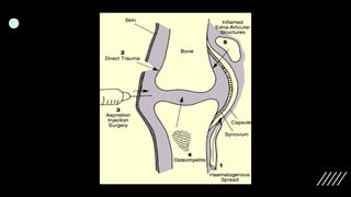
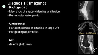
P Y O G E N I C
A R T H R I T I S
B Y: M U H A M M A D A N A S
Pyogenic Arthritis:
is an inflammatory joint disease caused by bacterial, viral,
and fungal infection
Route of infection
• dissemination of pathogens via the blood, from distant site….
(most common)
• dissemination from an acute osteomylitic focus
• dissemination from adjacent soft tissue infection,
• entry via penetrating trauma
• entry via iatrogenic means
What causes Pyogenic Arthritis?
• Staphylococci. ( S. aureus) >>>>> Most Common.
• Haemophilus influenzae. ( in Children)
• Gram negative bacilli.
• Streptococci.
• Gonococci.
• Viruses.
The Infectious agent can enter the body in a number of
ways, such as:
• A broken bone that goes through the skin (open fracture)
• An infection that spreads from another place on the body, such
as the skin or genitals
• An infected wound
• Foreign object that goes through the skin
• Injury that breaks the skin
Pathology
• There is an acute synovitis with a purulent joint effusion and
Synovial membrane becomes edematous, swollen and
hyperemic, and produces increase amount of cloudy exudates
contains leukocytes and bacteria
• As infection spread through the joint, articular cartilage is
destroyed by bacterial and cellular enzymes.
• If the infection is not arrested the cartilage may be completely
destroyed.
• Pus may burst out of the joint to form abscesses and sinuses.
• The joint may be become pathologically dislocated
• Who is at risk for Pyogenic arthritis?
• A systemic blood-borne infection
• IV drug use
• Osteoarthritis
• Past history of septic arthritis
• Rheumatoid arthritis
• Other factors that may increase the risk for Pyogenic arthritis
include:
• HIV
• Old age
• Suppressed immune system
Clinical presentation/ symptoms:
• The most common joints affected by septic arthritis are the
knee, hip, shoulder, elbow, wrist, and finger. Most often, only
one joint is affected. Symptoms can occur a bit differently in
each person, but common symptoms include:
• Fever
• Joint pain
• Joint swelling
• Inspection: Erythema, Effusion, Jt tend to be in Max.Vol
position
• Palpation: Tender, Warm
• Motion: Inability to tolerate weight, Inability to tolerate PROM
Diagnosis ( Imagimg)
• Radiograph :
• May show Jt space widening or effusion
• Periarticular osteopenia
• Ultrasound:
• For confirmation of effusion in large Jt’s
• For guiding aspirations
• MRI:
• detects jt effusion
Diagnosis (Studies)
• CBC: high WBC with left shift
• ESR: High ( > 30)
• CRP: High ( > 5)
• Joiny fluid Aspirate:
• gold standard for treatment and allows directed antibiotic treatment
Should be analysed for
• Cell count
• Gram stain
• Culture
• Glucose level
• Crystal analysis
Complications:
• Dislocation: a tense effusion may cause dislocation
• Epiphyseal destruction: in neglected infants the largely
cartilaginous epiphysis may be destroyed ,leaving an unstable
pseudarthrosis.
• Growth disturbance: physeal damage may result in
shortening or deformity
• Ankylosis: if articular cartilage is eroded healing may lead to
ankylosis
• Secondary osteoarthritis
• Osteomyleitis/abcess/sinus
Treatment
• General Measures:
The first priority is to aspirate the joint and examine the fluid, treatment is
then started without further delay.
• Analgesics and splinting of the involved joint in the position of maximal
comfort alleviate pain.
• Fluid replacement and nutritional support may be required.
• Other foci of infection and any coexisting medical conditions must be
identified and treated appropriately
• Intravenous antibiotics should be given empirically and started as soon
as joint fluid and blood sample have been taken for culture
• If gram –positive organisms are identified ,Flucloxacillin is
suitable . If in doubt ,a third generation cephalosporin will cover
both game+ and gram- organisms.
• Children less than 4 yr( if suspicion of H.Infl) treated with
Ampicillin.
• Once the bacterial sensitivity is known the appropriate drug is
substituted.
• Intravenous administration is continued for several weeks and is
followed by oral antibiotics for a further 2 or 3 weeks.
Pyogenic_Arthritis and its management ppt

Pyogenic_Arthritis and its management ppt

  • 2.
    P Y OG E N I C A R T H R I T I S B Y: M U H A M M A D A N A S
  • 3.
    Pyogenic Arthritis: is aninflammatory joint disease caused by bacterial, viral, and fungal infection Route of infection • dissemination of pathogens via the blood, from distant site…. (most common) • dissemination from an acute osteomylitic focus • dissemination from adjacent soft tissue infection, • entry via penetrating trauma • entry via iatrogenic means
  • 5.
    What causes PyogenicArthritis? • Staphylococci. ( S. aureus) >>>>> Most Common. • Haemophilus influenzae. ( in Children) • Gram negative bacilli. • Streptococci. • Gonococci. • Viruses.
  • 6.
    The Infectious agentcan enter the body in a number of ways, such as: • A broken bone that goes through the skin (open fracture) • An infection that spreads from another place on the body, such as the skin or genitals • An infected wound • Foreign object that goes through the skin • Injury that breaks the skin
  • 7.
    Pathology • There isan acute synovitis with a purulent joint effusion and Synovial membrane becomes edematous, swollen and hyperemic, and produces increase amount of cloudy exudates contains leukocytes and bacteria • As infection spread through the joint, articular cartilage is destroyed by bacterial and cellular enzymes. • If the infection is not arrested the cartilage may be completely destroyed. • Pus may burst out of the joint to form abscesses and sinuses. • The joint may be become pathologically dislocated
  • 8.
    • Who isat risk for Pyogenic arthritis? • A systemic blood-borne infection • IV drug use • Osteoarthritis • Past history of septic arthritis • Rheumatoid arthritis • Other factors that may increase the risk for Pyogenic arthritis include: • HIV • Old age • Suppressed immune system
  • 9.
    Clinical presentation/ symptoms: •The most common joints affected by septic arthritis are the knee, hip, shoulder, elbow, wrist, and finger. Most often, only one joint is affected. Symptoms can occur a bit differently in each person, but common symptoms include: • Fever • Joint pain • Joint swelling • Inspection: Erythema, Effusion, Jt tend to be in Max.Vol position • Palpation: Tender, Warm • Motion: Inability to tolerate weight, Inability to tolerate PROM
  • 10.
    Diagnosis ( Imagimg) •Radiograph : • May show Jt space widening or effusion • Periarticular osteopenia • Ultrasound: • For confirmation of effusion in large Jt’s • For guiding aspirations • MRI: • detects jt effusion
  • 11.
    Diagnosis (Studies) • CBC:high WBC with left shift • ESR: High ( > 30) • CRP: High ( > 5) • Joiny fluid Aspirate: • gold standard for treatment and allows directed antibiotic treatment Should be analysed for • Cell count • Gram stain • Culture • Glucose level • Crystal analysis
  • 12.
    Complications: • Dislocation: atense effusion may cause dislocation • Epiphyseal destruction: in neglected infants the largely cartilaginous epiphysis may be destroyed ,leaving an unstable pseudarthrosis. • Growth disturbance: physeal damage may result in shortening or deformity • Ankylosis: if articular cartilage is eroded healing may lead to ankylosis • Secondary osteoarthritis • Osteomyleitis/abcess/sinus
  • 13.
    Treatment • General Measures: Thefirst priority is to aspirate the joint and examine the fluid, treatment is then started without further delay. • Analgesics and splinting of the involved joint in the position of maximal comfort alleviate pain. • Fluid replacement and nutritional support may be required. • Other foci of infection and any coexisting medical conditions must be identified and treated appropriately • Intravenous antibiotics should be given empirically and started as soon as joint fluid and blood sample have been taken for culture
  • 14.
    • If gram–positive organisms are identified ,Flucloxacillin is suitable . If in doubt ,a third generation cephalosporin will cover both game+ and gram- organisms. • Children less than 4 yr( if suspicion of H.Infl) treated with Ampicillin. • Once the bacterial sensitivity is known the appropriate drug is substituted. • Intravenous administration is continued for several weeks and is followed by oral antibiotics for a further 2 or 3 weeks.