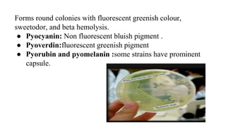
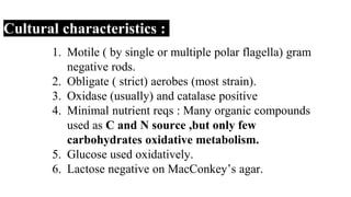
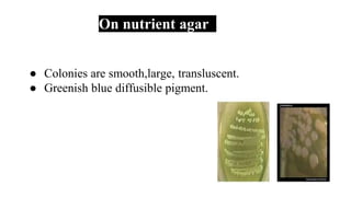
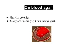
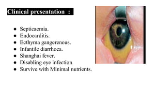
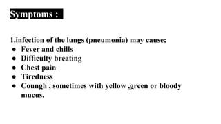
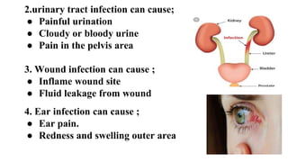
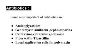
Pseudomonas aeruginosa
CONTENT:
Taxonomy,
Introduction
Pathogenesis
Morphology
Cultural
Characteristics
Clinical symptoms
Diagnosis
Treatment
By,
M.Malavika
II M.Sc Microbiology
Medical Microbiology
Under guidance of
Mrs.S.Selvajayathi,
Asst.Professor,
Dept of Microbiology,
Tirupur kumaran college for
women,TN,India.
Taxonomy :
Class : Gammaproteobacteia
Order : Pseudomonalea
Family : Pseudomonaceae
Genus : Pseudomonas
Spices : Pseudomonas aeruginosa
Introduction :
1. More than 140 species, most of which are
saprophytic.
2. Known to cause disease in humans are assoicated
with opportunistic infection which are,
○ P.aeruginosa.
○ P.maltophilia
○ P.cepacian
○ P.putida
● Only two species P.mallei and P.pseudomallei
produce specific human disease.
● P.aeruginosa is ubiquitous free-living bacterium
and is found in most moist environments,cause
disease in hospitalised patients ( cancer and burns).
Main features of P. aeruginosa biofilms and the respective consequences
on the infection progression. IMAGE Courtesy:
https://ibecbarcelona.eu/
Pathogenicity :
● Blue pus.
● Causing the nosocomial infection.
● Suppuratibe otitis.
● Localised and genralised infection.
● Urinary tract infection.
● Latrogenic meningitis.
Antigenic structure
Enzyme and toxins
Pili and non pilu adherin
Capsule (alginate glycalyx) seen
in culture from patients with
cystic fibrosis
LPs : entotoxin, multiple
immunotype
Pyocyanin :it also induce IL-8
production
Pyoverdin : a sideophore
Protease
Serine protease
Netalloproteaseand alkaline
Proteas cause tissue damage and he
bacteria spread.
PhospholipaseC : a hemolysis
ExotoxinA : causes tissue necrosis
ExoenzymeS and T : cytotoxic to h
cell.
Morphology :
● They are slender and gran negative bacillus 1.5-
3microbes×0.5microbes.
● Monoflgellar
● Non-capsulated but many strains have mucoid
silmelayer.
● Isolates from cystic fibrosis patients have abudunce
of extracelluar polysacccharide composed of
alginate polymers.
Forms round colonies with fluorescent greenish colour,
sweetodor, and beta hemolysis.
● Pyocyanin: Non fluorescent bluish pigment .
● Pyoverdin:fluorescent greenish pigment
● Pyorubin and pyomelanin :some strains have prominent
capsule.
Cultural characteristics :
1. Motile ( by single or multiple polar flagella) gram
negative rods.
2. Obligate ( strict) aerobes (most strain).
3. Oxidase (usually) and catalase positive
4. Minimal nutrient reqs : Many organic compounds
used as C and N source ,but only few
carbohydrates oxidative metabolism.
5. Glucose used oxidatively.
6. Lactose negative on MacConkey’s agar.
On nutrient agar
● Colonies are smooth,large, transluscent.
● Greenish blue diffusible pigment.
On blood agar
● Grayish colonies
● Many are haemolytis ( beta hemolysis)
● Non- lactose fermenting colourless colonies.
On EMB
● Non- lactose fermenting.
Colonies forming :
P.aeruginosa isolates produce three colony types:
1. Natural isolates from or water typically produce a
small ,rough colony.
2. Another one smooth colonies witb flat edges and
elevated appearance.
3. Another one urinary tract secretions in a mucoid
appearance, which is atttributed to the production of
alginate slime.
4. smooth and mucoid colonies as a play a role in
colonization and virulence.
Pigment production :
● Pyrcyanine (blue-green)
● Pyoverdin ( fluorescent yellow- greenish
pigment)
● Pyorubrin (red)
● Pyomelanin ( brown)
P.aeruginosa strains produce two soluble
pigment:
● Pyaverdin
● Pyocyanin ( “form pyocyaneus”)
Clinical presentation :
● Septicaemia.
● Endocarditis.
● Ecthyma gangerenous.
● Infantile diarrhoea.
● Shanghai fever.
● Disabling eye infection.
● Survive with Minimal nutrients.
Symptoms :
1.
1.infection of the lungs (pneumonia) may cause;
● Fever and chills
● Difficulty breating
● Chest pain
● Tiredness
● Coungh , sometimes with yellow ,green or bloody
mucus.
2.urinary tract infection can cause;
● Painful urination
● Cloudy or bloody urine
● Pain in the pelvis area
3. Wound infection can cause ;
● Inflame wound site
● Fluid leakage from wound
4. Ear infection can cause ;
● Ear pain.
● Redness and swelling outer area
Lab Diagnosis :
Specimen:
1.wound discharge
2.sputum
3.Blood
4.Urine
5.CSF.
Microscopy : Gramstain ;
Gram negative rod undistinguished form
enterobacterium.
Diagnosis :
● Diagnosis of P.aeruginosa infection depends upon
isolation and laboratory identification of the
bacterium.
● It grows well on most laboratory media and
commonly is isolated on blood agar or eosin-
methylthionine blue agar.
● It is identified on the basis of its gram morphology,
in ability to ferment lactose, a positive oxidase
reaction, its fruity odor, and its ability to grow at
42°c.
● Fluorescence under ultraviolet light isbhelpful in early
identification of P.aeruginosa colonies and may also help
identify its presence in wounds.
Biochemical test:
● Indole test : Negative.
● Methylred test : Negative.
● VP test : Negative.
● Citrate test : positive.
● Urease test : Negative.
● Oxidase test : positive
● Catalase test : positive.
Prevention :
● Most hospital have progrms for the prevention of nosocomial
infection.
● Patients with cystic fibrosis may be given periodic doses of
antibiotocs to prevent episodes of pseudomonas infection.
● Skin infection can be prevented by avoiding hot tub with cloudy
water, public swimming pool at the end of the day, removing net
swimming suit as soon as possible ,taking bath after sharing a hot
tub or using a public pool, abd also using appropriate amount of
chlorine in the water.
Treatment :
● P.aeruginosa is commonly resistant to antibiotics .
● Many strains are susceptible to Gentamicin, tbramycin,
colistin and amikacin,but sone resistant form have
developed.
● Infection are usually treated woth two antibiotics at once
the combination of gentamicin and carbonicillin is
frequently used to the at severe pseudomonas infection.
● Most antibiotics are administered intravenously or
oraly for 2-6 weeks.
● Eye infection requires link cal application of antibiotic
drops.
● Several types of vaccines are being tested but but none
is currently available for general use.
Antibiotics :
● Aminoglycosides
● Gentamycin,amikacin ,cephalosporins
● Cefotaxime,ceftazidims,ofloxacin
● Piperacillin,Ticarcillin
● Local application colistin, polymyxin
REFERENCE :
1. http://textbookofbacteriology.net/pseudom
onas.ntml.
2. http://www.classcontral.com/tag/microbiolo
gy.
3. https://course.org/course?query=microbiol
ogy.
4. http:/en.m.wikipedia.org/wiki/.pseudomona
saeruginosa.
THANK YOU
❤
❤

Pseudomonas aeruginosa-M

  • 1.
    Pseudomonas aeruginosa CONTENT: Taxonomy, Introduction Pathogenesis Morphology Cultural Characteristics Clinical symptoms Diagnosis Treatment By, M.Malavika IIM.Sc Microbiology Medical Microbiology Under guidance of Mrs.S.Selvajayathi, Asst.Professor, Dept of Microbiology, Tirupur kumaran college for women,TN,India.
  • 2.
    Taxonomy : Class :Gammaproteobacteia Order : Pseudomonalea Family : Pseudomonaceae Genus : Pseudomonas Spices : Pseudomonas aeruginosa
  • 3.
    Introduction : 1. Morethan 140 species, most of which are saprophytic. 2. Known to cause disease in humans are assoicated with opportunistic infection which are, ○ P.aeruginosa. ○ P.maltophilia ○ P.cepacian ○ P.putida
  • 4.
    ● Only twospecies P.mallei and P.pseudomallei produce specific human disease. ● P.aeruginosa is ubiquitous free-living bacterium and is found in most moist environments,cause disease in hospitalised patients ( cancer and burns).
  • 5.
    Main features ofP. aeruginosa biofilms and the respective consequences on the infection progression. IMAGE Courtesy: https://ibecbarcelona.eu/
  • 6.
    Pathogenicity : ● Bluepus. ● Causing the nosocomial infection. ● Suppuratibe otitis. ● Localised and genralised infection. ● Urinary tract infection. ● Latrogenic meningitis.
  • 7.
    Antigenic structure Enzyme andtoxins Pili and non pilu adherin Capsule (alginate glycalyx) seen in culture from patients with cystic fibrosis LPs : entotoxin, multiple immunotype Pyocyanin :it also induce IL-8 production Pyoverdin : a sideophore Protease Serine protease Netalloproteaseand alkaline Proteas cause tissue damage and he bacteria spread. PhospholipaseC : a hemolysis ExotoxinA : causes tissue necrosis ExoenzymeS and T : cytotoxic to h cell.
  • 8.
    Morphology : ● Theyare slender and gran negative bacillus 1.5- 3microbes×0.5microbes. ● Monoflgellar ● Non-capsulated but many strains have mucoid silmelayer. ● Isolates from cystic fibrosis patients have abudunce of extracelluar polysacccharide composed of alginate polymers.
  • 9.
    Forms round colonieswith fluorescent greenish colour, sweetodor, and beta hemolysis. ● Pyocyanin: Non fluorescent bluish pigment . ● Pyoverdin:fluorescent greenish pigment ● Pyorubin and pyomelanin :some strains have prominent capsule.
  • 10.
    Cultural characteristics : 1.Motile ( by single or multiple polar flagella) gram negative rods. 2. Obligate ( strict) aerobes (most strain). 3. Oxidase (usually) and catalase positive 4. Minimal nutrient reqs : Many organic compounds used as C and N source ,but only few carbohydrates oxidative metabolism. 5. Glucose used oxidatively. 6. Lactose negative on MacConkey’s agar.
  • 11.
    On nutrient agar ●Colonies are smooth,large, transluscent. ● Greenish blue diffusible pigment.
  • 12.
    On blood agar ●Grayish colonies ● Many are haemolytis ( beta hemolysis)
  • 13.
    ● Non- lactosefermenting colourless colonies.
  • 14.
    On EMB ● Non-lactose fermenting.
  • 15.
    Colonies forming : P.aeruginosaisolates produce three colony types: 1. Natural isolates from or water typically produce a small ,rough colony. 2. Another one smooth colonies witb flat edges and elevated appearance. 3. Another one urinary tract secretions in a mucoid appearance, which is atttributed to the production of alginate slime. 4. smooth and mucoid colonies as a play a role in colonization and virulence.
  • 16.
    Pigment production : ●Pyrcyanine (blue-green) ● Pyoverdin ( fluorescent yellow- greenish pigment) ● Pyorubrin (red) ● Pyomelanin ( brown) P.aeruginosa strains produce two soluble pigment: ● Pyaverdin ● Pyocyanin ( “form pyocyaneus”)
  • 17.
    Clinical presentation : ●Septicaemia. ● Endocarditis. ● Ecthyma gangerenous. ● Infantile diarrhoea. ● Shanghai fever. ● Disabling eye infection. ● Survive with Minimal nutrients.
  • 19.
    Symptoms : 1. 1.infection ofthe lungs (pneumonia) may cause; ● Fever and chills ● Difficulty breating ● Chest pain ● Tiredness ● Coungh , sometimes with yellow ,green or bloody mucus.
  • 20.
    2.urinary tract infectioncan cause; ● Painful urination ● Cloudy or bloody urine ● Pain in the pelvis area 3. Wound infection can cause ; ● Inflame wound site ● Fluid leakage from wound 4. Ear infection can cause ; ● Ear pain. ● Redness and swelling outer area
  • 21.
    Lab Diagnosis : Specimen: 1.wounddischarge 2.sputum 3.Blood 4.Urine 5.CSF. Microscopy : Gramstain ; Gram negative rod undistinguished form enterobacterium.
  • 22.
    Diagnosis : ● Diagnosisof P.aeruginosa infection depends upon isolation and laboratory identification of the bacterium. ● It grows well on most laboratory media and commonly is isolated on blood agar or eosin- methylthionine blue agar. ● It is identified on the basis of its gram morphology, in ability to ferment lactose, a positive oxidase reaction, its fruity odor, and its ability to grow at 42°c.
  • 24.
    ● Fluorescence underultraviolet light isbhelpful in early identification of P.aeruginosa colonies and may also help identify its presence in wounds. Biochemical test: ● Indole test : Negative. ● Methylred test : Negative. ● VP test : Negative. ● Citrate test : positive. ● Urease test : Negative. ● Oxidase test : positive ● Catalase test : positive.
  • 25.
    Prevention : ● Mosthospital have progrms for the prevention of nosocomial infection. ● Patients with cystic fibrosis may be given periodic doses of antibiotocs to prevent episodes of pseudomonas infection. ● Skin infection can be prevented by avoiding hot tub with cloudy water, public swimming pool at the end of the day, removing net swimming suit as soon as possible ,taking bath after sharing a hot tub or using a public pool, abd also using appropriate amount of chlorine in the water.
  • 26.
    Treatment : ● P.aeruginosais commonly resistant to antibiotics . ● Many strains are susceptible to Gentamicin, tbramycin, colistin and amikacin,but sone resistant form have developed. ● Infection are usually treated woth two antibiotics at once the combination of gentamicin and carbonicillin is frequently used to the at severe pseudomonas infection.
  • 28.
    ● Most antibioticsare administered intravenously or oraly for 2-6 weeks. ● Eye infection requires link cal application of antibiotic drops. ● Several types of vaccines are being tested but but none is currently available for general use.
  • 29.
    Antibiotics : ● Aminoglycosides ●Gentamycin,amikacin ,cephalosporins ● Cefotaxime,ceftazidims,ofloxacin ● Piperacillin,Ticarcillin ● Local application colistin, polymyxin
  • 30.
    REFERENCE : 1. http://textbookofbacteriology.net/pseudom onas.ntml. 2.http://www.classcontral.com/tag/microbiolo gy. 3. https://course.org/course?query=microbiol ogy. 4. http:/en.m.wikipedia.org/wiki/.pseudomona saeruginosa.
  • 31.