Blood plasma is the liquid component of blood, while serum is blood plasma without clotting factors. To prepare serum, whole blood is collected in red-topped tubes and allowed to clot at room temperature for 15-30 minutes. It is then centrifuged to separate the clot from the resulting supernatant, which is serum. For plasma preparation, whole blood is collected in anticoagulant-treated tubes and centrifuged to separate cells from the supernatant, which is plasma. Both serum and plasma samples should be aliquoted and stored at -20°C or lower to avoid freeze-thaw cycles.
Estimation of Blood Urea Nitrogen by Dr. TehmasTehmas Ahmad
Lecture/Demonstration of Biochemistry Practical of Blood Urea Nitrogen estimation in serum Delivered on 11-04-2018 to 2nd year MBBS students of Bannu Medical College, Bannu.
Estimation of Blood Urea Nitrogen by Dr. TehmasTehmas Ahmad
Lecture/Demonstration of Biochemistry Practical of Blood Urea Nitrogen estimation in serum Delivered on 11-04-2018 to 2nd year MBBS students of Bannu Medical College, Bannu.
Capsule is an layer around the bacteria cell which gives bacteria the protection and pathogenicity. Staining such an layer is difficult with the normal stains so it is necessary to stain the background and the cell itself which makes the capsule appear colourless.
Oxidase Test Microbiology - Principle, Procedure, Limitations, Results, QC - in lab #Oxidase Test
As the channel name suggests, our channel will be a perfect lounge for the malayali medicos..we wil be covering videos which will be like lecture classes related to the subjects biochemistry and microbiology in which we are specialised.. It will be a better learning experience for the students especially for those who are not able to understand and follow the normal classes in college..we assure the students that you will get a basic idea regarding the topic and extra reading can be done from the reference textbooks...
If you like my video
#like
#comment
#subscribe my channel
don't forget to subscribe my channel
Qualification
Maneesha M Joseph
MSc MLT (Microbiology)
Assistant Professor
Baby memorial college of allied Health science
Kozhikode
Our Partner Channel
Health & Voyage channel link - https://youtu.be/nzKqRVjlwc0
#Oxidase Test
#Medical
#Microbiology
# malayalam lecturer
#Mallu Medicos Lounge
#MalluMedicosLounge
#MLT
Each of the letters in “IMViC” stands for one of these tests. “I” is for indole; “M” is for methyl red; “V” is for Voges-Proskauer, and “C” is for citrate, lowercase “i” is added for the ease of pronunciation. IMViC is an acronym that stands for four different tests
Indole test
Methyl red test
Voges-Proskauer test
Citrate utilization test
Staphylococcus aureus,a bunch of grapes
commonly found on the skin or in the nose of even healthy individuals
cause skin infections but can cause pneumonia, heart valve infections, and bone infections.
Neisseria meninigitidis-brain infection,meningococcalSelvajeyanthi S
Neisseria meninigitidis
brain infection,meningococcal disease
meningococcemia-a life-threatening sepsis.
high mortality and morbidity,meningococcal disease needs immediate medical attention
initial adherence to the nasopharyngeal (nose and throat) mucosa to invasion of the deeper mucosal layers
This presentation explores a brief idea about the structural and functional attributes of nucleotides, the structure and function of genetic materials along with the impact of UV rays and pH upon them.
Deep Behavioral Phenotyping in Systems Neuroscience for Functional Atlasing a...Ana Luísa Pinho
Functional Magnetic Resonance Imaging (fMRI) provides means to characterize brain activations in response to behavior. However, cognitive neuroscience has been limited to group-level effects referring to the performance of specific tasks. To obtain the functional profile of elementary cognitive mechanisms, the combination of brain responses to many tasks is required. Yet, to date, both structural atlases and parcellation-based activations do not fully account for cognitive function and still present several limitations. Further, they do not adapt overall to individual characteristics. In this talk, I will give an account of deep-behavioral phenotyping strategies, namely data-driven methods in large task-fMRI datasets, to optimize functional brain-data collection and improve inference of effects-of-interest related to mental processes. Key to this approach is the employment of fast multi-functional paradigms rich on features that can be well parametrized and, consequently, facilitate the creation of psycho-physiological constructs to be modelled with imaging data. Particular emphasis will be given to music stimuli when studying high-order cognitive mechanisms, due to their ecological nature and quality to enable complex behavior compounded by discrete entities. I will also discuss how deep-behavioral phenotyping and individualized models applied to neuroimaging data can better account for the subject-specific organization of domain-general cognitive systems in the human brain. Finally, the accumulation of functional brain signatures brings the possibility to clarify relationships among tasks and create a univocal link between brain systems and mental functions through: (1) the development of ontologies proposing an organization of cognitive processes; and (2) brain-network taxonomies describing functional specialization. To this end, tools to improve commensurability in cognitive science are necessary, such as public repositories, ontology-based platforms and automated meta-analysis tools. I will thus discuss some brain-atlasing resources currently under development, and their applicability in cognitive as well as clinical neuroscience.
Slide 1: Title Slide
Extrachromosomal Inheritance
Slide 2: Introduction to Extrachromosomal Inheritance
Definition: Extrachromosomal inheritance refers to the transmission of genetic material that is not found within the nucleus.
Key Components: Involves genes located in mitochondria, chloroplasts, and plasmids.
Slide 3: Mitochondrial Inheritance
Mitochondria: Organelles responsible for energy production.
Mitochondrial DNA (mtDNA): Circular DNA molecule found in mitochondria.
Inheritance Pattern: Maternally inherited, meaning it is passed from mothers to all their offspring.
Diseases: Examples include Leber’s hereditary optic neuropathy (LHON) and mitochondrial myopathy.
Slide 4: Chloroplast Inheritance
Chloroplasts: Organelles responsible for photosynthesis in plants.
Chloroplast DNA (cpDNA): Circular DNA molecule found in chloroplasts.
Inheritance Pattern: Often maternally inherited in most plants, but can vary in some species.
Examples: Variegation in plants, where leaf color patterns are determined by chloroplast DNA.
Slide 5: Plasmid Inheritance
Plasmids: Small, circular DNA molecules found in bacteria and some eukaryotes.
Features: Can carry antibiotic resistance genes and can be transferred between cells through processes like conjugation.
Significance: Important in biotechnology for gene cloning and genetic engineering.
Slide 6: Mechanisms of Extrachromosomal Inheritance
Non-Mendelian Patterns: Do not follow Mendel’s laws of inheritance.
Cytoplasmic Segregation: During cell division, organelles like mitochondria and chloroplasts are randomly distributed to daughter cells.
Heteroplasmy: Presence of more than one type of organellar genome within a cell, leading to variation in expression.
Slide 7: Examples of Extrachromosomal Inheritance
Four O’clock Plant (Mirabilis jalapa): Shows variegated leaves due to different cpDNA in leaf cells.
Petite Mutants in Yeast: Result from mutations in mitochondrial DNA affecting respiration.
Slide 8: Importance of Extrachromosomal Inheritance
Evolution: Provides insight into the evolution of eukaryotic cells.
Medicine: Understanding mitochondrial inheritance helps in diagnosing and treating mitochondrial diseases.
Agriculture: Chloroplast inheritance can be used in plant breeding and genetic modification.
Slide 9: Recent Research and Advances
Gene Editing: Techniques like CRISPR-Cas9 are being used to edit mitochondrial and chloroplast DNA.
Therapies: Development of mitochondrial replacement therapy (MRT) for preventing mitochondrial diseases.
Slide 10: Conclusion
Summary: Extrachromosomal inheritance involves the transmission of genetic material outside the nucleus and plays a crucial role in genetics, medicine, and biotechnology.
Future Directions: Continued research and technological advancements hold promise for new treatments and applications.
Slide 11: Questions and Discussion
Invite Audience: Open the floor for any questions or further discussion on the topic.
Earliest Galaxies in the JADES Origins Field: Luminosity Function and Cosmic ...Sérgio Sacani
We characterize the earliest galaxy population in the JADES Origins Field (JOF), the deepest
imaging field observed with JWST. We make use of the ancillary Hubble optical images (5 filters
spanning 0.4−0.9µm) and novel JWST images with 14 filters spanning 0.8−5µm, including 7 mediumband filters, and reaching total exposure times of up to 46 hours per filter. We combine all our data
at > 2.3µm to construct an ultradeep image, reaching as deep as ≈ 31.4 AB mag in the stack and
30.3-31.0 AB mag (5σ, r = 0.1” circular aperture) in individual filters. We measure photometric
redshifts and use robust selection criteria to identify a sample of eight galaxy candidates at redshifts
z = 11.5 − 15. These objects show compact half-light radii of R1/2 ∼ 50 − 200pc, stellar masses of
M⋆ ∼ 107−108M⊙, and star-formation rates of SFR ∼ 0.1−1 M⊙ yr−1
. Our search finds no candidates
at 15 < z < 20, placing upper limits at these redshifts. We develop a forward modeling approach to
infer the properties of the evolving luminosity function without binning in redshift or luminosity that
marginalizes over the photometric redshift uncertainty of our candidate galaxies and incorporates the
impact of non-detections. We find a z = 12 luminosity function in good agreement with prior results,
and that the luminosity function normalization and UV luminosity density decline by a factor of ∼ 2.5
from z = 12 to z = 14. We discuss the possible implications of our results in the context of theoretical
models for evolution of the dark matter halo mass function.
Comparing Evolved Extractive Text Summary Scores of Bidirectional Encoder Rep...University of Maribor
Slides from:
11th International Conference on Electrical, Electronics and Computer Engineering (IcETRAN), Niš, 3-6 June 2024
Track: Artificial Intelligence
https://www.etran.rs/2024/en/home-english/
Cancer cell metabolism: special Reference to Lactate PathwayAADYARAJPANDEY1
Normal Cell Metabolism:
Cellular respiration describes the series of steps that cells use to break down sugar and other chemicals to get the energy we need to function.
Energy is stored in the bonds of glucose and when glucose is broken down, much of that energy is released.
Cell utilize energy in the form of ATP.
The first step of respiration is called glycolysis. In a series of steps, glycolysis breaks glucose into two smaller molecules - a chemical called pyruvate. A small amount of ATP is formed during this process.
Most healthy cells continue the breakdown in a second process, called the Kreb's cycle. The Kreb's cycle allows cells to “burn” the pyruvates made in glycolysis to get more ATP.
The last step in the breakdown of glucose is called oxidative phosphorylation (Ox-Phos).
It takes place in specialized cell structures called mitochondria. This process produces a large amount of ATP. Importantly, cells need oxygen to complete oxidative phosphorylation.
If a cell completes only glycolysis, only 2 molecules of ATP are made per glucose. However, if the cell completes the entire respiration process (glycolysis - Kreb's - oxidative phosphorylation), about 36 molecules of ATP are created, giving it much more energy to use.
IN CANCER CELL:
Unlike healthy cells that "burn" the entire molecule of sugar to capture a large amount of energy as ATP, cancer cells are wasteful.
Cancer cells only partially break down sugar molecules. They overuse the first step of respiration, glycolysis. They frequently do not complete the second step, oxidative phosphorylation.
This results in only 2 molecules of ATP per each glucose molecule instead of the 36 or so ATPs healthy cells gain. As a result, cancer cells need to use a lot more sugar molecules to get enough energy to survive.
introduction to WARBERG PHENOMENA:
WARBURG EFFECT Usually, cancer cells are highly glycolytic (glucose addiction) and take up more glucose than do normal cells from outside.
Otto Heinrich Warburg (; 8 October 1883 – 1 August 1970) In 1931 was awarded the Nobel Prize in Physiology for his "discovery of the nature and mode of action of the respiratory enzyme.
WARNBURG EFFECT : cancer cells under aerobic (well-oxygenated) conditions to metabolize glucose to lactate (aerobic glycolysis) is known as the Warburg effect. Warburg made the observation that tumor slices consume glucose and secrete lactate at a higher rate than normal tissues.
Multi-source connectivity as the driver of solar wind variability in the heli...Sérgio Sacani
The ambient solar wind that flls the heliosphere originates from multiple
sources in the solar corona and is highly structured. It is often described
as high-speed, relatively homogeneous, plasma streams from coronal
holes and slow-speed, highly variable, streams whose source regions are
under debate. A key goal of ESA/NASA’s Solar Orbiter mission is to identify
solar wind sources and understand what drives the complexity seen in the
heliosphere. By combining magnetic feld modelling and spectroscopic
techniques with high-resolution observations and measurements, we show
that the solar wind variability detected in situ by Solar Orbiter in March
2022 is driven by spatio-temporal changes in the magnetic connectivity to
multiple sources in the solar atmosphere. The magnetic feld footpoints
connected to the spacecraft moved from the boundaries of a coronal hole
to one active region (12961) and then across to another region (12957). This
is refected in the in situ measurements, which show the transition from fast
to highly Alfvénic then to slow solar wind that is disrupted by the arrival of
a coronal mass ejection. Our results describe solar wind variability at 0.5 au
but are applicable to near-Earth observatories.
Multi-source connectivity as the driver of solar wind variability in the heli...
Preparation of serum and plasma .pptx
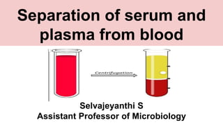
1. Separation of serum and
plasma from blood
Selvajeyanthi S
Assistant Professor of Microbiology
2. Blood Plasma :
liquid component
of blood
Serum : Blood
serum is blood
plasma without
fibrinogen or the
other clotting
factors and serum
is clearer than
plasme because of
fewer proteins
3. Serum preparation
● Collect whole blood in a covered test tube.
If commercially available tubes are to be
used, the researcher should use the red
topped tubes.
● These are available from Becton Dickinson
(BD). BD’s trade name for the blood
handling tubes is Vacutainer.
● After collection of the whole blood, allow
the blood to clot by leaving it undisturbed
at room temperature. This usually takes 15–
30 minutes.
● Remove the clot by centrifuging at 1,000–
2,000 x g for 10 minutes in a refrigerated
centrifuge.
4. ● The resulting supernatant is designated serum. Following centrifugation, it is important to
immediately transfer the liquid component (serum) into a clean polypropylene tube using a
Pasteur pipette.
● The samples should be maintained at 2–8°C while handling. If the serum is not analyzed
immediately, the serum should be apportioned into 0.5 ml aliquots, stored, and transported at
–20°C or lower.
● It is important to avoid freeze-thaw cycles because this is detrimental to many serum
components. Samples which are hemolyzed, icteric or lipemic can invalidate certain tests.
5. Plasma Preparation
● Collect whole blood into commercially
available anticoagulant-treated tubes e.g.,
EDTA-treated (lavender tops) or citrate-
treated (light blue tops). Heparinized tubes
(green tops) are indicated for some
applications; however, heparin can often
be contaminated with endotoxin, which
can stimulate white blood cells to release
cytokines.
● Cells are removed from plasma by
centrifugation for 10 minutes at 1,000–
2,000 x g using a refrigerated centrifuge.
Centrifugation for 15 minutes at 2,000 x g
depletes platelets in the plasma sample.
6. ● The resulting supernatant is designated plasma. Following
centrifugation, it is important to immediately transfer the liquid
component (plasma) into a clean polypropylene tube using a
Pasteur pipette.
● The samples should be maintained at 2–8°C while handling. If the
plasma is not analyzed immediately, the plasma should be
apportioned into 0.5 ml aliquots, stored, and transported at –20°C
or lower.
● It is important to avoid freeze-thaw cycles.
● Samples which are hemolyzed, icteric, or lipemic can invalidate
certain tests.
● There are other commercially available tubes for blood sample
collection.