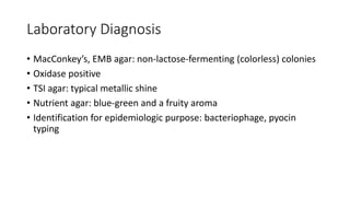
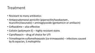
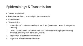
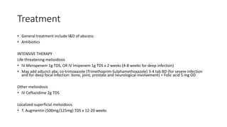
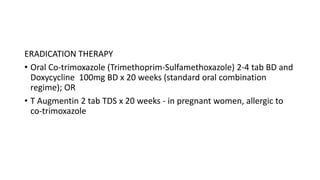
Pseudomonas Aeruginosa
• Species from genera Pseudomonas, Burkholderia and
Stenotrophomonas were all previously classified in the Pseudomonas
genus.
• P.aeruginosa also known as Burkhoderia aeruginosa
• P. cepacia renamed Burkhoderia cepacia
• P. maltophilia renamed Xanthomonas maltophilia, now named
Stenotrophomonas maltophilia
• P. pseudomallei also known as Burkhoderia pseudomallei
Epidemiology & Transmission
• Found mainly in soil and water
• Present as normal flora of colon in 10% of people
• Can colonize upper respiratory tract of hospitalized patients
• Contaminating respiratory therapy and anaesthesia equipment, intravenous fluid,
distilled water due to its ability to grow in simple aqueous solution
• Primarily an opportunistic pathogen causing infection:
o In hospitalized patient (e.g. in burn patient), in whom skin host defense destroyed
o In chronic respiratory disease patient (e.g. cystic fibrosis), in whom normal clearance
mechanism impaired
o In immunosuppressed patient
o In patient with neutrophil count <500/uL
o In patient with indwelling catheter
Properties
• Gram negative rod
• Strict aerobe
• Non-fermenters (derived energy only by oxidation of sugar rather by
fermentation)
• Oxidase-positive (oxidation involves electron transport by cytochrome c)
• Able to grow in water containing traces of nutrients (e.g. tap water) –
favors their persistence in hospital environment
• Remarkable ability to withstand disinfectant  hospital acquired infection
• Found growing in hexachlorophene-containing soap solution, in antiseptic
and in detergent
• Produces 2 pigments
1. Pyocyanin – color the pus in a wound blue
2. Pyoverdin (fluorescein) – a yellow-green pigment that fluoresces
under UV light – can be used in early detection of skin infection in
burn patient
• P aeruginosa the only Pseudomonas species synthesizes pyocyanin
• P aeruginosa isolated from cystic fibrosis patient have a prominent
slime layer (glycocalyx) , giving colonies a very mucoid appearance
• Slime layer mediates adherence of the organism to mucous
membranes of respiratory tract and prevents antibody from binding
to the organism
Virulence Factor
• Endotoxin
- Causes symptoms of sepsis and septic shock
• Exotoxin
- Exotoxin A causes tissue necrosis
- Inhibits eukaryotic protein synthesis by ADP ribosylation of elongation
factor-2 (same mechanism as diphteria exotoxin)
• Enzymes
- Elastase and proteases – histotoxic and facilitates invasion of organism into
bloodstream
- Pyocyanin – damages the cilia and mucosal cells of the respiratory tract
• “Type III secretion system”
- Significantly more virulent than those that do not
- Transfer the exotoxin grom the bacterium directly into adjacent
human cell, which allows toxin to avoid neutralizing antibody
- The secretion system mediated by transport pump in bacterial cell
membrane
- 4 exoenzyme known to be transported
- Exo S most clearly associated with virulence, most important mode of
action is ADP-ribosylation of a Ras protein -> damage to cytoskeleton
Clinical Picture
• Urinary tract infection
• Pneumonia especially in cystic fibrosis patient
• Wound infection especially in burn patient
• Hospital acquired pneumonia – especially ventilator-associated pneumonia
• Sepsis
• Ecthyma gangrenosum – spread to skin causing black, necrotic lesion
• Endocarditis in IVDU
• Severe otitis media (malignant otitis media) and skin lesion (e.g. folliculitis) – in
users of swimming pool and hot tub with inadequate chlorination
• Osteomyelitis of the foot – most common organism, occurs in those who sustain
puncture wounds through soles of gym shoes
• Corneal infection – in contact lenses users
Laboratory Diagnosis
• MacConkey’s, EMB agar: non-lactose-fermenting (colorless) colonies
• Oxidase positive
• TSI agar: typical metallic shine
• Nutrient agar: blue-green and a fruity aroma
• Identification for epidemiologic purpose: bacteriophage, pyocin
typing
Treatment
• Resistant to many antibiotics
• Antipseudomonal penicillin (piperacillin/tazobactam ,
ticarcillin/clavunate) + aminoglycoside (gentamicin or amikacin)
• Ceftazidime – also effective
• Colistin (polymyxin E) – highly resistant stains
• Ciprofloxacin – drug of choice for UTI
• Trimethoprim-sulfamethoxazole (co-trimoxazole) – infections caused
by B.cepacian, S.maltophilia
Burkholderia pseudomallei
Epidemiology & Transmission
• Causes meliodosis
• Rare disease found primarily in Southeast Asia
• Found in soil
• Transmission
1. Inhalation of contaminated dust particles (increased cases during rainy
season)
2. Direct contact with contaminated soil and water through penetrating
wounds, existing skin abrasions, burns
3. Aspiration of contaminated water
4. Ingestion of contaminated water
Clinical Features
• Incubation period 1-21 days (mean 9 days) but can be as long as
months and even years
• Present acutely with rapid progression and death or run a chronic and
relapsing course
Common Presentation
• Pneumonia
- Most common presentation + multiple abcesses in liver, spleen and
prostate
• Septicaemia
⁻ Metastatic foci of infection are established rapidly during
bacteraemia, particularly in the lungs (multifocal pneumonia, which
may cavitate), liver, spleen and kidneys (multiple abscesses), skin and
soft tissues (cellulitis, pustules), bones and joints, lymph nodes and
prostate, although any site may be affected.
Other presentations
• Soft tissue infection: cellulitis, fasciitis, skin abscess/ulcer
• Intra abdominal: single or multiple abscesses in the liver, spleen, kidney or
pancreas
• Bone and joint infection: osteomyelitis, septic arthritis (musculoskeletal
meliodosis)
• Genitourinary: prostatic abscess
• CNS infection: cerebral abscess, meningoencephalitis, encephalomyelitis
• Facial: suppurative parotitis
• Ocular infection: conjunctival ulcer, hypopyon, orbital cellulitis
• Asymptomatic infection (positive serology up to 50% of healthy adults
in endemic countries)
• Overall mortality of bacteraemic meliodosis approaches 100% if
untreated, reduced to 37-54% with optimal management and
aggressive intensive care
• Localised meliodosis – lower mortality rate (4-5%)
Diagnosis
• Culture. Collected from:
- Blood
- Respiratory samples e.g. sputum
- Pus
- Tissue
- Wound swab
- Urine
• Serology- IgM antibody
• Molecular diagnosis – PCR-RT
• Gram negative rod
• Safety pin appearance on M/E
• Obligate aerobe
Treatment
• General treatment include I&D of abscess
• Antibiotics
INTENSIVE THERAPY
Life threatening melioidosis
• IV Meropenem 1g TDS, OR IV Imipenem 1g TDS x 2 weeks (4-8 weeks for deep infection)
• May add adjunct abx; co-trimoxazole (Trimethoprim-Sulphamethoxazole) 3-4 tab BD (for severe infection
and for deep focal infection: bone, joint, prostate and neurological involvement) + Folic acid 5 mg OD
Other melioidosis
• IV Ceftazidime 2g TDS
Localized superficial melioidosis
• T. Augmentin (500mg/125mg) TDS x 12-20 weeks
ERADICATION THERAPY
• Oral Co-trimoxazole (Trimethoprim-Sulfamethoxazole) 2-4 tab BD and
Doxycycline 100mg BD x 20 weeks (standard oral combination
regime); OR
• T Augmentin 2 tab TDS x 20 weeks - in pregnant women, allergic to
co-trimoxazole

Pseudomonas Aeruginosa.pptx

  • 1.
  • 2.
    • Species fromgenera Pseudomonas, Burkholderia and Stenotrophomonas were all previously classified in the Pseudomonas genus. • P.aeruginosa also known as Burkhoderia aeruginosa • P. cepacia renamed Burkhoderia cepacia • P. maltophilia renamed Xanthomonas maltophilia, now named Stenotrophomonas maltophilia • P. pseudomallei also known as Burkhoderia pseudomallei
  • 3.
    Epidemiology & Transmission •Found mainly in soil and water • Present as normal flora of colon in 10% of people • Can colonize upper respiratory tract of hospitalized patients • Contaminating respiratory therapy and anaesthesia equipment, intravenous fluid, distilled water due to its ability to grow in simple aqueous solution • Primarily an opportunistic pathogen causing infection: o In hospitalized patient (e.g. in burn patient), in whom skin host defense destroyed o In chronic respiratory disease patient (e.g. cystic fibrosis), in whom normal clearance mechanism impaired o In immunosuppressed patient o In patient with neutrophil count <500/uL o In patient with indwelling catheter
  • 4.
    Properties • Gram negativerod • Strict aerobe • Non-fermenters (derived energy only by oxidation of sugar rather by fermentation) • Oxidase-positive (oxidation involves electron transport by cytochrome c) • Able to grow in water containing traces of nutrients (e.g. tap water) – favors their persistence in hospital environment • Remarkable ability to withstand disinfectant  hospital acquired infection • Found growing in hexachlorophene-containing soap solution, in antiseptic and in detergent
  • 5.
    • Produces 2pigments 1. Pyocyanin – color the pus in a wound blue 2. Pyoverdin (fluorescein) – a yellow-green pigment that fluoresces under UV light – can be used in early detection of skin infection in burn patient • P aeruginosa the only Pseudomonas species synthesizes pyocyanin • P aeruginosa isolated from cystic fibrosis patient have a prominent slime layer (glycocalyx) , giving colonies a very mucoid appearance • Slime layer mediates adherence of the organism to mucous membranes of respiratory tract and prevents antibody from binding to the organism
  • 6.
    Virulence Factor • Endotoxin -Causes symptoms of sepsis and septic shock • Exotoxin - Exotoxin A causes tissue necrosis - Inhibits eukaryotic protein synthesis by ADP ribosylation of elongation factor-2 (same mechanism as diphteria exotoxin) • Enzymes - Elastase and proteases – histotoxic and facilitates invasion of organism into bloodstream - Pyocyanin – damages the cilia and mucosal cells of the respiratory tract
  • 7.
    • “Type IIIsecretion system” - Significantly more virulent than those that do not - Transfer the exotoxin grom the bacterium directly into adjacent human cell, which allows toxin to avoid neutralizing antibody - The secretion system mediated by transport pump in bacterial cell membrane - 4 exoenzyme known to be transported - Exo S most clearly associated with virulence, most important mode of action is ADP-ribosylation of a Ras protein -> damage to cytoskeleton
  • 8.
    Clinical Picture • Urinarytract infection • Pneumonia especially in cystic fibrosis patient • Wound infection especially in burn patient • Hospital acquired pneumonia – especially ventilator-associated pneumonia • Sepsis • Ecthyma gangrenosum – spread to skin causing black, necrotic lesion • Endocarditis in IVDU • Severe otitis media (malignant otitis media) and skin lesion (e.g. folliculitis) – in users of swimming pool and hot tub with inadequate chlorination • Osteomyelitis of the foot – most common organism, occurs in those who sustain puncture wounds through soles of gym shoes • Corneal infection – in contact lenses users
  • 9.
    Laboratory Diagnosis • MacConkey’s,EMB agar: non-lactose-fermenting (colorless) colonies • Oxidase positive • TSI agar: typical metallic shine • Nutrient agar: blue-green and a fruity aroma • Identification for epidemiologic purpose: bacteriophage, pyocin typing
  • 10.
    Treatment • Resistant tomany antibiotics • Antipseudomonal penicillin (piperacillin/tazobactam , ticarcillin/clavunate) + aminoglycoside (gentamicin or amikacin) • Ceftazidime – also effective • Colistin (polymyxin E) – highly resistant stains • Ciprofloxacin – drug of choice for UTI • Trimethoprim-sulfamethoxazole (co-trimoxazole) – infections caused by B.cepacian, S.maltophilia
  • 11.
  • 12.
    Epidemiology & Transmission •Causes meliodosis • Rare disease found primarily in Southeast Asia • Found in soil • Transmission 1. Inhalation of contaminated dust particles (increased cases during rainy season) 2. Direct contact with contaminated soil and water through penetrating wounds, existing skin abrasions, burns 3. Aspiration of contaminated water 4. Ingestion of contaminated water
  • 13.
    Clinical Features • Incubationperiod 1-21 days (mean 9 days) but can be as long as months and even years • Present acutely with rapid progression and death or run a chronic and relapsing course
  • 14.
    Common Presentation • Pneumonia -Most common presentation + multiple abcesses in liver, spleen and prostate • Septicaemia ⁻ Metastatic foci of infection are established rapidly during bacteraemia, particularly in the lungs (multifocal pneumonia, which may cavitate), liver, spleen and kidneys (multiple abscesses), skin and soft tissues (cellulitis, pustules), bones and joints, lymph nodes and prostate, although any site may be affected.
  • 15.
    Other presentations • Softtissue infection: cellulitis, fasciitis, skin abscess/ulcer • Intra abdominal: single or multiple abscesses in the liver, spleen, kidney or pancreas • Bone and joint infection: osteomyelitis, septic arthritis (musculoskeletal meliodosis) • Genitourinary: prostatic abscess • CNS infection: cerebral abscess, meningoencephalitis, encephalomyelitis • Facial: suppurative parotitis • Ocular infection: conjunctival ulcer, hypopyon, orbital cellulitis
  • 16.
    • Asymptomatic infection(positive serology up to 50% of healthy adults in endemic countries) • Overall mortality of bacteraemic meliodosis approaches 100% if untreated, reduced to 37-54% with optimal management and aggressive intensive care • Localised meliodosis – lower mortality rate (4-5%)
  • 17.
    Diagnosis • Culture. Collectedfrom: - Blood - Respiratory samples e.g. sputum - Pus - Tissue - Wound swab - Urine • Serology- IgM antibody • Molecular diagnosis – PCR-RT
  • 18.
    • Gram negativerod • Safety pin appearance on M/E • Obligate aerobe
  • 19.
    Treatment • General treatmentinclude I&D of abscess • Antibiotics INTENSIVE THERAPY Life threatening melioidosis • IV Meropenem 1g TDS, OR IV Imipenem 1g TDS x 2 weeks (4-8 weeks for deep infection) • May add adjunct abx; co-trimoxazole (Trimethoprim-Sulphamethoxazole) 3-4 tab BD (for severe infection and for deep focal infection: bone, joint, prostate and neurological involvement) + Folic acid 5 mg OD Other melioidosis • IV Ceftazidime 2g TDS Localized superficial melioidosis • T. Augmentin (500mg/125mg) TDS x 12-20 weeks
  • 20.
    ERADICATION THERAPY • OralCo-trimoxazole (Trimethoprim-Sulfamethoxazole) 2-4 tab BD and Doxycycline 100mg BD x 20 weeks (standard oral combination regime); OR • T Augmentin 2 tab TDS x 20 weeks - in pregnant women, allergic to co-trimoxazole