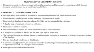
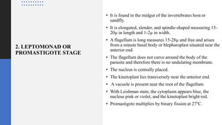
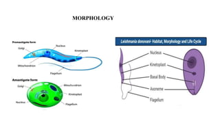
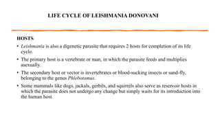
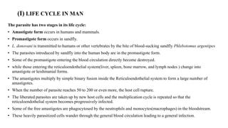
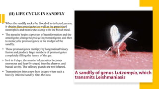
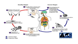
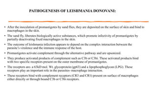
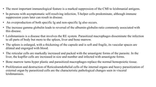
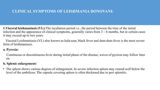
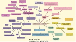
3. SIR WILLIAM LEISHMAN
• 1900- Sir William Leishman
discovered L.donovani in spleen
smears of a soldier who died of
fever Dum Dum, India
• This disease was known locally
as Dum Dum Fever or Kala azar
4. CHARLES DONOVAN
• Charles Donovan recognized these
symptoms in other kala azar patients
and published his discovery a few weeks
after Leishman
• After examining the Parasite using
Leishman's stain, these amastigotes
were known as Leishman Donvan
bodies
6. LEISHMANIA DONOVANI
• The species of Leishmania donovani was reported
simultaneously by Leishmania from London
and Donovan from Madras in (1903) hence the
name Leishmania donovani.
• L. donovani causes a malaria-like fever-oriental disease in
the man called kala-azar, Dumdum fever, or Black fever.
• L. donovani, the causative organism of Visceral
leishmaniasis (kala-azar) in Africa, the Middle East,
Mediterranean coasts, Asia, and South America.
• It is endemic in Asia, Africa, the Americas, and the
Mediterranean region.
8. MORPHOLOGY OF LEISHMANIA DONOVANI
• The parasite occurs in two forms or stages, leishmanial or amastigote and leptomonad or promastigote, which alternate
between a vertebrate (man) and an invertebrate (sandfly) host.
1. LEISHMANIAL OR AMASTIGOTE STAGE
• This stage occurs intracellularly in blood cells or reticuloendothelial cells of the vertebrate hosts or man.
• It is microscopic, rounded, or oval in shape measuring 2-4 micrometer in length.
• There is no free flagellum, it is greatly reduced, fibril-like, and lies embedded in the cytoplasm.
• A flagellar stage of amastigote is known as LD bodies.
• The nucleus is central or eccentric.
• The cell membrane is delicate and can be demonstrated only in a fresh specimen.
• Kinetoplast is rod-shaped or dot-like and lies at the right angle to the nucleus.
• The axoneme(rhizoplat) is a delicate filament extending from the kinetoplast to the margin of the body. It represents the foot
of the flagellum.
• They are stained well with Giemsa or Wright stain.
• In a Giemsa stained preparation, the cytoplasm surrounded by a limiting membrane appears pale blue. The nucleus relatively
is larger and stained red. The kinetoplast stained deep red.
• Amastigote divides by binary fission at 37°C.
9. 2. LEPTOMONAD OR
PROMASTIGOTE STAGE
• It is found in the midgut of the invertebrates host or
sandfly.
• It is elongated, slender, and spindle-shaped measuring 15-
20µ in length and 1-2µ in width.
• A flagellum is long measures 15-28µ and free and arises
from a minute basal body or blepharoplast situated near the
anterior end.
• The flagellum does not curve around the body of the
parasite and therefore there is no undulating membrane.
• The nucleus is centrally placed.
• The kinetoplast lies transversely near the anterior end.
• A vacuole is present near the root of the flagellum
• With Leishman stain, the cytoplasm appears blue, the
nucleus pink or violet, and the kinetoplast bright red.
• Promastigote multiplies by binary fission at 27°C.
11. LIFE CYCLE OF LEISHMANIA DONOVANI
HOSTS
• Leishmania is also a digenetic parasite that requires 2 hosts for completion of its life
cycle.
• The primary host is a vertebrate or man, in which the parasite feeds and multiplies
asexually.
• The secondary host or vector is invertebrates or blood-sucking insects or sand-fly,
belonging to the genus Phlebotomus.
• Some mammals like dogs, jackals, gerbils, and squirrels also serve as reservoir hosts in
which the parasite does not undergo any change but simply waits for its introduction into
the human host.
12. (I) LIFE CYCLE IN MAN
The parasite has two stages in its life cycle:
• Amastigote form occurs in humans and mammals.
• Promastigote form occurs in sandfly.
• L. donovani is transmitted to humans or other vertebrates by the bite of blood-sucking sandfly Phlebotomus argentipes
• The parasites introduced by sandfly into the human body are in the promastigote form.
• Some of the promastigote entering the blood circulation directly become destroyed.
• while those entering the reticuloendothelial system(liver, spleen, bone marrow, and lymph nodes ) change into
amastigote or leishmanial forms.
• The amastigotes multiply by simple binary fusion inside the Reticuloendothelial system to form a large number of
amastigotes.
• When the number of parasite reaches 50 to 200 or even more, the host cell rupture.
• The liberated parasites are taken up by new host cells and the multiplication cycle is repeated so that the
reticuloendothelial system becomes progressively infected.
• Some of the free amastigotes are phagocytosed by the neutrophils and monocytes(macrophages) in the bloodstream.
• These heavily parasitized cells wander through the general blood circulation leading to a general infection.
13. (II) LIFE CYCLE IN SANDFLY
• When the sandfly sucks the blood of an infected person,
it obtains free amastigotes as well as the parasitized
neutrophils and monocytes along with the blood-meal.
• The parasite begins a process of transformation and the
amastigotes change to procyclin promastigotes and then
to metacyclic promastigotes in the midgut of the
sandfly.
• These promastigotes multiply by longitudinal binary
fusion and produce large numbers of promastigotes
completely filling the lumen of the gut.
• In 6 to 9 days, the number of parasites becomes
enormous and heavily spread into the pharynx and
buccal cavity. The salivary glands are not infected.
• Transmission into a new host occurs when such a
heavily infected sandfly bites the host.
14.
15. MODE OF TRANSMISSION:
• The infection is transmitted to Human mainly by the bite of vector sandfly of
genus Phlebotomus and genus Lutzomyia.
• Less frequently the infection is transmitted by:
• Blood transfusion, congenital infection, accidental inoculation of cultured promastigotes in
the lab workers and sexual intercourse.
• Males are affected more due to increase exposure through the occupation and leisure
activities.
16. PATHOGENESIS OF LEISHMANIA DONOVANI:
• After the inoculation of promastigotes by sand flies, they are deposited on the surface of skin and bind to
macrophages in the skin.
• The sand fly, liberates biologically active substances, which promote infectivity of promastigotes by
partially deactivating fixed macrophages in the skin.
• The outcome of leishmania infection appears to depend on the complex interaction between the
parasite’s virulence and the immune response of the host.
• Promastigotes activate complement through the alternative pathway and are opsonized.
• They produce activated products of complement such as C3b or C3bi. These activated products bind
with two specific receptors present on the outer membrane of promastigotes.
• The receptors are- a 63kD mol. Wt. glycoprotein (gp63) and a lipophosphoglycan (LPG). These
receptors play an important role in the parasites- macrophage interaction.
• These receptors bind with complement receptors (CR3 and CR1) present on surface of macrophages
either directly or through bound C3b or C3bi receptors.
17. • The most important immunological feature is a marked suppression of the CMI to leishmanial antigens.
• In persons with asymptomatic self-resolving infection, T-helper cells predominate, although immune
suppression years later can result in disease.
• An overproduction of both specific Ig and non-specific Ig also occurs.
• The increase gamma globulin leads to reversal of the albumin-globulin ratio commonly associated with
this disease.
• Leishmaniasis is a disease that involves the RE system. Parasitized macrophages disseminate the infection
to all parts of body but more to the spleen, liver and bone marrow.
• The spleen is enlarged, with a thickening of the capsule and is soft and fragile, its vascular spaces are
dilated and engorged with blood.
• The reticular cells are markedly increased and packed with the amastigote forms of the parasite. In the
liver, the kupffer cells are increased in size and number and infected with amastigote forms.
• Bone marrow turns hyper plastic and parasitized macrophages replace the normal hemopoietic tissue.
• Proliferation and destruction of Reticuloendothelial cells of the internal organs and heavy parasitization of
external organ by parasitized cells are the characteristic pathological changes seen in visceral
leishmaniasis.
18. CLINICAL SYMPTOMS OF LEISHMANIA DONOVANI:
1.Visceral leishmaniasis (VL):The incubation period i.e., the period between the time of the initial
infection and the appearance of clinical symptoms, generally varies from 3 – 6 months, but in certain cases
it may exceed up to two years.
Visceral Leishmaniasis (VL) also known as kala-azar, black fever and dum-dum fever is the most severe
form of leishmaniasis.
a. Pyrexia:
Continuous or discontinuous fever during initial phase of the disease, waves of pyrexia may follow later
on.
b. Splenic enlargement:
• The spleen shows various degrees of enlargement. In severe infection spleen may extend well below the
level of the umbilicus. The capsule covering spleen is often thickened due to peri splenitis.
19. c. Liver enlargement:
• Liver is less frequently enlarged than the spleen in kala-azar. The Kuffer’s cells are greatly enlarged both in size and
number as their cytoplasm gets packed with the parasites. Normally jaundice does not appear in kala-azar unless
liver is greatly damage.
d. Changes in bone marrow:
• Leishmania infection causes hyperplasia (abnormal increase in the number of normal cells) and a profound
disturbance in the haemopoietic activities of the bone marrow. Leucopenia (neutropenia) and Mono cytosis occurs
which reduces the resistance of the body against other infections.
e. Skin:
• In kala-azar patient the skin over the entire body become dry, rough, harsh and often darkened The hair tends to be
brittle and falls out. In few cases cutaneous lesions may appear.
f. Anemia in kala-azar:
• Profound anemia may occur in kala-azar. The possible reason for this is haemolysis and destruction of RBC’s in the
spleen of the patient.
g. Changes in lymph nodes:
• The lymphatic glands are frequently enlarged. Parasites have been observed in the lymph nodes from cases
occurring in China and Mediterranee.
h. Changes in intestine:
• Intestinal lesion in kala-azar patients may appear as a secondary infection.
20. 2. Post kala-azar Dermal
Leishmaniasis (PKDL)
Post-kala-azar dermal
leishmaniasis (also known as "Post-
kala-azar dermatosis") found
mainly on the face, arms, and upper
part of the trunk
21. 3. Cutaneous leishmaniasis (CL):
• Cutaneous leishmaniasis is the most common form of leishmaniasis affecting
humans.
• It is a skin infection caused by a single-celled parasite that is transmitted by the bite of
a phlebotomine sand fly.
• There are about thirty species of Leishmania that may cause cutaneous leishmaniasis.
• This disease is considered to be a zoonosis (an infectious disease that is naturally
transmissible from animals to humans), with the exception of Leishmania tropica —
which is often an anthroponotic disease (an infectious disease that is naturally
transmissible from humans to vertebrate animals)
22. 4. Lupoid leishmaniasis
• Lupoid leishmaniasis is a unique form of cutaneous
leishmaniasis characterized by unusual clinical features and a
chronic relapsing course, mostly caused by infection with
Leishmania tropics.
• In this clinical form, 1-2 yr after healing of the acute lesion, new
papules and nodules appear at the margin of the remaining scar.
23. 5. Mucocutaneous leishmaniasis
• mucocutaneous leishmaniasis a disease endemic in South and Central A
merica caused by Leishmania Vannia, marked by ulceration of the mucou
s membranes of the nose, mouth, and pharynx; widespread destruction of
soft tissues in nasal and oral regions may occur.
• Called also espundia. Treatment consists of injections of pentavalent anti
monial compounds.
24. LABORATORY DIAGNOSIS OF
LEISHMANIASIS:
a) Specimens:
• Splenic aspiration-Splenic aspiration is
the removal of fluid from the spleen,
often with the use of a fine-needle
25. • Bone marrow aspirations-Bone
marrow aspiration is a procedure that
involves taking a sample of the liquid
part of the soft tissue inside
your bones.
26.
27. • Peripheral blood-Penumbra’s
Indigo Aspiration System can
be used to remove emboli and thrombi from
vessels of the peripheral arterial and venous
systems, and for treatment of pulmonary
embolism.
28. b) Microscopy: smear preparation
• The amastigotes of Leishmania donovani is
known as LD bodies
• LD bodies can be demonstrated in the
smears of bone marrow, liver, lymph node
and peripheral blood smear stained with
Leishman, Giemza or eight stains.
• Brown Hopps staining is a recent method.
• LD bodies are seen within macrophages.
• Some of LD bodies can also be
demonstrated free released from the cells
ruptures during making of the smear.
29. c) Culture
• About 1-2 ml of blood (also splenic and
bone marrow aspiration, other tissue and
buffy coats of blood) is taken aseptically
and diluted with 10ml of citrated saline
solution.
• The cells are then either allowed to settle in
a cool incubator (22°C) overnight or
centrifuged.
• At the end of each week, a drop of
condensation fluid is examined for
promastigote forms.
• In a positive culture, motile promastigotes
can be demonstrated microscopically in a
few days to 4 weeks.
30. d)Immunological diagnosis
• Specific serological test:
Direct agglutination test,
ELISA
• Direct agglutination test:
Direct agglutination is based
on agglutination of the
trypsenized whole
promastigote is useful in
endemic regions
• Its sensitivity ranges from 91-
100% and specificity 72-
100%
31. • ELISA: Enzyme Linked
Immunosorbent assay is an important
Sero diagnostic tool for leishmaniasis
• It is highly sensitivity test and it is
specificity depends upon the antigen
used
32. PREVENTION AND CONTROL
OF LEISHMANIA DONOVANI:
• Reservoir control
• Active and passive case detection
• Treatment of those found infected including PKDL
• Killing of infected dogs in case of zoonotic kala-azar
• Vector control
• Reduction of sand fly population by insecticides mainly
DDT, dieldrin, malathion.
• Concomitantly prevent VL and other vector borne disease,
such as malaria and JE
• Health education to community about cause, MOT of
leishmaniasis
• Using insect repellent, bed nets and window mess
• Keeping environment clean
33. TREATMENT
• The mortality in untreated patients is high, from 90 to 95
per cent in adults and from 75 to 85 per cent in children.
The patient usually die within two years.
• Death usually occurs from complications, anemia or
toxemia. Proper treatment through systematic
chemotherapy, along with high protein and vitamin diet
has reduced the mortality rate considerably.
• Following drugs are used to treat the kala-azar patient-
• 1. Pentavalent antimony compounds like urea stibamine,
nostrum, neostibosan, aminostiburea, sodium-antimony-
gluconate etc. are the drugs of choice.
• 2. Pentamidine isethionate-a synthetic non-metallic
compound is also being recommended.
35. MULTIPLE CHOISE QUESTIONS
1. Kala azar is caused by
(a) Leishmania tropics
(b) Leishmania donovani
(c) Leishmania orientalism
(d) Trypanosoma Gambians
Ans:(b) Leishmania donovani
2.Leishmania is cultured in ________________ media?
(a). Chocolate agar
(b). NNN
(c). Tellurite
(d). Sabourauds
Ans:(b). NNN
36. 3.The flageller stage of amastigote is known as
(a). LD bodies
(b).Charles bodies
(c). Fibril like
(d). Oriental bodies
Ans:(a). LD bodies
4.Amastigote divides by binary fission at
(a).45°c
(b).27°c
(c). 65°
(d).37°c
Ans:(d). 37°c
5.Visceral Leishmaniasis (VL) also know as
(a). Dum Dum fever
(b). Kala azar
(c).black fever
(d). All of the above
Ans:(d). All of the above
37. 6. Post kala azar Leishmaniasis found mainly
(a).Face
(b).Mouth
(c). Leg
(d). Hand
Ans:(a).Face
7.which system used to remove emboli and thrombi from vessels of the peripheral
(a). Metric system
(b). Indigo aspiration system
(c).STS system
(d). Techno system
8.which assay is an important sero diagnostic tool for leishmaniasis
(a). Direct agglutination
(b). Widel
(c).ELISA
(d). Western blot
Ans: (c).ELISA
38. 9. LEISHMANIA DONOVANI death usually occurs from
(a). Complication
(b). Anemia
(C).toximia
(d). All the above
Ans:(d). All the above
10. LEISHMANIA DONOVANI required life cycle.How many host are include
(a). Two
(b). Three
(c).four
(d). Five
Ans:(a). Two