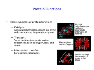
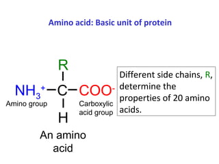
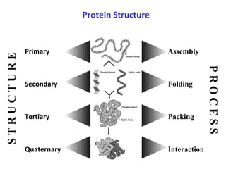
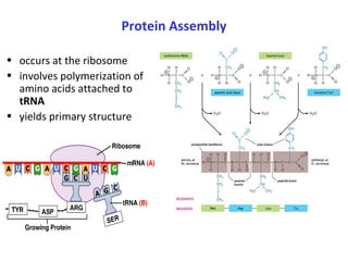
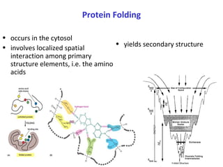
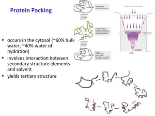
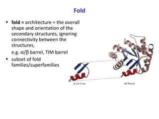
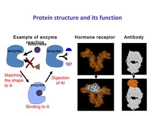
Proteins have three main functions: catalysis, transport, and information transfer. They are made of amino acids that polymerize and fold into complex three-dimensional structures, from primary to quaternary levels, that determine their unique functions. Protein structure and function are closely linked, as the structure allows proteins to bind specifically to other molecules and carry out reactions.
Tertiary Structure basically of Hydrophobic interactions, (interactions in side chains), hydrogen bonding, salt bridges, Vander Waals interactions.
e.g. Globular proteins & Fibrous Proteins
Folding depends upon sequence of Amino Acids not the Composition. Folding starts with the secondary structure and ends at quaternary structure.
Denaturation occur at secondary, tertiary & quaternary level but not at primary level.
Gives in detail primary, secondary, tertiary and Quaternary structure of proteins. Gives classification of secondary structure: alpha helix, beta pleated sheet and different types of tight turns and explains most commonly found tight turn in proteins i.e. beta turn. Briefs about the Ramachandran plot of proteins, dihedral or torsion angles and explains why glycine and proline act as alpha helix breakers. Explains tertiary structure of proteins and different covalent and non covalent bonds in the tertiary structure and relative importance of these bonding interactions. Details about the quaternary structure of proteins and explains why hemoglobin is a quaternary protein and insulin is not.
I have tried to make a precise presentation on protein transport, targeting and sorting into organelle's other than nucleus. Hope this might help you. Comments are welcome.
Introduction-Cell wall and functions
Gram +ve and -ve cell wall
Bacterial cell wall - structure
Peptidoglycan-Composition and Structure
Types of polysaccharidesBacterial cell wall
Functions of polysaccharides in Bacterial cell wall
This presentation gives an overview of Lipid Rafts, how it was discovered, its importance and the future research in this area,Feel free to comment and ask any questions
SDS-Polyacrylamide Gel Electrophoresis
What is SDS?
Preparation of Gel
Process of SDS-PAGE
Visualization of protein bands
SDS-PAGE is differentiated into two systems.
*continuous sds-page
*discontinuous sds-page.
Polyacrylamide is used to form a gel, a matrix of a pores which allow the molecules migrate at different rates.
Negatively charged detergent sodium dodecyl sulfate.
Used to denature and linearize the proteins
Coated the proteins with negatively charged.
SDS-page is a technique that used to separate proteins according to their molecular size through the gel.
Proteins are unfolded and migrate from cathode to anode terminal at different rates.
Molecular weight is determined by compare the result with a standard curve of relative motility of standard proteins.
Visualizes the band under UV light.
Types of stains;
Coomassie Blue;
* Coomassie Brilliant Blue staining The Coomassie dyes R-250 and G-250 bind to proteins stoichiometrically through their sulfonic acid groups.
* . The interactions between dye and protein are Van der Waals and ionic. The sulfonic acid groups interact with positive amine groups. Therefore coomassie dye binds to wide range of proteins.
* Limited to ~100ng of protein.
Silver stain;
*most sensitive test
*detection limit 0.1-1.0ng of protein
The size of pores is determined by the concentration of acrylamide.
The higher the concentration, the smaller the size of pores.
Discontinuos sds-page consist of two different gels.
*stacking gel -4%of acrylamide
*separating gel-range from 5-15% of acrylamide.
Presentation given by Dr. Karthikeyan at Department of Biochemistry, Maulana Azad Medical College.
Addition:
There are certain proteins which are degraded by proteasome without ubiquitin tag. one such example is ornithine decarboxylase - rate limiting enzyme of polyamine synthesis.
Proteins depending upon their physical and chemical structure and location inside the cell, they perform various functions. Proteins are grouped as follows, based on their metabolic function they perform.
الروابط التي تعمل على تثبيت أوضاع السلسلة الببتيدية
The disulphide (-s-s-) bond
Electrostatic interaction
Hydrogen bond
Van Der Waals Interaction
In this presentation i have explained about all the super secondary structure their types and their functions . The ppt has been made in such a way that it will clear out our basic concepts first and then it will go higher. I hope you like it
Proteins are naturally occurring polymers made up of amino acids and linked together by peptide bonds.
Proteins are the most abundant organic molecules in the living system.
The term "protein" is derived from the Greek word proteios, meaning holding the first place.
These are nitrogenous organic compounds that have large molecules weight of one or more long chains of amino acids.
Proteins are made from 20 ɑ-amino acids. (chains of amino acids)
A single unit of amino acid is known as a monomer. When many monomers combine together, they form polymers.
RECOMBINATION MOLECULAR BIOLOGY PPT UPDATED new.pptxSabahat Ali
This ppt is about recombination and where it occurs. Types of recombination and models of recombination along with many factors in prokaryotic and eukaryotic recombination
Seminar of U.V. Spectroscopy by SAMIR PANDASAMIR PANDA
Spectroscopy is a branch of science dealing the study of interaction of electromagnetic radiation with matter.
Ultraviolet-visible spectroscopy refers to absorption spectroscopy or reflect spectroscopy in the UV-VIS spectral region.
Ultraviolet-visible spectroscopy is an analytical method that can measure the amount of light received by the analyte.
Multi-source connectivity as the driver of solar wind variability in the heli...Sérgio Sacani
The ambient solar wind that flls the heliosphere originates from multiple
sources in the solar corona and is highly structured. It is often described
as high-speed, relatively homogeneous, plasma streams from coronal
holes and slow-speed, highly variable, streams whose source regions are
under debate. A key goal of ESA/NASA’s Solar Orbiter mission is to identify
solar wind sources and understand what drives the complexity seen in the
heliosphere. By combining magnetic feld modelling and spectroscopic
techniques with high-resolution observations and measurements, we show
that the solar wind variability detected in situ by Solar Orbiter in March
2022 is driven by spatio-temporal changes in the magnetic connectivity to
multiple sources in the solar atmosphere. The magnetic feld footpoints
connected to the spacecraft moved from the boundaries of a coronal hole
to one active region (12961) and then across to another region (12957). This
is refected in the in situ measurements, which show the transition from fast
to highly Alfvénic then to slow solar wind that is disrupted by the arrival of
a coronal mass ejection. Our results describe solar wind variability at 0.5 au
but are applicable to near-Earth observatories.
Earliest Galaxies in the JADES Origins Field: Luminosity Function and Cosmic ...Sérgio Sacani
We characterize the earliest galaxy population in the JADES Origins Field (JOF), the deepest
imaging field observed with JWST. We make use of the ancillary Hubble optical images (5 filters
spanning 0.4−0.9µm) and novel JWST images with 14 filters spanning 0.8−5µm, including 7 mediumband filters, and reaching total exposure times of up to 46 hours per filter. We combine all our data
at > 2.3µm to construct an ultradeep image, reaching as deep as ≈ 31.4 AB mag in the stack and
30.3-31.0 AB mag (5σ, r = 0.1” circular aperture) in individual filters. We measure photometric
redshifts and use robust selection criteria to identify a sample of eight galaxy candidates at redshifts
z = 11.5 − 15. These objects show compact half-light radii of R1/2 ∼ 50 − 200pc, stellar masses of
M⋆ ∼ 107−108M⊙, and star-formation rates of SFR ∼ 0.1−1 M⊙ yr−1
. Our search finds no candidates
at 15 < z < 20, placing upper limits at these redshifts. We develop a forward modeling approach to
infer the properties of the evolving luminosity function without binning in redshift or luminosity that
marginalizes over the photometric redshift uncertainty of our candidate galaxies and incorporates the
impact of non-detections. We find a z = 12 luminosity function in good agreement with prior results,
and that the luminosity function normalization and UV luminosity density decline by a factor of ∼ 2.5
from z = 12 to z = 14. We discuss the possible implications of our results in the context of theoretical
models for evolution of the dark matter halo mass function.
Richard's entangled aventures in wonderlandRichard Gill
Since the loophole-free Bell experiments of 2020 and the Nobel prizes in physics of 2022, critics of Bell's work have retreated to the fortress of super-determinism. Now, super-determinism is a derogatory word - it just means "determinism". Palmer, Hance and Hossenfelder argue that quantum mechanics and determinism are not incompatible, using a sophisticated mathematical construction based on a subtle thinning of allowed states and measurements in quantum mechanics, such that what is left appears to make Bell's argument fail, without altering the empirical predictions of quantum mechanics. I think however that it is a smoke screen, and the slogan "lost in math" comes to my mind. I will discuss some other recent disproofs of Bell's theorem using the language of causality based on causal graphs. Causal thinking is also central to law and justice. I will mention surprising connections to my work on serial killer nurse cases, in particular the Dutch case of Lucia de Berk and the current UK case of Lucy Letby.
Introduction:
RNA interference (RNAi) or Post-Transcriptional Gene Silencing (PTGS) is an important biological process for modulating eukaryotic gene expression.
It is highly conserved process of posttranscriptional gene silencing by which double stranded RNA (dsRNA) causes sequence-specific degradation of mRNA sequences.
dsRNA-induced gene silencing (RNAi) is reported in a wide range of eukaryotes ranging from worms, insects, mammals and plants.
This process mediates resistance to both endogenous parasitic and exogenous pathogenic nucleic acids, and regulates the expression of protein-coding genes.
What are small ncRNAs?
micro RNA (miRNA)
short interfering RNA (siRNA)
Properties of small non-coding RNA:
Involved in silencing mRNA transcripts.
Called “small” because they are usually only about 21-24 nucleotides long.
Synthesized by first cutting up longer precursor sequences (like the 61nt one that Lee discovered).
Silence an mRNA by base pairing with some sequence on the mRNA.
Discovery of siRNA?
The first small RNA:
In 1993 Rosalind Lee (Victor Ambros lab) was studying a non- coding gene in C. elegans, lin-4, that was involved in silencing of another gene, lin-14, at the appropriate time in the
development of the worm C. elegans.
Two small transcripts of lin-4 (22nt and 61nt) were found to be complementary to a sequence in the 3' UTR of lin-14.
Because lin-4 encoded no protein, she deduced that it must be these transcripts that are causing the silencing by RNA-RNA interactions.
Types of RNAi ( non coding RNA)
MiRNA
Length (23-25 nt)
Trans acting
Binds with target MRNA in mismatch
Translation inhibition
Si RNA
Length 21 nt.
Cis acting
Bind with target Mrna in perfect complementary sequence
Piwi-RNA
Length ; 25 to 36 nt.
Expressed in Germ Cells
Regulates trnasposomes activity
MECHANISM OF RNAI:
First the double-stranded RNA teams up with a protein complex named Dicer, which cuts the long RNA into short pieces.
Then another protein complex called RISC (RNA-induced silencing complex) discards one of the two RNA strands.
The RISC-docked, single-stranded RNA then pairs with the homologous mRNA and destroys it.
THE RISC COMPLEX:
RISC is large(>500kD) RNA multi- protein Binding complex which triggers MRNA degradation in response to MRNA
Unwinding of double stranded Si RNA by ATP independent Helicase
Active component of RISC is Ago proteins( ENDONUCLEASE) which cleave target MRNA.
DICER: endonuclease (RNase Family III)
Argonaute: Central Component of the RNA-Induced Silencing Complex (RISC)
One strand of the dsRNA produced by Dicer is retained in the RISC complex in association with Argonaute
ARGONAUTE PROTEIN :
1.PAZ(PIWI/Argonaute/ Zwille)- Recognition of target MRNA
2.PIWI (p-element induced wimpy Testis)- breaks Phosphodiester bond of mRNA.)RNAse H activity.
MiRNA:
The Double-stranded RNAs are naturally produced in eukaryotic cells during development, and they have a key role in regulating gene expression .
Cancer cell metabolism: special Reference to Lactate PathwayAADYARAJPANDEY1
Normal Cell Metabolism:
Cellular respiration describes the series of steps that cells use to break down sugar and other chemicals to get the energy we need to function.
Energy is stored in the bonds of glucose and when glucose is broken down, much of that energy is released.
Cell utilize energy in the form of ATP.
The first step of respiration is called glycolysis. In a series of steps, glycolysis breaks glucose into two smaller molecules - a chemical called pyruvate. A small amount of ATP is formed during this process.
Most healthy cells continue the breakdown in a second process, called the Kreb's cycle. The Kreb's cycle allows cells to “burn” the pyruvates made in glycolysis to get more ATP.
The last step in the breakdown of glucose is called oxidative phosphorylation (Ox-Phos).
It takes place in specialized cell structures called mitochondria. This process produces a large amount of ATP. Importantly, cells need oxygen to complete oxidative phosphorylation.
If a cell completes only glycolysis, only 2 molecules of ATP are made per glucose. However, if the cell completes the entire respiration process (glycolysis - Kreb's - oxidative phosphorylation), about 36 molecules of ATP are created, giving it much more energy to use.
IN CANCER CELL:
Unlike healthy cells that "burn" the entire molecule of sugar to capture a large amount of energy as ATP, cancer cells are wasteful.
Cancer cells only partially break down sugar molecules. They overuse the first step of respiration, glycolysis. They frequently do not complete the second step, oxidative phosphorylation.
This results in only 2 molecules of ATP per each glucose molecule instead of the 36 or so ATPs healthy cells gain. As a result, cancer cells need to use a lot more sugar molecules to get enough energy to survive.
introduction to WARBERG PHENOMENA:
WARBURG EFFECT Usually, cancer cells are highly glycolytic (glucose addiction) and take up more glucose than do normal cells from outside.
Otto Heinrich Warburg (; 8 October 1883 – 1 August 1970) In 1931 was awarded the Nobel Prize in Physiology for his "discovery of the nature and mode of action of the respiratory enzyme.
WARNBURG EFFECT : cancer cells under aerobic (well-oxygenated) conditions to metabolize glucose to lactate (aerobic glycolysis) is known as the Warburg effect. Warburg made the observation that tumor slices consume glucose and secrete lactate at a higher rate than normal tissues.
2. Protein Functions
• Three examples of protein functions
– Catalysis:
Almost all chemical reactions in a living
cell are catalyzed by protein enzymes.
– Transport:
Some proteins transports various
substances, such as oxygen, ions, and
so on.
– Information transfer:
For example, hormones.
Alcohol
dehydrogenase
oxidizes
alcohols to
aldehydes or
ketones
Haemoglobin
carries oxygen
Insulin controls
the amount of
sugar in the
blood
3. Amino acid: Basic unit of protein
COO-
NH3
+
C
R
H
An amino
acid
Different side chains, R,
determine the
properties of 20 amino
acids.
Amino group Carboxylic
acid group
4. Each protein has a unique structure!
Amino acid sequence
NLKTEWPELVGKSVEEAK
KVILQDKPEAQIIVLPVGTI
VTMEYRIDRVRLFVDKLD
Folding!
6. Protein Assembly
• occurs at the ribosome
• involves polymerization of
amino acids attached to
tRNA
• yields primary structure
7. Primary Structure
• linear
• ordered
• 1 dimensional
• sequence of amino acid
polymer
• by convention, written
from amino end to
carboxyl end
• a perfectly linear amino
acid polymer is neither
functional nor
energetically favorable
folding!
primary structure of human insulin
CHAIN 1: GIVEQ CCTSI CSLYQ LENYC N
CHAIN 2: FVNQH LCGSH LVEAL YLVCG ERGFF YTPKT
8. Protein Folding
• yields secondary structure
• occurs in the cytosol
• involves localized spatial
interaction among primary
structure elements, i.e. the amino
acids
9. Secondary Structure
• non-linear
• 3 dimensional
• localized to regions of an
amino acid chain
• formed and stabilized by
hydrogen bonding,
electrostatic and van der
Waals interactions
10. Protein Packing
• occurs in the cytosol (~60% bulk
water, ~40% water of
hydration)
• involves interaction between
secondary structure elements
and solvent
• yields tertiary structure
12. Protein Interaction
• occurs in the cytosol, in close proximity to other folded and
packed proteins
• involves interaction among tertiary structure elements of
separate polymer chains
13. Class/Motif
• class = secondary structure
composition,
e.g. all α, all β, α/β , α+β
• motif = small, specific combinations of
secondary structure elements,
e.g. β-α-β loop
• both subset of fold
α/
β
14. Fold
• fold = architecture = the overall
shape and orientation of the
secondary structures, ignoring
connectivity between the
structures,
e.g. α/β barrel, TIM barrel
• subset of fold
families/superfamilies
15. Fold families/Superfamilies
• fold families = categorization that
takes into account topology and
previous subsets as well as
empirical/biological properties, e.g.
flavodoxin
• superfamilies = in addition to fold
families, includes
evolutionary/ancestral properties
CLASS: α+β
FOLD: sandwich
FOLD FAMILY: flavodoxin
16. Hierarchical nature of protein structure
Primary structure (Amino acid sequence)
↓
Secondary structure ( α-helix, β-sheet )
↓
Tertiary structure ( Three-dimensional structure
formed by assembly of secondary structures )
↓
Quaternary structure ( Structure formed by more
than one polypeptide chains )
17. Protein structure and its function
enzyme A
B
A
Binding to A
Digestion
of A!
enzyme
Matching
the shape
to A
Hormone receptor AntibodyExample of enzyme
reaction
enzyme
substrates
18. Summary
• Proteins are key players in our living systems.
• Proteins are polymers consisting of 20 kinds of amino acids.
• Each protein folds into a unique three-dimensional structure
defined by its amino acid sequence.
• Protein structure has a hierarchical nature.
• Protein structure is closely related to its function.
• Protein structure prediction is a grand challenge of
computational biology.