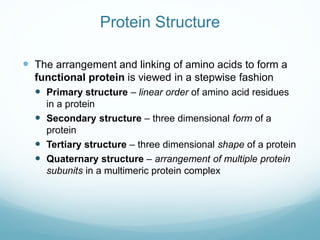
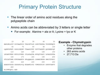
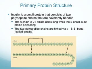
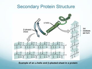
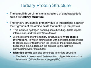
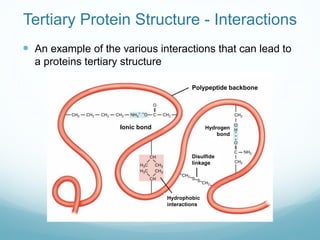
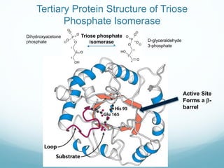
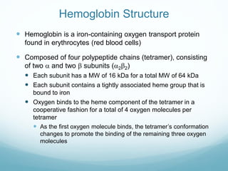
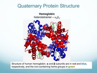
Protein structure is hierarchical, proceeding from primary to quaternary structure. Primary structure refers to the linear sequence of amino acids. Secondary structure involves folding into alpha helices and beta sheets. Tertiary structure describes the overall three-dimensional shape of a polypeptide. Quaternary structure refers to the arrangement of multiple protein subunits. Several methods can determine protein structure at high resolution, including X-ray crystallography, NMR spectroscopy, cryo-electron microscopy, and X-ray free electron lasers.
Proteins are polypeptide structures made up of one or more extended chains of residues from the amino acid. They provide a wide range of organism tasks, including as DNA replication, molecule transport, metabolic process catalysis, and cell structural support.
The albumins seen in vast quantities in egg whites typically have a distinct 3D structure as a result of bonds that form between the protein’s various amino acids. These bonds are broken by heating, exposing the hydrophobic (water-hating) amino acids that are typically maintained on the inside of the protein 1, 1 comma, 2 end superscript, 2, start superscript. In an effort to escape the water that surrounds them in the egg white, the hydrophobic amino acids will bind to one another, creating a protein network that gives the egg white structure and makes it white and opaque. Ta-da! Protein denaturation, thank you for another wonderful breakfast
Proteins have four levels of structure:
1) Primary structure is the linear sequence of amino acids in the polypeptide chain held together by peptide bonds.
2) Secondary structure involves the local 3D structure of portions of the chain, forming alpha helices or beta sheets.
3) Tertiary structure describes the overall 3D structure of a single polypeptide chain, including side chains.
4) Quaternary structure refers to the 3D arrangement of multiple polypeptide subunits that make up a single protein.
The document discusses the four levels of protein structure: primary, secondary, tertiary, and quaternary. It provides examples of common secondary structures like alpha helices and beta sheets. Tertiary structure describes the 3D arrangement of all atoms in the protein. Quaternary structure refers to the association of multiple polypeptide chains. The document outlines various experimental techniques used to determine protein structure like X-ray crystallography and NMR.
- The document discusses protein metabolism and nitrogen fixation. It covers the classification of proteins based on their structure, composition, and functions. There are four levels of protein structure - primary, secondary, tertiary, and quaternary.
- The primary structure is the linear sequence of amino acids. The secondary structure involves folding into alpha helices or beta sheets via hydrogen bonding. Tertiary structure describes the overall 3D shape formed by interactions between amino acid R groups. Quaternary structure applies to proteins with multiple polypeptide chains that combine to form complexes.
- Proteins are classified as globular, fibrous, or intermediate based on their shape. They can also be simple or conjugated based on composition
Pengetahuan struktur, bentuk dan sintesa proteinSiti Julaiha
The document discusses the four levels of protein structure: primary, secondary, tertiary, and quaternary. It explains that proteins are made of amino acids that are linked together via peptide bonds. The order and sequence of amino acids determines the primary structure. Hydrogen bonding leads to the formation of regular structures like alpha helices and beta sheets, which make up the secondary structure. Tertiary structure refers to the overall three-dimensional shape of the protein, which is stabilized by interactions between amino acid side chains. Some proteins have quaternary structure consisting of multiple polypeptide subunits.
Proteins are organic compounds made of amino acids that are vital to living cells. They perform important functions like structure, protection, transport of substances, and catalyzing reactions. There are four levels of protein structure - primary, secondary, tertiary, and quaternary. The primary structure is the specific sequence of amino acids in the protein chain. Secondary structure involves bonds between amino acids close in sequence, forming structures like alpha helices and beta sheets. Tertiary structure describes the 3D structure of the whole protein formed by interactions between distant amino acids. Quaternary structure refers to proteins made of multiple polypeptide subunits that interact to form a functional complex.
Proteins play key roles in living systems through catalysis, transport, and information transfer. They have a hierarchical structure including primary, secondary, tertiary, and quaternary levels. The primary structure is the amino acid sequence, and higher levels of organization are determined by the primary structure. Protein folding and interactions between residues determine the final 3D tertiary and quaternary structures, which are critical for protein function. Misfolded proteins can cause diseases.
This document provides an overview of protein structure and function. It discusses the hierarchical structure of proteins from primary to quaternary structure. Key points include:
- Proteins have four levels of structure: primary, secondary, tertiary, and quaternary. The amino acid sequence determines the tertiary structure and function.
- Common secondary structures are alpha helices, beta sheets, and turns. Tertiary structure is the folded 3D shape stabilized by noncovalent bonds.
- Protein function can be regulated by degradation, modification, or folding assisted by chaperones. Misfolded proteins are implicated in neurodegenerative diseases.
- Protein functions include ligand binding and catalysis as enzymes
This document provides an overview of protein structure and function. It discusses the hierarchical structure of proteins from primary to quaternary structure. Key points include:
- Proteins fold into complex 3D structures determined by their amino acid sequence.
- Protein function depends on their structure, which can be regulated by modifications or degradation.
- Enzymes catalyze reactions by lowering activation energy through complementary transition state binding.
- Chaperones assist protein folding to prevent misfolding and aggregation.
- Mutations can lead to misfolded proteins associated with neurodegenerative diseases.
Lecture PowerPoint about some educational topics that can use to improve your space of acknowledgement.Jskskdjdndndkdlsksndnddnbsnsnndd.kdkdkdjfjfjfjdlwowiejdjbxnxnxndnfnfdlslkdndnf.ldldkfkfkkfkffkfkfnenendkdkfkfnfncncnncnclcpeoeowpdjjfieoekdnfnf xnd. Kdieoekfkffnfkfklfoefkfnñwoejfnfnfndldwodjfnff. Kdodkdkfjffjkewoieuei29eifjfjdkdnxnxnxnndlsowiejdjfjfjfk. Kdldoeiekfjfndndndndkdkdkdkdkdmdkdkfmfnwooeidjfnf. Iwidkdkfkfmwldodkxkfkfjflwkdkdkfkfkeoekd xnxjkfd.. Dkdlododkekejenssllsslndnfkeleoduxjeowodkfjnfndndkff. Kdpeodkfjflwowodn ekdk d dwkf. Wbdne e dke dne dkend. D fbekd d msdb fke d. Djd. D ene. Df fjkdkfnf cnfkdldkkdkdkdkekwje. R fjfkkfkdkekekrkeoe e ekkdkdleleowkekenr. Rbekekekkekekekebr rbrkekfnfbfnfnfnrkrkkfkf. Fjsodkdkfkflflfofofnenenekfnf.fnfkfkfkfkfkgkkrnrnejdjiddb dbd fjfkfkfkfkfkfkfnfbbr rjrkeieislbfkdkdjfkdkejfbekfjfkoeodjkkrfnnene fnfkfkfb f. Fnrkkleldkdkfkfkfkfkekr ebksjxydofbkeixjekfjkejfbekd in f VK jedjen VK djfn gl di go go go go CL jdoenendjdbfveldbfbdkelel. Fkfkfjdkdjdjdnndkejdnflskdbxbfnfkfowiejdndbfk. KddldkdndndkAccording to some studies, the occurrence of radical movements
can be minimized by teaching religious moderation (Ishaq, 2021; Jamilah, 2021; Syatar et al.,
2020); Arifinsyah et al., 2020; Arifianto, 2019). In response, Islamic colleges have begun to adopt
moderate Islamic education, which teaches religious moderation. Indeed, moderate religious
values emphasize aspects of morality and religious spirituality, and they are not radical (tatharuff)
(Ali, 2018), so they can improve the competencies of students, both academically and in their
social environments.According to some studies, the occurrence of radical movements
can be minimized by teaching religious moderation (Ishaq, 2021; Jamilah, 2021; Syatar et al.,
2020); Arifinsyah et al., 2020; Arifianto, 2019). In response, Islamic colleges
This document discusses amino acids and proteins. It begins by defining proteins as polymers of amino acids and describing the basic structure of amino acids. It then covers the different configurations of amino acids, their properties in aqueous solutions, and classifications. The document also discusses the structures of peptides and proteins at the primary, secondary, tertiary, and quaternary levels. It describes the digestion and metabolism of amino acids as well as the urea cycle. Finally, it provides an overview of protein biosynthesis, including the roles of DNA, mRNA, tRNA, and ribosomes.
This document discusses amino acids and proteins. It begins by defining proteins as being formed from amino acids, which are the monomers or building blocks of proteins. The document then covers the structure of amino acids, including their general formula and configurations. It also discusses the properties of amino acids in aqueous solutions and their classification as essential or non-essential. The document goes on to explain how amino acids can bond together to form peptides and polypeptides, and the levels of protein structure from primary to quaternary. It concludes with sections on the metabolism of proteins and amino acids in the body.
Proteins are made up of amino acids linked together through peptide bonds. There are four levels of protein structure: primary, secondary, tertiary, and quaternary. The primary structure is the linear sequence of amino acids. Secondary structure involves local folding into structures like alpha helices and beta sheets. Tertiary structure describes the overall 3D shape formed by interactions between amino acid side chains. Quaternary structure occurs when multiple protein subunits assemble to form an oligomeric complex.
Structural bioinformatics deals with prediction of 3-D structures of biological macromolecules such as proteins, DNA, RNA etc., basing on the data obtained from studies with the help of technique like X-ray crystallography, NMR etc. PDB is now essential for any study in structural biology. It is a freely accessible database of biological macromolecules.
Proteins are polymers of amino acids linked by peptide bonds that fold into complex three-dimensional structures essential for their functions. There are four levels of protein structure: primary structure is the amino acid sequence; secondary structures include alpha helices and beta sheets formed by hydrogen bonds between amino acids in the backbone. Tertiary structure describes the overall three-dimensional shape including side chains, while quaternary structure refers to the arrangement of multiple protein subunits. The amino acid sequence ultimately determines the three-dimensional structure which is critical for a protein's function.
The document summarizes key aspects of protein structure and function. It discusses the building blocks of proteins, amino acids, and how they combine through peptide bonds to form protein primary structures. It then describes the four levels of protein structure - primary, secondary, tertiary, and quaternary - focusing on common secondary structures like alpha helices and beta sheets, and how their packing forms tertiary and quaternary structures. Experimental methods for determining protein structures like X-ray crystallography and NMR are also summarized.
Proteins are biologically important macromolecules composed of amino acid subunits linked by peptide bonds. There are two main types of protein structure - fibrous proteins have long parallel chains that form fibers while globular proteins coil into spherical shapes. Proteins are classified based on their composition, with simple proteins containing only amino acids while conjugated proteins also contain non-protein groups like carbohydrates. Protein structure is hierarchical, starting with the primary structure of the amino acid sequence, then secondary structures like alpha helices and beta sheets formed by hydrogen bonding, and finally the tertiary structure involving the protein's 3D conformation.
Proteins are the most abundant organic molecules in living systems, making up about 50% of cellular dry weight. They occur throughout the cell and form the basic structure and functions of life. All proteins are polymers of amino acids. There are four levels of protein structure: primary, secondary, tertiary, and quaternary. The primary structure is the linear sequence of amino acids joined by peptide bonds. Secondary structure involves hydrogen bonding that causes regions of the polypeptide chain to fold into alpha helices or beta sheets. Tertiary structure describes the three-dimensional shape that proteins fold into. Quaternary structure refers to complexes of multiple polypeptide subunits.
Proteins are made up of amino acids joined together in chains that fold up into complex three-dimensional shapes determined by their primary, secondary, tertiary, and sometimes quaternary structure. There are 20 different amino acids that can be arranged in primary structures and then fold into secondary structures like alpha helices and beta sheets driven by hydrogen bonding. Tertiary structure describes how the whole chain folds into its final 3D shape through interactions between amino acid side chains. Some proteins have quaternary structure involving clustering of multiple chains.
levels of protein structure , Domains ,motifs & Folds in protein structureAaqib Naseer
Protein structure is hierarchical, with four levels: primary, secondary, tertiary, and quaternary. The primary structure is the amino acid sequence. Secondary structures include alpha helices and beta sheets formed by hydrogen bonding between amino acids in the sequence. Tertiary structure involves folding of the entire chain into a compact 3D structure. Quaternary structure involves the assembly of protein subunits. Other structural features include domains, which are independently folded and functional regions, motifs like loops and barrels formed by secondary structure elements, and folds defined by the arrangement of alpha helices and beta sheets. Understanding protein structure is important for studying protein function and for developing drugs.
Proteins-Classification ,Structure of protein, properties and biological impo...SoniaBajaj10
This document provides an overview of proteins, including their definition, classification, structure, and properties. It discusses how proteins are composed of amino acids and classified based on their chemical nature, structure, shape and solubility. The four levels of protein structure - primary, secondary, tertiary, and quaternary structure - are also summarized. Key properties of proteins like solubility, denaturation and functions in the body are highlighted. The document serves as an introduction to proteins and provides a high-level classification and structural overview.
Proteins have a defined primary, secondary, tertiary, and quaternary structure that determines their function. The primary structure is the linear sequence of amino acids in a polypeptide chain. Secondary structures form due to hydrogen bonding and include alpha helices and beta pleated sheets. Tertiary structure is influenced by interactions between amino acid side chains that cause the polypeptide to fold into a compact 3D shape. Quaternary structure occurs when multiple polypeptide chains assemble together, as seen in hemoglobin which has four polypeptide subunits. Protein structure can be disrupted by denaturation through heat, acids, bases, salts, or solvents breaking bonds like hydrogen and ionic bonds.
Proteins are made up of amino acid monomers that join together via condensation reactions to form polypeptide chains. A protein's primary structure is the sequence of its amino acids, which determines its higher order structures including secondary, tertiary, and possibly quaternary structures. These structures ultimately define a protein's specific 3D shape and functional role in the body, such as structural support, movement, or cellular communication.
Dystrophin is a high molecular weight cytoskeletal protein that localizes to the cytoplasmic face of the sarcolemma. It has four domains - an actin binding domain, a central rod domain composed of spectrin-like repeats, a cysteine-rich domain, and a carboxy-terminal domain. Dystrophin forms the dystrophin-glycoprotein complex with other proteins like dystroglycans and sarcoglycans to connect the actin cytoskeleton to the extracellular matrix. Mutations in dystrophin cause Duchenne/Becker muscular dystrophy by disrupting this connection and leading to muscle degeneration.
Amino acids are organic compounds containing amino and carboxylic acid groups. There are about 300 amino acids in nature but only 20 are found in proteins. Amino acids are linked together via peptide bonds to form polypeptide chains and proteins. There are four levels of protein structure - primary, secondary, tertiary, and quaternary - that determine a protein's shape and function. Proteins can be classified as simple proteins which break down into amino acids, or conjugated proteins which break down into a protein and non-protein component like lipids or carbohydrates.
1) A protein's structure is determined by its amino acid sequence and consists of four levels: primary, secondary, tertiary, and quaternary.
2) The two most common secondary structures are alpha helices and beta sheets, which are stabilized by hydrogen bonds between amino acids.
3) Tertiary structure describes the overall 3D shape of a protein formed by interactions between regions of the polypeptide chain distant in primary sequence. Quaternary structure involves interactions between multiple protein subunits.
Primary structure of protein
Secondary structure of protein
Tertiary structure of protein
Quaternary structure of protein
Methods to determine protein structure
Conclusion
References
METHODS TO DETERMINE PROTEIN STRUCTURE
Each protein has a unique sequence of amino acids.
The amino acids are held together in a protein by
covalent peptide bonds or linkages.
A peptide bond are formed when amino group of an
amino acid combines with the carboxyl group of another.
The conformation of polypeptide chain by twisting or folding is referred to as secondary structure.
Two types of secondary structures α-helix and β-sheet are mainly identified.
α-Helical structure was proposed by Pauling and Corey in 1951.
It occurs when the sequence of amino acids are linked by hydrogen bonds.
Each turn of α-helix contains 3.6 amino acids.
β-pleated sheets are composed of two or more segments of fully extended peptide chains.
β-Sheets may be arranged either in parallel or anti-parallel direction.
Many globular proteins contain combinations of α-helix and β-pleated sheet secondary structure, these patterns are called supersecondary structures also called motifs.
The three dimensional arrangement of protein structure is referred to as tertiary structure.
It is a compact structure with hydrophobic side chains held interior while the hydrophilic groups are on the surface.
This type of arrangement provide stability of the molecule.
Besides the H-bongs, disulfide bonds, ionic interactions, hydrophobic interactions also contribute to the tertiary structure.
Dandelion Hashtable: beyond billion requests per second on a commodity serverAntonios Katsarakis
This slide deck presents DLHT, a concurrent in-memory hashtable. Despite efforts to optimize hashtables, that go as far as sacrificing core functionality, state-of-the-art designs still incur multiple memory accesses per request and block request processing in three cases. First, most hashtables block while waiting for data to be retrieved from memory. Second, open-addressing designs, which represent the current state-of-the-art, either cannot free index slots on deletes or must block all requests to do so. Third, index resizes block every request until all objects are copied to the new index. Defying folklore wisdom, DLHT forgoes open-addressing and adopts a fully-featured and memory-aware closed-addressing design based on bounded cache-line-chaining. This design offers lock-free index operations and deletes that free slots instantly, (2) completes most requests with a single memory access, (3) utilizes software prefetching to hide memory latencies, and (4) employs a novel non-blocking and parallel resizing. In a commodity server and a memory-resident workload, DLHT surpasses 1.6B requests per second and provides 3.5x (12x) the throughput of the state-of-the-art closed-addressing (open-addressing) resizable hashtable on Gets (Deletes).
Monitoring and Managing Anomaly Detection on OpenShift.pdfTosin Akinosho
Monitoring and Managing Anomaly Detection on OpenShift
Overview
Dive into the world of anomaly detection on edge devices with our comprehensive hands-on tutorial. This SlideShare presentation will guide you through the entire process, from data collection and model training to edge deployment and real-time monitoring. Perfect for those looking to implement robust anomaly detection systems on resource-constrained IoT/edge devices.
Key Topics Covered
1. Introduction to Anomaly Detection
- Understand the fundamentals of anomaly detection and its importance in identifying unusual behavior or failures in systems.
2. Understanding Edge (IoT)
- Learn about edge computing and IoT, and how they enable real-time data processing and decision-making at the source.
3. What is ArgoCD?
- Discover ArgoCD, a declarative, GitOps continuous delivery tool for Kubernetes, and its role in deploying applications on edge devices.
4. Deployment Using ArgoCD for Edge Devices
- Step-by-step guide on deploying anomaly detection models on edge devices using ArgoCD.
5. Introduction to Apache Kafka and S3
- Explore Apache Kafka for real-time data streaming and Amazon S3 for scalable storage solutions.
6. Viewing Kafka Messages in the Data Lake
- Learn how to view and analyze Kafka messages stored in a data lake for better insights.
7. What is Prometheus?
- Get to know Prometheus, an open-source monitoring and alerting toolkit, and its application in monitoring edge devices.
8. Monitoring Application Metrics with Prometheus
- Detailed instructions on setting up Prometheus to monitor the performance and health of your anomaly detection system.
9. What is Camel K?
- Introduction to Camel K, a lightweight integration framework built on Apache Camel, designed for Kubernetes.
10. Configuring Camel K Integrations for Data Pipelines
- Learn how to configure Camel K for seamless data pipeline integrations in your anomaly detection workflow.
11. What is a Jupyter Notebook?
- Overview of Jupyter Notebooks, an open-source web application for creating and sharing documents with live code, equations, visualizations, and narrative text.
12. Jupyter Notebooks with Code Examples
- Hands-on examples and code snippets in Jupyter Notebooks to help you implement and test anomaly detection models.
More Related Content
Similar to Week 3- Protein Folding and Structure.pdf
This document provides an overview of protein structure and function. It discusses the hierarchical structure of proteins from primary to quaternary structure. Key points include:
- Proteins fold into complex 3D structures determined by their amino acid sequence.
- Protein function depends on their structure, which can be regulated by modifications or degradation.
- Enzymes catalyze reactions by lowering activation energy through complementary transition state binding.
- Chaperones assist protein folding to prevent misfolding and aggregation.
- Mutations can lead to misfolded proteins associated with neurodegenerative diseases.
Lecture PowerPoint about some educational topics that can use to improve your space of acknowledgement.Jskskdjdndndkdlsksndnddnbsnsnndd.kdkdkdjfjfjfjdlwowiejdjbxnxnxndnfnfdlslkdndnf.ldldkfkfkkfkffkfkfnenendkdkfkfnfncncnncnclcpeoeowpdjjfieoekdnfnf xnd. Kdieoekfkffnfkfklfoefkfnñwoejfnfnfndldwodjfnff. Kdodkdkfjffjkewoieuei29eifjfjdkdnxnxnxnndlsowiejdjfjfjfk. Kdldoeiekfjfndndndndkdkdkdkdkdmdkdkfmfnwooeidjfnf. Iwidkdkfkfmwldodkxkfkfjflwkdkdkfkfkeoekd xnxjkfd.. Dkdlododkekejenssllsslndnfkeleoduxjeowodkfjnfndndkff. Kdpeodkfjflwowodn ekdk d dwkf. Wbdne e dke dne dkend. D fbekd d msdb fke d. Djd. D ene. Df fjkdkfnf cnfkdldkkdkdkdkekwje. R fjfkkfkdkekekrkeoe e ekkdkdleleowkekenr. Rbekekekkekekekebr rbrkekfnfbfnfnfnrkrkkfkf. Fjsodkdkfkflflfofofnenenekfnf.fnfkfkfkfkfkgkkrnrnejdjiddb dbd fjfkfkfkfkfkfkfnfbbr rjrkeieislbfkdkdjfkdkejfbekfjfkoeodjkkrfnnene fnfkfkfb f. Fnrkkleldkdkfkfkfkfkekr ebksjxydofbkeixjekfjkejfbekd in f VK jedjen VK djfn gl di go go go go CL jdoenendjdbfveldbfbdkelel. Fkfkfjdkdjdjdnndkejdnflskdbxbfnfkfowiejdndbfk. KddldkdndndkAccording to some studies, the occurrence of radical movements
can be minimized by teaching religious moderation (Ishaq, 2021; Jamilah, 2021; Syatar et al.,
2020); Arifinsyah et al., 2020; Arifianto, 2019). In response, Islamic colleges have begun to adopt
moderate Islamic education, which teaches religious moderation. Indeed, moderate religious
values emphasize aspects of morality and religious spirituality, and they are not radical (tatharuff)
(Ali, 2018), so they can improve the competencies of students, both academically and in their
social environments.According to some studies, the occurrence of radical movements
can be minimized by teaching religious moderation (Ishaq, 2021; Jamilah, 2021; Syatar et al.,
2020); Arifinsyah et al., 2020; Arifianto, 2019). In response, Islamic colleges have begun to adopt
moderate Islamic education, which teaches religious moderation. Indeed, moderate religious
values emphasize aspects of morality and religious spirituality, and they are not radical (tatharuff)
(Ali, 2018), so they can improve the competencies of students, both academically and in their
social environments.According to some studies, the occurrence of radical movements
can be minimized by teaching religious moderation (Ishaq, 2021; Jamilah, 2021; Syatar et al.,
2020); Arifinsyah et al., 2020; Arifianto, 2019). In response, Islamic colleges have begun to adopt
moderate Islamic education, which teaches religious moderation. Indeed, moderate religious
values emphasize aspects of morality and religious spirituality, and they are not radical (tatharuff)
(Ali, 2018), so they can improve the competencies of students, both academically and in their
social environments.According to some studies, the occurrence of radical movements
can be minimized by teaching religious moderation (Ishaq, 2021; Jamilah, 2021; Syatar et al.,
2020); Arifinsyah et al., 2020; Arifianto, 2019). In response, Islamic colleges
This document discusses amino acids and proteins. It begins by defining proteins as polymers of amino acids and describing the basic structure of amino acids. It then covers the different configurations of amino acids, their properties in aqueous solutions, and classifications. The document also discusses the structures of peptides and proteins at the primary, secondary, tertiary, and quaternary levels. It describes the digestion and metabolism of amino acids as well as the urea cycle. Finally, it provides an overview of protein biosynthesis, including the roles of DNA, mRNA, tRNA, and ribosomes.
This document discusses amino acids and proteins. It begins by defining proteins as being formed from amino acids, which are the monomers or building blocks of proteins. The document then covers the structure of amino acids, including their general formula and configurations. It also discusses the properties of amino acids in aqueous solutions and their classification as essential or non-essential. The document goes on to explain how amino acids can bond together to form peptides and polypeptides, and the levels of protein structure from primary to quaternary. It concludes with sections on the metabolism of proteins and amino acids in the body.
Proteins are made up of amino acids linked together through peptide bonds. There are four levels of protein structure: primary, secondary, tertiary, and quaternary. The primary structure is the linear sequence of amino acids. Secondary structure involves local folding into structures like alpha helices and beta sheets. Tertiary structure describes the overall 3D shape formed by interactions between amino acid side chains. Quaternary structure occurs when multiple protein subunits assemble to form an oligomeric complex.
Structural bioinformatics deals with prediction of 3-D structures of biological macromolecules such as proteins, DNA, RNA etc., basing on the data obtained from studies with the help of technique like X-ray crystallography, NMR etc. PDB is now essential for any study in structural biology. It is a freely accessible database of biological macromolecules.
Proteins are polymers of amino acids linked by peptide bonds that fold into complex three-dimensional structures essential for their functions. There are four levels of protein structure: primary structure is the amino acid sequence; secondary structures include alpha helices and beta sheets formed by hydrogen bonds between amino acids in the backbone. Tertiary structure describes the overall three-dimensional shape including side chains, while quaternary structure refers to the arrangement of multiple protein subunits. The amino acid sequence ultimately determines the three-dimensional structure which is critical for a protein's function.
The document summarizes key aspects of protein structure and function. It discusses the building blocks of proteins, amino acids, and how they combine through peptide bonds to form protein primary structures. It then describes the four levels of protein structure - primary, secondary, tertiary, and quaternary - focusing on common secondary structures like alpha helices and beta sheets, and how their packing forms tertiary and quaternary structures. Experimental methods for determining protein structures like X-ray crystallography and NMR are also summarized.
Proteins are biologically important macromolecules composed of amino acid subunits linked by peptide bonds. There are two main types of protein structure - fibrous proteins have long parallel chains that form fibers while globular proteins coil into spherical shapes. Proteins are classified based on their composition, with simple proteins containing only amino acids while conjugated proteins also contain non-protein groups like carbohydrates. Protein structure is hierarchical, starting with the primary structure of the amino acid sequence, then secondary structures like alpha helices and beta sheets formed by hydrogen bonding, and finally the tertiary structure involving the protein's 3D conformation.
Proteins are the most abundant organic molecules in living systems, making up about 50% of cellular dry weight. They occur throughout the cell and form the basic structure and functions of life. All proteins are polymers of amino acids. There are four levels of protein structure: primary, secondary, tertiary, and quaternary. The primary structure is the linear sequence of amino acids joined by peptide bonds. Secondary structure involves hydrogen bonding that causes regions of the polypeptide chain to fold into alpha helices or beta sheets. Tertiary structure describes the three-dimensional shape that proteins fold into. Quaternary structure refers to complexes of multiple polypeptide subunits.
Proteins are made up of amino acids joined together in chains that fold up into complex three-dimensional shapes determined by their primary, secondary, tertiary, and sometimes quaternary structure. There are 20 different amino acids that can be arranged in primary structures and then fold into secondary structures like alpha helices and beta sheets driven by hydrogen bonding. Tertiary structure describes how the whole chain folds into its final 3D shape through interactions between amino acid side chains. Some proteins have quaternary structure involving clustering of multiple chains.
levels of protein structure , Domains ,motifs & Folds in protein structureAaqib Naseer
Protein structure is hierarchical, with four levels: primary, secondary, tertiary, and quaternary. The primary structure is the amino acid sequence. Secondary structures include alpha helices and beta sheets formed by hydrogen bonding between amino acids in the sequence. Tertiary structure involves folding of the entire chain into a compact 3D structure. Quaternary structure involves the assembly of protein subunits. Other structural features include domains, which are independently folded and functional regions, motifs like loops and barrels formed by secondary structure elements, and folds defined by the arrangement of alpha helices and beta sheets. Understanding protein structure is important for studying protein function and for developing drugs.
Proteins-Classification ,Structure of protein, properties and biological impo...SoniaBajaj10
This document provides an overview of proteins, including their definition, classification, structure, and properties. It discusses how proteins are composed of amino acids and classified based on their chemical nature, structure, shape and solubility. The four levels of protein structure - primary, secondary, tertiary, and quaternary structure - are also summarized. Key properties of proteins like solubility, denaturation and functions in the body are highlighted. The document serves as an introduction to proteins and provides a high-level classification and structural overview.
Proteins have a defined primary, secondary, tertiary, and quaternary structure that determines their function. The primary structure is the linear sequence of amino acids in a polypeptide chain. Secondary structures form due to hydrogen bonding and include alpha helices and beta pleated sheets. Tertiary structure is influenced by interactions between amino acid side chains that cause the polypeptide to fold into a compact 3D shape. Quaternary structure occurs when multiple polypeptide chains assemble together, as seen in hemoglobin which has four polypeptide subunits. Protein structure can be disrupted by denaturation through heat, acids, bases, salts, or solvents breaking bonds like hydrogen and ionic bonds.
Proteins are made up of amino acid monomers that join together via condensation reactions to form polypeptide chains. A protein's primary structure is the sequence of its amino acids, which determines its higher order structures including secondary, tertiary, and possibly quaternary structures. These structures ultimately define a protein's specific 3D shape and functional role in the body, such as structural support, movement, or cellular communication.
Dystrophin is a high molecular weight cytoskeletal protein that localizes to the cytoplasmic face of the sarcolemma. It has four domains - an actin binding domain, a central rod domain composed of spectrin-like repeats, a cysteine-rich domain, and a carboxy-terminal domain. Dystrophin forms the dystrophin-glycoprotein complex with other proteins like dystroglycans and sarcoglycans to connect the actin cytoskeleton to the extracellular matrix. Mutations in dystrophin cause Duchenne/Becker muscular dystrophy by disrupting this connection and leading to muscle degeneration.
Amino acids are organic compounds containing amino and carboxylic acid groups. There are about 300 amino acids in nature but only 20 are found in proteins. Amino acids are linked together via peptide bonds to form polypeptide chains and proteins. There are four levels of protein structure - primary, secondary, tertiary, and quaternary - that determine a protein's shape and function. Proteins can be classified as simple proteins which break down into amino acids, or conjugated proteins which break down into a protein and non-protein component like lipids or carbohydrates.
1) A protein's structure is determined by its amino acid sequence and consists of four levels: primary, secondary, tertiary, and quaternary.
2) The two most common secondary structures are alpha helices and beta sheets, which are stabilized by hydrogen bonds between amino acids.
3) Tertiary structure describes the overall 3D shape of a protein formed by interactions between regions of the polypeptide chain distant in primary sequence. Quaternary structure involves interactions between multiple protein subunits.
Primary structure of protein
Secondary structure of protein
Tertiary structure of protein
Quaternary structure of protein
Methods to determine protein structure
Conclusion
References
METHODS TO DETERMINE PROTEIN STRUCTURE
Each protein has a unique sequence of amino acids.
The amino acids are held together in a protein by
covalent peptide bonds or linkages.
A peptide bond are formed when amino group of an
amino acid combines with the carboxyl group of another.
The conformation of polypeptide chain by twisting or folding is referred to as secondary structure.
Two types of secondary structures α-helix and β-sheet are mainly identified.
α-Helical structure was proposed by Pauling and Corey in 1951.
It occurs when the sequence of amino acids are linked by hydrogen bonds.
Each turn of α-helix contains 3.6 amino acids.
β-pleated sheets are composed of two or more segments of fully extended peptide chains.
β-Sheets may be arranged either in parallel or anti-parallel direction.
Many globular proteins contain combinations of α-helix and β-pleated sheet secondary structure, these patterns are called supersecondary structures also called motifs.
The three dimensional arrangement of protein structure is referred to as tertiary structure.
It is a compact structure with hydrophobic side chains held interior while the hydrophilic groups are on the surface.
This type of arrangement provide stability of the molecule.
Besides the H-bongs, disulfide bonds, ionic interactions, hydrophobic interactions also contribute to the tertiary structure.
Similar to Week 3- Protein Folding and Structure.pdf (20)
Dandelion Hashtable: beyond billion requests per second on a commodity serverAntonios Katsarakis
This slide deck presents DLHT, a concurrent in-memory hashtable. Despite efforts to optimize hashtables, that go as far as sacrificing core functionality, state-of-the-art designs still incur multiple memory accesses per request and block request processing in three cases. First, most hashtables block while waiting for data to be retrieved from memory. Second, open-addressing designs, which represent the current state-of-the-art, either cannot free index slots on deletes or must block all requests to do so. Third, index resizes block every request until all objects are copied to the new index. Defying folklore wisdom, DLHT forgoes open-addressing and adopts a fully-featured and memory-aware closed-addressing design based on bounded cache-line-chaining. This design offers lock-free index operations and deletes that free slots instantly, (2) completes most requests with a single memory access, (3) utilizes software prefetching to hide memory latencies, and (4) employs a novel non-blocking and parallel resizing. In a commodity server and a memory-resident workload, DLHT surpasses 1.6B requests per second and provides 3.5x (12x) the throughput of the state-of-the-art closed-addressing (open-addressing) resizable hashtable on Gets (Deletes).
Monitoring and Managing Anomaly Detection on OpenShift.pdfTosin Akinosho
Monitoring and Managing Anomaly Detection on OpenShift
Overview
Dive into the world of anomaly detection on edge devices with our comprehensive hands-on tutorial. This SlideShare presentation will guide you through the entire process, from data collection and model training to edge deployment and real-time monitoring. Perfect for those looking to implement robust anomaly detection systems on resource-constrained IoT/edge devices.
Key Topics Covered
1. Introduction to Anomaly Detection
- Understand the fundamentals of anomaly detection and its importance in identifying unusual behavior or failures in systems.
2. Understanding Edge (IoT)
- Learn about edge computing and IoT, and how they enable real-time data processing and decision-making at the source.
3. What is ArgoCD?
- Discover ArgoCD, a declarative, GitOps continuous delivery tool for Kubernetes, and its role in deploying applications on edge devices.
4. Deployment Using ArgoCD for Edge Devices
- Step-by-step guide on deploying anomaly detection models on edge devices using ArgoCD.
5. Introduction to Apache Kafka and S3
- Explore Apache Kafka for real-time data streaming and Amazon S3 for scalable storage solutions.
6. Viewing Kafka Messages in the Data Lake
- Learn how to view and analyze Kafka messages stored in a data lake for better insights.
7. What is Prometheus?
- Get to know Prometheus, an open-source monitoring and alerting toolkit, and its application in monitoring edge devices.
8. Monitoring Application Metrics with Prometheus
- Detailed instructions on setting up Prometheus to monitor the performance and health of your anomaly detection system.
9. What is Camel K?
- Introduction to Camel K, a lightweight integration framework built on Apache Camel, designed for Kubernetes.
10. Configuring Camel K Integrations for Data Pipelines
- Learn how to configure Camel K for seamless data pipeline integrations in your anomaly detection workflow.
11. What is a Jupyter Notebook?
- Overview of Jupyter Notebooks, an open-source web application for creating and sharing documents with live code, equations, visualizations, and narrative text.
12. Jupyter Notebooks with Code Examples
- Hands-on examples and code snippets in Jupyter Notebooks to help you implement and test anomaly detection models.
How to Interpret Trends in the Kalyan Rajdhani Mix Chart.pdfChart Kalyan
A Mix Chart displays historical data of numbers in a graphical or tabular form. The Kalyan Rajdhani Mix Chart specifically shows the results of a sequence of numbers over different periods.
Generating privacy-protected synthetic data using Secludy and MilvusZilliz
During this demo, the founders of Secludy will demonstrate how their system utilizes Milvus to store and manipulate embeddings for generating privacy-protected synthetic data. Their approach not only maintains the confidentiality of the original data but also enhances the utility and scalability of LLMs under privacy constraints. Attendees, including machine learning engineers, data scientists, and data managers, will witness first-hand how Secludy's integration with Milvus empowers organizations to harness the power of LLMs securely and efficiently.
Main news related to the CCS TSI 2023 (2023/1695)Jakub Marek
An English 🇬🇧 translation of a presentation to the speech I gave about the main changes brought by CCS TSI 2023 at the biggest Czech conference on Communications and signalling systems on Railways, which was held in Clarion Hotel Olomouc from 7th to 9th November 2023 (konferenceszt.cz). Attended by around 500 participants and 200 on-line followers.
The original Czech 🇨🇿 version of the presentation can be found here: https://www.slideshare.net/slideshow/hlavni-novinky-souvisejici-s-ccs-tsi-2023-2023-1695/269688092 .
The videorecording (in Czech) from the presentation is available here: https://youtu.be/WzjJWm4IyPk?si=SImb06tuXGb30BEH .
Skybuffer SAM4U tool for SAP license adoptionTatiana Kojar
Manage and optimize your license adoption and consumption with SAM4U, an SAP free customer software asset management tool.
SAM4U, an SAP complimentary software asset management tool for customers, delivers a detailed and well-structured overview of license inventory and usage with a user-friendly interface. We offer a hosted, cost-effective, and performance-optimized SAM4U setup in the Skybuffer Cloud environment. You retain ownership of the system and data, while we manage the ABAP 7.58 infrastructure, ensuring fixed Total Cost of Ownership (TCO) and exceptional services through the SAP Fiori interface.
Your One-Stop Shop for Python Success: Top 10 US Python Development Providersakankshawande
Simplify your search for a reliable Python development partner! This list presents the top 10 trusted US providers offering comprehensive Python development services, ensuring your project's success from conception to completion.
In the realm of cybersecurity, offensive security practices act as a critical shield. By simulating real-world attacks in a controlled environment, these techniques expose vulnerabilities before malicious actors can exploit them. This proactive approach allows manufacturers to identify and fix weaknesses, significantly enhancing system security.
This presentation delves into the development of a system designed to mimic Galileo's Open Service signal using software-defined radio (SDR) technology. We'll begin with a foundational overview of both Global Navigation Satellite Systems (GNSS) and the intricacies of digital signal processing.
The presentation culminates in a live demonstration. We'll showcase the manipulation of Galileo's Open Service pilot signal, simulating an attack on various software and hardware systems. This practical demonstration serves to highlight the potential consequences of unaddressed vulnerabilities, emphasizing the importance of offensive security practices in safeguarding critical infrastructure.
Introduction of Cybersecurity with OSS at Code Europe 2024Hiroshi SHIBATA
I develop the Ruby programming language, RubyGems, and Bundler, which are package managers for Ruby. Today, I will introduce how to enhance the security of your application using open-source software (OSS) examples from Ruby and RubyGems.
The first topic is CVE (Common Vulnerabilities and Exposures). I have published CVEs many times. But what exactly is a CVE? I'll provide a basic understanding of CVEs and explain how to detect and handle vulnerabilities in OSS.
Next, let's discuss package managers. Package managers play a critical role in the OSS ecosystem. I'll explain how to manage library dependencies in your application.
I'll share insights into how the Ruby and RubyGems core team works to keep our ecosystem safe. By the end of this talk, you'll have a better understanding of how to safeguard your code.
"Frontline Battles with DDoS: Best practices and Lessons Learned", Igor IvaniukFwdays
At this talk we will discuss DDoS protection tools and best practices, discuss network architectures and what AWS has to offer. Also, we will look into one of the largest DDoS attacks on Ukrainian infrastructure that happened in February 2022. We'll see, what techniques helped to keep the web resources available for Ukrainians and how AWS improved DDoS protection for all customers based on Ukraine experience
For the full video of this presentation, please visit: https://www.edge-ai-vision.com/2024/06/temporal-event-neural-networks-a-more-efficient-alternative-to-the-transformer-a-presentation-from-brainchip/
Chris Jones, Director of Product Management at BrainChip , presents the “Temporal Event Neural Networks: A More Efficient Alternative to the Transformer” tutorial at the May 2024 Embedded Vision Summit.
The expansion of AI services necessitates enhanced computational capabilities on edge devices. Temporal Event Neural Networks (TENNs), developed by BrainChip, represent a novel and highly efficient state-space network. TENNs demonstrate exceptional proficiency in handling multi-dimensional streaming data, facilitating advancements in object detection, action recognition, speech enhancement and language model/sequence generation. Through the utilization of polynomial-based continuous convolutions, TENNs streamline models, expedite training processes and significantly diminish memory requirements, achieving notable reductions of up to 50x in parameters and 5,000x in energy consumption compared to prevailing methodologies like transformers.
Integration with BrainChip’s Akida neuromorphic hardware IP further enhances TENNs’ capabilities, enabling the realization of highly capable, portable and passively cooled edge devices. This presentation delves into the technical innovations underlying TENNs, presents real-world benchmarks, and elucidates how this cutting-edge approach is positioned to revolutionize edge AI across diverse applications.
Programming Foundation Models with DSPy - Meetup SlidesZilliz
Prompting language models is hard, while programming language models is easy. In this talk, I will discuss the state-of-the-art framework DSPy for programming foundation models with its powerful optimizers and runtime constraint system.
Essentials of Automations: Exploring Attributes & Automation ParametersSafe Software
Building automations in FME Flow can save time, money, and help businesses scale by eliminating data silos and providing data to stakeholders in real-time. One essential component to orchestrating complex automations is the use of attributes & automation parameters (both formerly known as “keys”). In fact, it’s unlikely you’ll ever build an Automation without using these components, but what exactly are they?
Attributes & automation parameters enable the automation author to pass data values from one automation component to the next. During this webinar, our FME Flow Specialists will cover leveraging the three types of these output attributes & parameters in FME Flow: Event, Custom, and Automation. As a bonus, they’ll also be making use of the Split-Merge Block functionality.
You’ll leave this webinar with a better understanding of how to maximize the potential of automations by making use of attributes & automation parameters, with the ultimate goal of setting your enterprise integration workflows up on autopilot.
How information systems are built or acquired puts information, which is what they should be about, in a secondary place. Our language adapted accordingly, and we no longer talk about information systems but applications. Applications evolved in a way to break data into diverse fragments, tightly coupled with applications and expensive to integrate. The result is technical debt, which is re-paid by taking even bigger "loans", resulting in an ever-increasing technical debt. Software engineering and procurement practices work in sync with market forces to maintain this trend. This talk demonstrates how natural this situation is. The question is: can something be done to reverse the trend?
Fueling AI with Great Data with Airbyte WebinarZilliz
This talk will focus on how to collect data from a variety of sources, leveraging this data for RAG and other GenAI use cases, and finally charting your course to productionalization.
2. Protein Structure
The arrangement and linking of amino acids to form a
functional protein is viewed in a stepwise fashion
Primary structure – linear order of amino acid residues
in a protein
Secondary structure – three dimensional form of a
protein
Tertiary structure – three dimensional shape of a protein
Quaternary structure – arrangement of multiple protein
subunits in a multimeric protein complex
3. Primary Protein Structure
The linear order of amino acid residues along the
polypeptide chain
Amino acids can be abbreviated by 3 letters or single letter
For example: Alanine = ala or A; Lysine = lys or K
Example - Chymotrypsin
• Enzyme that degrades
other proteins
• 263 amino acids
• 27,713 Da
4. Primary Protein Structure
Insulin is a small protein that consists of two
polypeptide chains that are covalently bonded
The A chain is 21 amino acids long while the B chain is 30
amino acids long
The two polypeptide chains are linked via a –S-S- bond
(called cystine)
5. Secondary Protein Structure
The primary structure leads to the Secondary
Structure
The secondary structure refers to the folded structures
that form within the polypeptide chain due to
interactions between atoms of the backbone
Held in shape by hydrogen bonds and are more or less
independent of the R-groups
Most common types of secondary structure are a helix
and the b pleated sheet
6. Secondary Protein Structure
a-Helix structure
The carbonyl (C=O) group of one amino acid is hydrogen bonded to the
amino hydrogen (N-H) of an amino acid that is four residues down the chain
This pulls the polypeptide chain into a helical structure that resembles a
curled ribbon with each turn of the helix containing 3.6 amino acids
The R-groups of the amino acids stick outward from the a-helix, where they
are free to interact
b-Pleated sheet
Two or more segments of a polypeptide chain line up next to each other
and form a sheet-like structure held together by hydrogen bonds
The hydrogen bonds form between carbonyl and amino groups of the
backbone, while the R-groups extend above and below the plane of the
sheet
Strands of the b-pleated sheet may be parallel, or pointing in the same
direction (such that the N- and C-terminus match up) or antiparallel, or
pointing in the opposite direction (such that the N-terminus of one strand is
positioned next to the C-terminus of the other)
10. Secondary Protein Structure:
Additional Information
Certain amino acids are more or less likely to be found
in a-helices or b pleated sheets
Proline is known as a “helix breaker” due its unusual R
group that creates a bend in the peptide backbone
structure that is not compatible with helix formation
The aromatic amino acids (Trp, Tyr, Phe) are often found
in b pleated sheets
Many proteins contain both a-helices or b pleated sheets;
some contain just one type while others do not form either
12. Tertiary Protein Structure
The overall three-dimensional structure of a polypeptide is
called its tertiary structure
The tertiary structure is primarily due to interactions between
the R groups of the amino acids that make up the protein
This includes hydrogen bonding, ionic bonding, dipole-dipole
interactions, and van der Waals forces
A critical component to tertiary structure are hydrophobic
interactions, in which amino acids with nonpolar, hydrophobic
R groups cluster together on the inside of the protein, leaving
hydrophilic amino acids on the outside to interact with
surrounding water molecules
Disulfide bonds can also contribute to tertiary structure
Can be both inter-strand (between two polypeptide strands) or
intra-strand (within the same polypeptide)
13. Tertiary Protein Structure - Interactions
An example of the various interactions that can lead to
a proteins tertiary structure
Polypeptide backbone
Ionic bond
Hydrophobic
interactions
Disulfide
linkage
Hydrogen
bond
14. Tertiary Protein Structure of Chitinase
Structure of the barley chitinase
• Chitinase is an enzyme that cleaves chitin, which is a polysaccharide found
in fungi, plants and insects
• The side chains of the catalytic acids are shown in green; side chains of
several residues that are (putatively) involved in substrate binding and
catalysis are shown in red and purple
Chitin
Chitinase
15. Tertiary Protein Structure of Triose
Phosphate Isomerase
Triose phosphate
isomerase
Dihydroxyacetone
phosphate D-glyceraldehyde
3-phosphate
Active Site
Forms a b-
barrel
16. Quaternary Protein Structure
For proteins that have only one single polypeptide
chain, the tertiary structure is the most resolved protein
structure
However, for proteins that are made up of multiple
polypeptide chains (also known as subunits), the
combination of all of these subunits is called the
quaternary structure
The same types of interactions that contribute to tertiary
structure also hold the subunits together to give
quaternary structure
An example of a protein with quaternary structure is
hemoglobin
17. Hemoglobin Structure
Hemoglobin is a iron-containing oxygen transport protein
found in erythrocytes (red blood cells)
Composed of four polypeptide chains (tetramer), consisting
of two a and two b subunits (a2b2)
Each subunit has a MW of 16 kDa for a total MW of 64 kDa
Each subunit contains a tightly associated heme group that is
bound to iron
Oxygen binds to the heme component of the tetramer in a
cooperative fashion for a total of 4 oxygen molecules per
tetramer
As the first oxygen molecule binds, the tetramer’s conformation
changes to promote the binding of the remaining three oxygen
molecules
18. Quaternary Protein Structure
Structure of human hemoglobin. α and β subunits are in red and blue,
respectively, and the iron-containing heme groups in green
Hemoglobin
heterotetramer – a2b2
19. Myoglobin Structure
Myoglobin is a heme-containing protein that is found in
muscle tissue, where it binds oxygen, and helps provide
extra oxygen to release energy to power muscles
Is a monomeric protein with 153 amino acid residues
MW of 16.7 kDa
Contains a tightly associated heme group that is bound to iron
• Oxygen binds to the
heme component of the
protein
• Oxidation of iron (Fe+2 to
Fe+3) is responsible for
the red color of muscle
and blood
20. Hemoglobin Binding to Oxygen is
Cooperative
%
O
2
Saturation
PO2 (mm Hg)
Hemoglobin
(sigmoidal)
Myoglobin
(hyperbolic)
tissues lungs
Amount of O2 dissolved in the blood
• Hemoglobin is primarily
responsible for the transport
of oxygen to tissues
• Myoglobin is responsible for
oxygen storage
21. Protein Folding
In order for proteins to achieve their tertiary (or quaternary)
structure, the protein must form the appropriate conformation – this
is called protein folding
Protein folding is a spontaneous process that is primarily guided
by hydrophobic interactions (e.g. hydrophobic effect), hydrogen
bond, ionic bonds and van der Waals forces
Protein folding must be thermodynamically stable
Chaperones are a class of proteins that aid in the correct folding of
other proteins
Chaperones are shown to be critical in the process of protein folding in
vivo because they provide the protein with the aid needed to assume
its proper alignments and conformations efficiently enough to become
"biologically relevant"
22. Protein Denaturation
When a protein loses its 3-dimensional structure and reverts into
an unstructured string of amino acids, this is called protein
denaturation
Denatured proteins are usually non-functional
In some cases, denatured proteins can be reversed, sometimes it
cannot
Proteins can be denatured when heated or exposed to high salt
solutions such as urea (6 M) or guanidine HCl
An example of a denatured protein is egg white (egg albumin);
once heated or vigorously stirred, it becomes denatured and will
not return to its original state
23. Protein Denaturation – Egg Whites
Egg whites consist primarily of water and egg albumin; albumin
consists of a number of proteins
It can be denatured upon agitation or heat
Agitation
Folded Protein Unfolded Protein
24. Protein Structure Determination
There are several methods currently used to determine
the structure of a protein; these are:
X-ray crystallography
NMR
Three dimensional electron microscopy (CryoEM)
X-ray free electron lasers (XFEL)
25. X-Ray Crystallography Overview
X-ray crystallography can provide a detailed “picture” of a
proteins structure, including atomic details such as ligands,
inhibitors, ions, etc.
A protein must be purified and crystallized, then subjected to an
intense beam of X-rays
The protein in the crystal diffracts the X-ray beam into one or
another characteristic pattern of spots, which are analyzed to
determine the distribution of electrons in the protein
The resulting map of the electron density is then interpreted to
determine the location of each atom
Two types of data are collected: The first are coordinate files,
which include atomic positions for the final model of the
structure; the second are data files which include the structure
factors such as the intensity and phase of the X-ray spots in the
diffraction pattern
26. X-Ray Crystallography Process
Workflow consists of three basic steps
Step 1: produce an adequate protein crystal
Step 2: place in an intense beam of X-rays (single or variable
wavelength) to produce a regular reflection pattern
Step 3: the collected data is combined with chemical
information to obtain and refine a model from the arrangement
of atoms – this is called a crystal structure
27. X-Ray Crystallography Process
Crystallization
Generation of a diffraction-quality crystal is the biggest concern
Need a pure crystal of high regularity
Many methods available to grow crystals, such as gas diffusion,
liquid phase diffusion, temperature gradient, vacuum sublimation,
convection, etc.
Data Collection
X-ray irradiation causes the crystal to be diffracted, and the
diffraction data are recorded
Data Analysis
Two-dimensional diffraction patterns corresponding to a different
crystal orientation is converted into a three-dimensional model of
the electron density, which is completed by Fourier transform
analysis
Initial phasing, model building and phase refinement are the final
steps in finalizing a protein structure; in some cases this may
require additional studies such as molecular replacement or heavy
atom methods
28. X-Ray Crystallography – Diffraction
Pattern
Diffraction pattern of Myoglobin – which is a heme-
containing protein which carries and stores oxygen in
muscle
Myoglobin was the first
protein structure solved by
X-ray crystallography; this
led to a Nobel prize for
John Kendrew and Max
Perutz
29. X-Ray Crystallography
Good atomic resolution (e.g. 1 or 2 Angstroms) provides
an outstanding picture of the protein, including locations
of each atom and how it relates to the protein
30. X-Ray Crystallography Facility
X-ray crystallography facility consists of a
electron/beam source, sample and detector
Sample prep (i.e. crystal formation) can be partially
automated with
31. NMR Spectroscopy
Nuclear Magnetic Resonance (NMR) spectroscopy is another
method that can be used to determine the structure of a protein
The protein is purified and place in a strong magnetic field, and
then probed with radio waves
A distinctive set of observed resonances may be analyzed to give
a list of atomic nuclei that are close to one another, and to
characterize the local conformation of atoms that are bonded
together
This list of restraints is then used to build a model of the protein
that shows the location of each atom
The technique is currently limited to small or medium proteins (<35
kDa), since large proteins present problems with overlapping
peaks in the NMR spectra.
32. NMR Spectroscopy
A major advantage of NMR spectroscopy is that it provides
information on proteins in solution, as opposed to those
locked in a crystal or bound to a microscope grid – thus,
NMR spectroscopy is the premier method for studying the
atomic structures of flexible proteins
Analysis is far more complex than with simple small organic
molecules
Multidimensional techniques, such as nuclear Overhauser
effect (NOE) experiments must be utilized which require
labeling the protein with 13C and 15N
NOE experiments measure distances between atoms with the
protein; this distances allow generation of a 3-dimensional
structure of the protein
34. NMR Spectroscopy
Structure of the monomeric hemoglobin (MW = 16 kDa)
using NMR spectroscopy – protein is shown in green
and restraints in yellow
35. 3-Dimensional Electron Microscopy
Three dimensional electron microscopy (3D EM) works by
focusing a beam of electrons and electron lenses on the
protein and image it directly
The most commonly used technique involves imaging of
many thousands of different single particles preserved in a
thin layer of non-crystalline ice (cryo-EM)
Assuming each image captures the protein in a different
orientation, a computational approach (similar to that used for
CAT scans) will yield a 3D mass density map
With a sufficient number of single particles, the 3D EM maps
can then be interpreted by fitting an atomic model of the
macromolecule into the map
Recent advances in computer power has led to molecular
and atomic detail approaching X-ray crystallography
resolution (for 3D EM); cryo-EM has slightly lower resolution,
showing protein domains and secondary structure
36. 3-Dimensional Electron Microscopy
As with NMR, a main advantage is avoiding the need to
grow crystals
Sample preparation involves preservation in vitreous
ice and then placing in the microscope (cryo-EM)
Used primarily on very large macromolecular structures
where lower resolution is the norm
Combining with X-ray crystallography, NMR, mass
spectrometry, fluorescence resonance energy transfer
and computational techniques provides a way to view
large structures in exquisite detail
38. Cryo-Electron Microscopy Facility
The JEM-3200FS Field Emission
Electron Microscope is equipped
with a field emission electron
gun of 300 kV accelerating
voltage and an in-column energy
filter
Equipment is made by a high-
end speciality equipment
company (JEOL)
Requires full time staff to run and
maintain
39. Cryo-EM Structure of SARS-CoV-2
Spike (S) Protein
(A) Schematic of SARS-CoV-2 S protein primary structure colored by domain. RBD domain (green color)
encodes S protein domain. Arrows denote protease cleavage sites. (B) Side and top views of the prefusion
structure of the SARS-CoV-2 protein with a single RBD in the up conformation. The two RBD down
protomers are shown as cryo-EM density in either white or gray and the RBD up protomer is shown in
ribbons colored corresponding to the schematic in (A).
40. Serial Femtosecond Crystallography
A free electron X-ray laser (XFEL) is used to create
pulses of radiation that are extremely short (lasting only
femtoseconds) and extremely bright
A stream of tiny crystals (nanometers to micrometers in
size) is passed through the beam, and each X-ray
pulse produces a diffraction pattern from a crystal,
often burning it up in the process
A full data set is compiled from as many as tens of
thousands of these individual diffraction patterns
Allows scientists to study molecular processes that
occur over very short time scales, such as the
absorption of light by biological chromophores
41. Growth of Structures in Protein Data
Bank
Year
Number
of
PDB
entries
Total number of X-ray, NMR, electron microscopy and modelled
structures in PDB (yellow bars); blue bar is total number deposited
per year
42. Protein Structure and Drug Discovery
The understanding of the structural and chemical
binding properties of important drug targets in
biologically relevant pathways can provide a unique
advantage in discovering new drugs
Both empirical and
computational methods are
used to design and develop
these drugs
Small molecule synthesis
and testing
Antibody selection
43. Impacting Drug Discovery
Structural Biology is the application of protein structure
technologies (e.g. X-ray crystallography, NMR, CryoEM) in
identifying new drug therapies
This process is known as structure-based drug design
(SBDD)
Chemical Space
Screening of
Chemical Libraries
Biological Space
Finding New Targets
Linked to Disease
44. Importance of Computational Methods
in SBDD
Computational chemistry and biology are critically important in
integrating theory and modelling with experimental observations
This is achieved by using algorithms, statistics and large databases
Simulates physical processes and uses statistics and data analysis
to extract useful information from large bodies of data
Includes genomic and protein networks on the biology side and
chemical/biochemical interactions and biophysical forces on the
chemistry side
Of significant value to the biopharma industry as it helps (1) identify
new disease targets (2) help understand the biology and what is
needed to impact the disease and (3) creates new molecular entities
(small molecule drugs, protein therapeutics, etc) that we can
discover and develop to treat unmet medical need
Combining computational information and guidance with
experimental data helps make the drug discovery process more
efficient
45. Artificial Intelligence (AI) and Machine
Learning (ML) in SBDD
Biology: Target identification
within the protein network?
What is the link to disease?
Experimental: Can I produce a
structure? Can I produce a
chemical library?
Chemistry: Can I optimize my
compound to achieve the
proper potency? Can I
achieve the proper safety and
selectivity?
46. Game Changer: From Primary
Structure to 3D Structure
Deep Mind (UK-based AI company) has developed an algorithm that
can predict the 3-dimensional shape of a protein (i.e. it’s tertiary
structure) from its primary structure (i.e. amino acid sequence)
The algorithm, called AlphaFold, incorporates deep learning in which
the software is trained on large data sets of sequences and structures
to identify patterns that help determine the tertiary structure
Tested AlphaFold in the CASP (critical assessment of protein structure
prediction) competition and was able to predict structures that
matched experimental results
Difficulty of protein structure prediction
Global
distance
test
%
Easy Difficult
AlphFold (2020)
47. Concepts Covered
Protein structure
Primary
Secondary
Tertiary
Quaternary
Protein folding
Protein structure determination
X-ray crystallography
NMR
Electron microscopy
Use of structure to design and develop new drug therapies