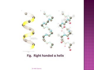
Proteins

 -Dr Nidhi Sharma
Protein Structure
Four levels of organization
  •Primary structure
  •Secondary structure
    oAlpha helix
    oBeta pleated sheets
  •Tertiary structure
  •Quaternary structure


                           Dr Nidhi Sharma
Protein Structure


               Dr Nidhi Sharma
•   Proteins are the major components of living
    organisms and perform a wide range of
    essential functions in cells.

•   While DNA is the information molecule, it is
    proteins that do the work of all cells -
    microbial, plant, animal.

•   Proteins regulate metabolic activity, catalyze
    biochemical reactions and maintain structural
    integrity of cells and organisms.


                                            Dr Nidhi Sharma
   Proteins can be classified in a variety of ways, including
     their biological function
Type:                           Example:
Enzymes- Catalyze biological
                                ß-galactosidase
reactions
Transport and Storage           Hemoglobin
                                Actin
Movement
                                And Myosin in muscles
                                Immunoglobulins
Immune Protection
                                (antibodies)
Regulatory Function within
                                Transcription Factors
cells
                                Insulin
Hormones
                                Estrogen
Structural                      Collagen
                                                        Dr Nidhi Sharma
   Proteins can be formed using 20 different building blocks
    called amino acids.

   Each of these amino acid building blocks has a different
    chemical structure and different properties.

   Each protein has a unique amino acid sequence that is
    genetically determined by the order of nucleotide bases
    in the DNA, the genetic code.

   Since each protein has different numbers and kinds of
    the twenty available amino acids, each protein has a
    unique chemical composition and structure


                        Dr Nidhi Sharma
A   change in just one amino acid can change the
  structure and function of a protein.
 For example, sickle cell anemia is a disease that
  results from an altered structure of the protein
  hemoglobin, resulting from a change of the sixth
  amino acid from glutamic acid to valine. (This is the
  result of a single base pair change at the DNA level.)
 This single amino acid change is enough to change
  the conformation of hemoglobin so that this protein
  clumps at lower oxygen concentrations and causes
  the characteristic sickle shaped red blood cells of the
  disease.


                     Dr Nidhi Sharma
Amino acid structure:
 Amino   acids are composed of carbon, hydrogen,
  oxygen, and nitrogen.

 Two  amino acids, cysteine and methionine, also
  contain sulfur.

 All amino acids have an amino group (NH2) and a
  carboxyl group (COOH) bonded to the same carbon
  atom, known as the alpha carbon.


                  Dr Nidhi Sharma
Dr Nidhi Sharma
   Amino acids differ in the side chain or R group that is
    bonded to the alpha carbon. Glycine, the simplest
    amino acid has a single hydrogen atom as its R group
    - Alanine has a methyl (-CH3) group.

   The chemical composition of the unique R groups is
    responsible for the important characteristics of
    amino acids such as chemical reactivity, ionic charge
    and relative hydrophobicity.


                      Dr Nidhi Sharma
   The amino acids are grouped according to their
    polarity and charge.

   They are divided into four categories, those with
    polar uncharged R groups, those with apolar
    (nonpolar) R groups, acidic (charged) and basic
    (charged) groups.


                    Dr Nidhi Sharma
Dr Nidhi Sharma
   The polar amino acids are soluble in water because
    their R groups can form hydrogen bonds with water.

   For example, serine, threonine and tyrosine all have
    hydroxyl groups (OH).


                     Dr Nidhi Sharma
   The acidic amino acids carry a net negative charge at
    neutral pH contain a second carboxyl group.

   These are aspartic acid and glutamic acid, also called
    aspartate and glutamate, respectively.


                      Dr Nidhi Sharma
   The basic amino acids have R groups with a net positive
    charge at pH 7.0.

   These include lysine, arginine and histidine.


                      Dr Nidhi Sharma
   There are eight amino acids with nonpolar R
    groups. As a group, these amino acids are less
    soluble in water than the polar amino acids.

   These are alanine, valine, leucine, isoleucine,
    tryptophan, phenylalanine, methionine, and
    proline

   If a protein has a greater percentage of nonpolar R
    groups, the protein will be more hydrophobic
    (water hating) in character.


                     Dr Nidhi Sharma
   A protein is formed by amino acid subunits linked
    together in a chain.

   The bond between two amino acids is formed by the
    removal of a H20 molecule from two different amino
    acids, forming a dipeptide.

   The bond between two amino acids is called a
    peptide bond and the chain of amino acids is called a
    peptide (20 amino acids or smaller) or a polypeptide.


                     Dr Nidhi Sharma
I
                  H


Dr Nidhi Sharma
   Primary Structure refers to the linear sequence of
    amino acids that make up the polypeptide chain.

   This sequence is determined by the genetic code, the
    sequence of nucleotide bases in the DNA.

   The sequence of amino acids determines the
    positioning of the different R groups relative to each
    other.

   This positioning therefore determines the way that the
    protein folds and the final structure of the molecule.
                      Dr Nidhi Sharma
 Fig. A pentapeptide. The chain starts at the
 amino acid end


              Dr Nidhi Sharma
   The secondary structure of protein molecules refers to
    the formation of a regular pattern of twists or kinks of
    the polypeptide chain.

   The regularity is due to hydrogen bonds forming
    between the atoms of the amino acid backbone of the
    polypeptide chain.

   The two most common types of secondary structure
    are called the alpha helix and ß pleated sheet.


                      Dr Nidhi Sharma
Fig. Right handed α helix


   Dr Nidhi Sharma
Fig. Antiparallel β pleated sheet


     Dr Nidhi Sharma
   Tertiary structure refers to the three dimensional
    globular structure formed by bending and twisting of
    the polypeptide chain.

   This process often means that the linear sequence of
    amino acids is folded into a compact globular structure.


                      Dr Nidhi Sharma
   The folding of the polypeptide chain is stabilized by
    multiple weak, noncovalent interactions. These
    interactions include:

       Hydrogen bonds that form when a Hydrogen atom
        is shared by two other atoms.
       Electrostatic interactions that occur between
        charged amino acid side chains. Electrostatic
        interactions are attractions between positive and
        negative sites on macromolecules.


                        Dr Nidhi Sharma
   Hydrophobic interactions: During folding of the
        polypeptide chain, amino acids with a polar (water
        soluble) side chain are often found on the surface of
        the molecule while amino acids with non polar
        (water insoluble) side chain are buried in the
        interior. This means that the folded protein is
        soluble in water or aqueous solutions.

   Covalent bonds may also contribute to tertiary
    structure.

   The amino acid, cysteine, has an SH group as part of
    its R group and therefore, the disulfide bond (S-S ) can
    form with an adjacent cysteine. For example, insulin
    has two polypeptide chains that are joined by two
    disulfide bonds.

                        Dr Nidhi Sharma
   Quaternary structure refers to the fact that some proteins
    contain more than one polypeptide chain, adding an
    additional level of structural organization: the association of
    the polypeptide chains.

   Each polypeptide chain in the protein is called a subunit.

   The subunits can be the same polypeptide chain or different
    ones. For example, the enzyme ß-galactosidase is a tetramer,
    meaning that it is composed of four subunits, and, in this
    case, the subunits are identical - each polypeptide chain has
    the same sequence of amino acids.


                        Dr Nidhi Sharma
   Hemoglobin, the oxygen carrying protein in the
    blood, is also a tetramer but it is composed of
    two polypeptide chains of one type (141 amino
    acids) and two of a different type (146 amino
    acids).

   In chemical shorthand, hemoglobin is referred to
    as a2ß2 .

   For some proteins, quaternary structure is
    required for full activity (function) of the protein.


                       Dr Nidhi Sharma
Fig. Four levels of organization of protein structure
           Dr Nidhi Sharma
Dr Nidhi Sharma

Protein structure

  • 1.
  • 2.
    Protein Structure Four levelsof organization •Primary structure •Secondary structure oAlpha helix oBeta pleated sheets •Tertiary structure •Quaternary structure Dr Nidhi Sharma
  • 3.
    Protein Structure Dr Nidhi Sharma
  • 4.
    Proteins are the major components of living organisms and perform a wide range of essential functions in cells. • While DNA is the information molecule, it is proteins that do the work of all cells - microbial, plant, animal. • Proteins regulate metabolic activity, catalyze biochemical reactions and maintain structural integrity of cells and organisms. Dr Nidhi Sharma
  • 5.
    Proteins can be classified in a variety of ways, including their biological function Type: Example: Enzymes- Catalyze biological ß-galactosidase reactions Transport and Storage Hemoglobin Actin Movement And Myosin in muscles Immunoglobulins Immune Protection (antibodies) Regulatory Function within Transcription Factors cells Insulin Hormones Estrogen Structural Collagen Dr Nidhi Sharma
  • 6.
    Proteins can be formed using 20 different building blocks called amino acids.  Each of these amino acid building blocks has a different chemical structure and different properties.  Each protein has a unique amino acid sequence that is genetically determined by the order of nucleotide bases in the DNA, the genetic code.  Since each protein has different numbers and kinds of the twenty available amino acids, each protein has a unique chemical composition and structure Dr Nidhi Sharma
  • 7.
    A change in just one amino acid can change the structure and function of a protein.  For example, sickle cell anemia is a disease that results from an altered structure of the protein hemoglobin, resulting from a change of the sixth amino acid from glutamic acid to valine. (This is the result of a single base pair change at the DNA level.)  This single amino acid change is enough to change the conformation of hemoglobin so that this protein clumps at lower oxygen concentrations and causes the characteristic sickle shaped red blood cells of the disease. Dr Nidhi Sharma
  • 8.
    Amino acid structure: Amino acids are composed of carbon, hydrogen, oxygen, and nitrogen.  Two amino acids, cysteine and methionine, also contain sulfur.  All amino acids have an amino group (NH2) and a carboxyl group (COOH) bonded to the same carbon atom, known as the alpha carbon. Dr Nidhi Sharma
  • 9.
  • 10.
    Amino acids differ in the side chain or R group that is bonded to the alpha carbon. Glycine, the simplest amino acid has a single hydrogen atom as its R group - Alanine has a methyl (-CH3) group.  The chemical composition of the unique R groups is responsible for the important characteristics of amino acids such as chemical reactivity, ionic charge and relative hydrophobicity. Dr Nidhi Sharma
  • 11.
    The amino acids are grouped according to their polarity and charge.  They are divided into four categories, those with polar uncharged R groups, those with apolar (nonpolar) R groups, acidic (charged) and basic (charged) groups. Dr Nidhi Sharma
  • 12.
  • 13.
    The polar amino acids are soluble in water because their R groups can form hydrogen bonds with water.  For example, serine, threonine and tyrosine all have hydroxyl groups (OH). Dr Nidhi Sharma
  • 14.
    The acidic amino acids carry a net negative charge at neutral pH contain a second carboxyl group.  These are aspartic acid and glutamic acid, also called aspartate and glutamate, respectively. Dr Nidhi Sharma
  • 15.
    The basic amino acids have R groups with a net positive charge at pH 7.0.  These include lysine, arginine and histidine. Dr Nidhi Sharma
  • 16.
    There are eight amino acids with nonpolar R groups. As a group, these amino acids are less soluble in water than the polar amino acids.  These are alanine, valine, leucine, isoleucine, tryptophan, phenylalanine, methionine, and proline  If a protein has a greater percentage of nonpolar R groups, the protein will be more hydrophobic (water hating) in character. Dr Nidhi Sharma
  • 17.
    A protein is formed by amino acid subunits linked together in a chain.  The bond between two amino acids is formed by the removal of a H20 molecule from two different amino acids, forming a dipeptide.  The bond between two amino acids is called a peptide bond and the chain of amino acids is called a peptide (20 amino acids or smaller) or a polypeptide. Dr Nidhi Sharma
  • 18.
    I H Dr Nidhi Sharma
  • 19.
    Primary Structure refers to the linear sequence of amino acids that make up the polypeptide chain.  This sequence is determined by the genetic code, the sequence of nucleotide bases in the DNA.  The sequence of amino acids determines the positioning of the different R groups relative to each other.  This positioning therefore determines the way that the protein folds and the final structure of the molecule. Dr Nidhi Sharma
  • 20.
     Fig. Apentapeptide. The chain starts at the amino acid end Dr Nidhi Sharma
  • 21.
    The secondary structure of protein molecules refers to the formation of a regular pattern of twists or kinks of the polypeptide chain.  The regularity is due to hydrogen bonds forming between the atoms of the amino acid backbone of the polypeptide chain.  The two most common types of secondary structure are called the alpha helix and ß pleated sheet. Dr Nidhi Sharma
  • 22.
    Fig. Right handedα helix Dr Nidhi Sharma
  • 23.
    Fig. Antiparallel βpleated sheet Dr Nidhi Sharma
  • 24.
    Tertiary structure refers to the three dimensional globular structure formed by bending and twisting of the polypeptide chain.  This process often means that the linear sequence of amino acids is folded into a compact globular structure. Dr Nidhi Sharma
  • 25.
    The folding of the polypeptide chain is stabilized by multiple weak, noncovalent interactions. These interactions include:  Hydrogen bonds that form when a Hydrogen atom is shared by two other atoms.  Electrostatic interactions that occur between charged amino acid side chains. Electrostatic interactions are attractions between positive and negative sites on macromolecules. Dr Nidhi Sharma
  • 26.
    Hydrophobic interactions: During folding of the polypeptide chain, amino acids with a polar (water soluble) side chain are often found on the surface of the molecule while amino acids with non polar (water insoluble) side chain are buried in the interior. This means that the folded protein is soluble in water or aqueous solutions.  Covalent bonds may also contribute to tertiary structure.  The amino acid, cysteine, has an SH group as part of its R group and therefore, the disulfide bond (S-S ) can form with an adjacent cysteine. For example, insulin has two polypeptide chains that are joined by two disulfide bonds. Dr Nidhi Sharma
  • 27.
    Quaternary structure refers to the fact that some proteins contain more than one polypeptide chain, adding an additional level of structural organization: the association of the polypeptide chains.  Each polypeptide chain in the protein is called a subunit.  The subunits can be the same polypeptide chain or different ones. For example, the enzyme ß-galactosidase is a tetramer, meaning that it is composed of four subunits, and, in this case, the subunits are identical - each polypeptide chain has the same sequence of amino acids. Dr Nidhi Sharma
  • 28.
    Hemoglobin, the oxygen carrying protein in the blood, is also a tetramer but it is composed of two polypeptide chains of one type (141 amino acids) and two of a different type (146 amino acids).  In chemical shorthand, hemoglobin is referred to as a2ß2 .  For some proteins, quaternary structure is required for full activity (function) of the protein. Dr Nidhi Sharma
  • 29.
    Fig. Four levelsof organization of protein structure Dr Nidhi Sharma
  • 30.