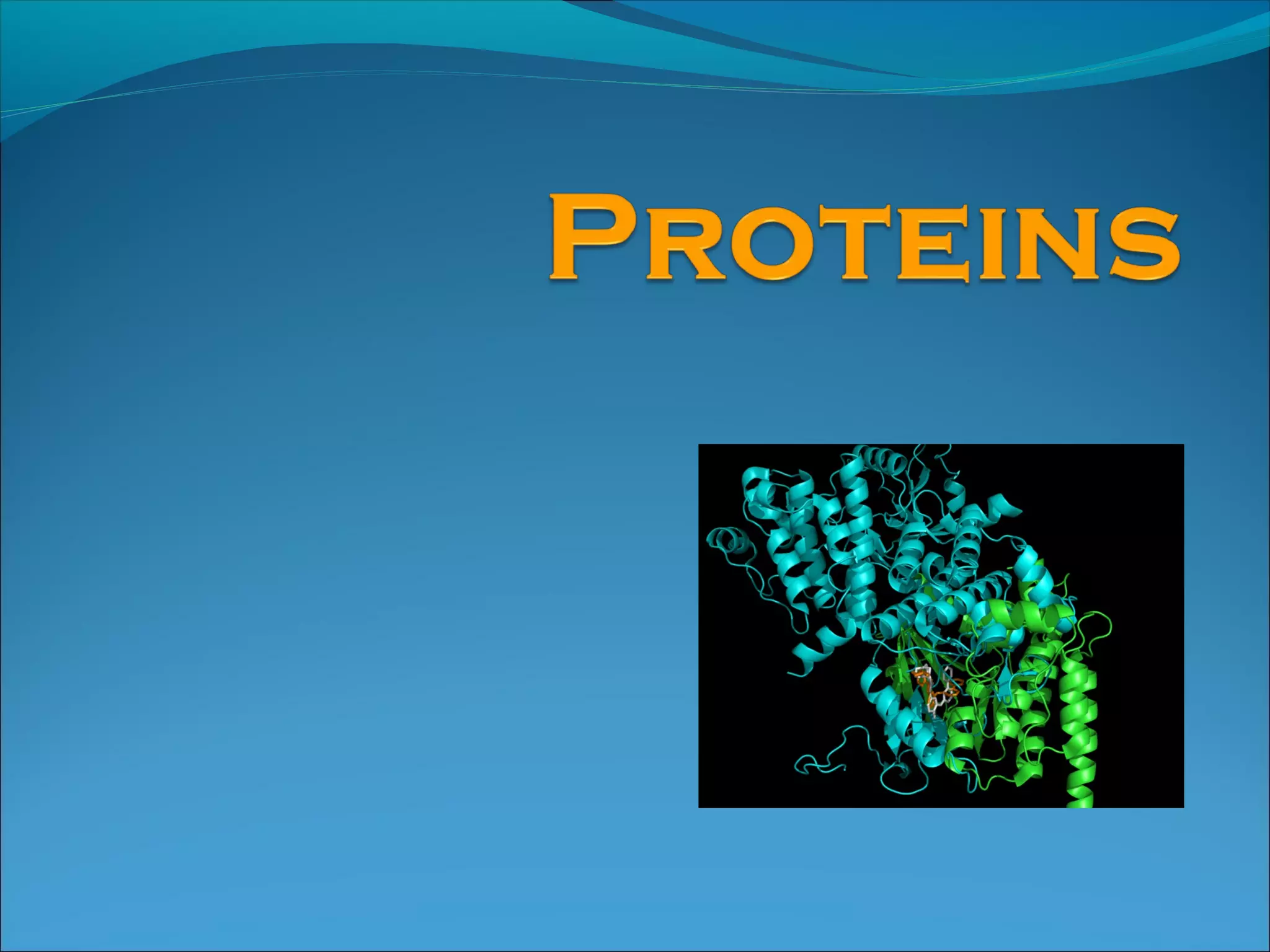
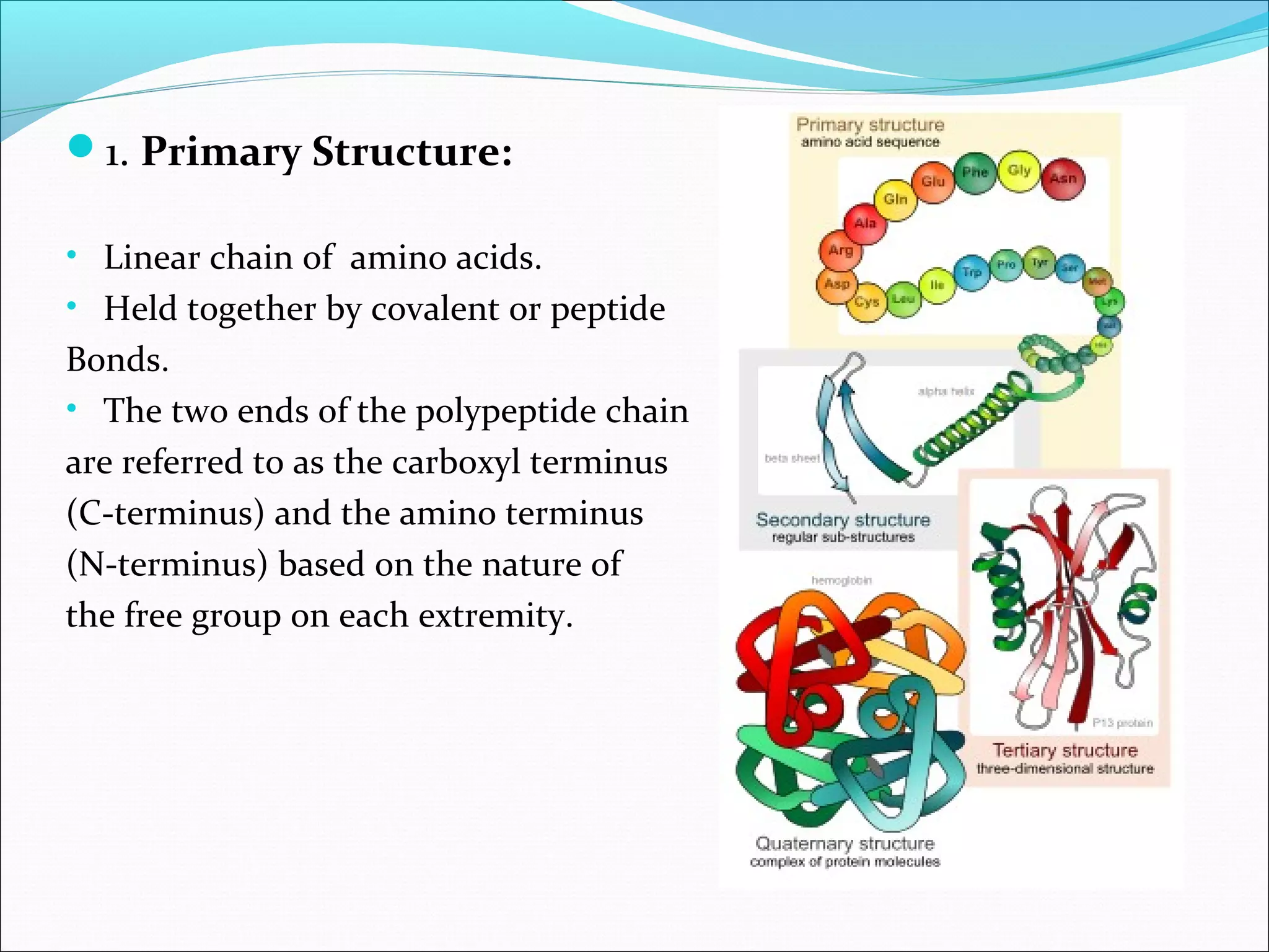
WHAT ARE PROTEINS ?
Proteins are biochemical compounds consisting of one or
more polypeptides typically folded into a globular or
fibrous form, facilitating a biological function.
It takes part in maintaining the structural Integrity of the
cell, transport and storage of small molecules , catalysis,
regulation, immune system.
Functions:
Enzymes: Accelerate biochemical reactions
Structural: Form biological structures
Transport: Carry biochemically important substances
Defense: Protect the body from foreign invaders
AMINO ACIDS:
Building units of proteins
There are about 300 amino acids occur in nature.
On the basis of nutritional value they have been
categorized as essential and nonessential amino acids
Essential: which cannot be synthesized by the body or
cannot be synthesized at an adequate rate or are in
insufficient quantity and are obtained through diet.
Adults require 9 (Phenylalanine Valine Tryptophan
Threonine Isoleucine Methionine Histidine Leucine
Lysine) while infants and children require 10 ( arginine)
The remaining amino acids are Non essential, i.e
those that can be synthesized by the body and are
different from essential amino acids that are obtained
from food
Anatomy of an amino acid
• Majority of amino acids has amphoteric character – functional group –
COOH is the reason of acidity and –NH2 group causes basic properties.
• In basic environment AA dissociate proton to form carboxyl anion –
COO-
. Basic surround defends –NH2 against dissociation.
• In acidic environment AA accept proton to form amonium cation –NH3
+
.
Acidic environment defends –COOH against dissociation.
AMPHOTERIC CHARACTER OF AMINO ACID
GENERAL PROPERTIES OF ΑLPHA-AMINO
ACIDS.
a)Polarity of amino acids:
Since amino acids are present in the Zwitterions form at
physiological pH where it carries a +ve charge on the α-amino
group and a –ve charge on the α-carboxylic group , thus
creating two opposite charges on both sides of the molecule
thus showing polar properties.
b)Isoelectric point pI :
It is that pH at which the amino acid net electrical charge is
equal to zero ,and thus cannot move in an electrical field.
Amino Acids Are Joined By
Peptide Bonds In Peptides
- α-carboxyl of one amino acid is joined to α-amino
of a second amino acid (with removal of water)
- only α-carboxyl and α-amino groups are used, not
R-group carboxyl or amino groups
Chemistry of peptide bond formation
Amino acids can be classified by R groups:
NONPOLAR, ALIPHATIC R GROUPS
The R groups in this class of amino acids are nonpolar
and hydrophobic.
AROMATIC R GROUPS
Phenylalanine, tyrosine, and tryptophan, with their
aromatic side chains, are relatively nonpolar
(hydrophobic).
POLAR, UNCHARGED R GROUPS
The R groups of these amino acids are more soluble in water, or more
hydrophilic, because they contain functional groups that form
hydrogen bonds with water.
This class of amino acids includes serine, threonine, cysteine,
asparagine, and glutamine.
POSITIVELY CHARGED (BASIC) R GROUPS
Amino acids with very polar side chains that render
them highly hydrophilic. Lysine and arginine have
relatively long side chains that terminate with groups
that are positively charged at neutral pH.
NEGATIVELY CHARGED (ACIDIC) R GROUPS
The two amino acids having R groups with a net
negative charge at pH 7.0 are aspartate and
glutamate, each of which has a second carboxyl
group.
Levels of Protein Structure
Primary structure = order of amino
acids in the protein chain
1. Primary Structure:
• Linear chain of amino acids.
• Held together by covalent or peptide
Bonds.
• The two ends of the polypeptide chain
are referred to as the carboxyl terminus
(C-terminus) and the amino terminus
(N-terminus) based on the nature of
the free group on each extremity.
Secondary structure = local folding
of residues into regular patterns
2. SECONDARY STRUCTURE:
• ‘local’ ordered structure via H- Bonds , mainly
within backbone.
• Includes the alpha helix and the beta strand
or beta sheets
Alpha helix (α-helix) is a right-handed coiled or spiral
conformation, in which every backbone N-H group
donates a hydrogen bond to the backbone C=O group of
the amino acid four residues earlier.
Three-dimensional arrangement of amino acids with the
polypeptide chain in a corkscrew shape
Looks like a coiled “telephone cord”
Beta sheets consist of beta strands connected laterally by at least two
or three backbone hydrogen bonds, forming a generally twisted,
pleated sheet.
A beta strand (also β strand) is a stretch of polypeptide chain
typically 3 to 10 amino acids long with backbone in an almost fully
extended conformation.
Hydrogen bonds form between chains
The beta sheets could be arranged either parallel or antiparallel
direction
 In an antiparallel
arrangement, the successive β
strands alternate directions so
that the N-terminus of one
strand is adjacent to the C-
terminus of the next.
Produces the strongest inter-
strand stability because it allows
the inter-strand hydrogen
bonds between carbonyls and
amines to be planar
In a parallel arrangement, all
of the N-termini of successive
strands are oriented in the same
direction;
Slightly less stable because it
introduces nonplanarity in the
inter-strand hydrogen bonding
pattern.
Tertiary structure = global folding of
a protein chain
3. TERTIARY STRUCTURE:
• 3D structure of protein, results by large number
of non-covalent interactions b/w amino acids.
• The alpha-helices and beta-sheets are folded
into a compact globule
• structure is stable only when the parts of a protein
domain are locked into place by specific tertiary
interactions, such as salt bridges, hydrogen bonds,
and the tight packing of side chains and disulfide bonds
This shape is held in place by bonds such as
weak Hydrogen bonds between amino acids that lie close to each
other,
strong ionic bonds between R groups with positive and negative
charges, and
disulfide bridges (strong covalent S-S bonds)
Quaternary structure = Higher-order
assembly of proteins
Levels of Protein Structure
4. QUATERNARY STRUCTURE
 It is the non-covalent interactions that bind
multiple polypeptides into a single, larger protein
Eg: Haemoglobin
2 α-globin and 2 β-globin polypeptides
Fibrous Proteins
Little or no tertiary structure.
Long parallel polypeptide chains.
Cross linkages at intervals forming long fibres or sheets.
Usually insoluble.
Many have structural roles.
E.g. keratin in hair and the outer layer of skin, collagen (a connective
tissue).
Globular Proteins
Have complex tertiary and sometimes quaternary structures.
Folded into spherical (globular) shapes.
Usually soluble as hydrophobic side chains in centre of structure.
Roles in metabolic reactions.
E.g. enzymes, haemoglobin in blood.
29
Disruption of secondary, tertiary and quaternary
protein structure by
Heat/organics :
Break apart H bonds and disrupt hydrophobic attractions
Acids/ bases:
Break H bonds between polar R groups and ionic bonds
Heavy metal ions :
React with S-S bonds to form solids
Agitation :
Stretches chains until bonds break
DENATURATION:
Protein Functions
Antibodies - are specialized proteins involved in defending the body
from antigens (foreign invaders). They travel through the blood
stream and are utilized by the immune system to identify and defend
against bacteria, viruses, and other foreign intruders. One way
antibodies destroy antigens is by immobilizing them so that they can
be destroyed by white blood cells.
Contractile Proteins - are responsible for movement. Examples
include actin and myosin. These proteins are involved in muscle
contraction and movement.
Enzymes - are proteins that facilitate biochemical reactions. They are
often referred to as catalysts because they speed up chemical
reactions. Examples include the enzymes lactase and pepsin. Lactase
breaks down the sugar lactose found in milk. Pepsin is a digestive
enzyme that works in the stomach to break down proteins in food.
Hormonal Proteins - are messenger proteins which help to
coordinate certain bodily activities. Examples include insulin,
oxytocin, and somatotropin. Insulin regulates glucose metabolism by
controlling the blood-sugar concentration. Oxytocin stimulates
contractions in females during childbirth. Somatotropin is a growth
hormone that stimulates protein production in muscle cells.
Structural Proteins - are fibrous and stringy and provide support.
Examples include keratin, collagen, and elastin. Keratins strengthen
protective coverings such as hair, quills, feathers, horns, and beaks.
Collagens and elastin provide support for connective tissues such as
tendons and ligaments.
Storage Proteins - store amino acids. Examples include ovalbumin
and casein. Ovalbumin is found in egg whites and casein is a milk-
based protein.
Transport Proteins - are carrier proteins which move molecules
from one place to another around the body. Examples include
hemoglobin and cytochromes. Hemoglobin transports oxygen
through the blood. Cytochromes operate in the electron transport
chain as electron carrier proteins.

Proteins.

  • 2.
    WHAT ARE PROTEINS? Proteins are biochemical compounds consisting of one or more polypeptides typically folded into a globular or fibrous form, facilitating a biological function. It takes part in maintaining the structural Integrity of the cell, transport and storage of small molecules , catalysis, regulation, immune system.
  • 3.
    Functions: Enzymes: Accelerate biochemicalreactions Structural: Form biological structures Transport: Carry biochemically important substances Defense: Protect the body from foreign invaders
  • 4.
    AMINO ACIDS: Building unitsof proteins There are about 300 amino acids occur in nature.
  • 5.
    On the basisof nutritional value they have been categorized as essential and nonessential amino acids Essential: which cannot be synthesized by the body or cannot be synthesized at an adequate rate or are in insufficient quantity and are obtained through diet. Adults require 9 (Phenylalanine Valine Tryptophan Threonine Isoleucine Methionine Histidine Leucine Lysine) while infants and children require 10 ( arginine) The remaining amino acids are Non essential, i.e those that can be synthesized by the body and are different from essential amino acids that are obtained from food
  • 6.
    Anatomy of anamino acid
  • 7.
    • Majority ofamino acids has amphoteric character – functional group – COOH is the reason of acidity and –NH2 group causes basic properties. • In basic environment AA dissociate proton to form carboxyl anion – COO- . Basic surround defends –NH2 against dissociation. • In acidic environment AA accept proton to form amonium cation –NH3 + . Acidic environment defends –COOH against dissociation. AMPHOTERIC CHARACTER OF AMINO ACID
  • 8.
    GENERAL PROPERTIES OFΑLPHA-AMINO ACIDS. a)Polarity of amino acids: Since amino acids are present in the Zwitterions form at physiological pH where it carries a +ve charge on the α-amino group and a –ve charge on the α-carboxylic group , thus creating two opposite charges on both sides of the molecule thus showing polar properties. b)Isoelectric point pI : It is that pH at which the amino acid net electrical charge is equal to zero ,and thus cannot move in an electrical field.
  • 9.
    Amino Acids AreJoined By Peptide Bonds In Peptides - α-carboxyl of one amino acid is joined to α-amino of a second amino acid (with removal of water) - only α-carboxyl and α-amino groups are used, not R-group carboxyl or amino groups
  • 10.
    Chemistry of peptidebond formation
  • 11.
    Amino acids canbe classified by R groups: NONPOLAR, ALIPHATIC R GROUPS The R groups in this class of amino acids are nonpolar and hydrophobic.
  • 12.
    AROMATIC R GROUPS Phenylalanine,tyrosine, and tryptophan, with their aromatic side chains, are relatively nonpolar (hydrophobic).
  • 13.
    POLAR, UNCHARGED RGROUPS The R groups of these amino acids are more soluble in water, or more hydrophilic, because they contain functional groups that form hydrogen bonds with water. This class of amino acids includes serine, threonine, cysteine, asparagine, and glutamine.
  • 14.
    POSITIVELY CHARGED (BASIC)R GROUPS Amino acids with very polar side chains that render them highly hydrophilic. Lysine and arginine have relatively long side chains that terminate with groups that are positively charged at neutral pH.
  • 15.
    NEGATIVELY CHARGED (ACIDIC)R GROUPS The two amino acids having R groups with a net negative charge at pH 7.0 are aspartate and glutamate, each of which has a second carboxyl group.
  • 16.
  • 17.
    Primary structure =order of amino acids in the protein chain
  • 18.
    1. Primary Structure: •Linear chain of amino acids. • Held together by covalent or peptide Bonds. • The two ends of the polypeptide chain are referred to as the carboxyl terminus (C-terminus) and the amino terminus (N-terminus) based on the nature of the free group on each extremity.
  • 19.
    Secondary structure =local folding of residues into regular patterns
  • 20.
    2. SECONDARY STRUCTURE: •‘local’ ordered structure via H- Bonds , mainly within backbone. • Includes the alpha helix and the beta strand or beta sheets
  • 21.
    Alpha helix (α-helix)is a right-handed coiled or spiral conformation, in which every backbone N-H group donates a hydrogen bond to the backbone C=O group of the amino acid four residues earlier. Three-dimensional arrangement of amino acids with the polypeptide chain in a corkscrew shape Looks like a coiled “telephone cord”
  • 22.
    Beta sheets consistof beta strands connected laterally by at least two or three backbone hydrogen bonds, forming a generally twisted, pleated sheet. A beta strand (also β strand) is a stretch of polypeptide chain typically 3 to 10 amino acids long with backbone in an almost fully extended conformation. Hydrogen bonds form between chains The beta sheets could be arranged either parallel or antiparallel direction
  • 23.
     In anantiparallel arrangement, the successive β strands alternate directions so that the N-terminus of one strand is adjacent to the C- terminus of the next. Produces the strongest inter- strand stability because it allows the inter-strand hydrogen bonds between carbonyls and amines to be planar In a parallel arrangement, all of the N-termini of successive strands are oriented in the same direction; Slightly less stable because it introduces nonplanarity in the inter-strand hydrogen bonding pattern.
  • 24.
    Tertiary structure =global folding of a protein chain
  • 25.
    3. TERTIARY STRUCTURE: •3D structure of protein, results by large number of non-covalent interactions b/w amino acids. • The alpha-helices and beta-sheets are folded into a compact globule • structure is stable only when the parts of a protein domain are locked into place by specific tertiary interactions, such as salt bridges, hydrogen bonds, and the tight packing of side chains and disulfide bonds This shape is held in place by bonds such as weak Hydrogen bonds between amino acids that lie close to each other, strong ionic bonds between R groups with positive and negative charges, and disulfide bridges (strong covalent S-S bonds)
  • 26.
    Quaternary structure =Higher-order assembly of proteins
  • 27.
    Levels of ProteinStructure 4. QUATERNARY STRUCTURE  It is the non-covalent interactions that bind multiple polypeptides into a single, larger protein Eg: Haemoglobin 2 α-globin and 2 β-globin polypeptides
  • 28.
    Fibrous Proteins Little orno tertiary structure. Long parallel polypeptide chains. Cross linkages at intervals forming long fibres or sheets. Usually insoluble. Many have structural roles. E.g. keratin in hair and the outer layer of skin, collagen (a connective tissue). Globular Proteins Have complex tertiary and sometimes quaternary structures. Folded into spherical (globular) shapes. Usually soluble as hydrophobic side chains in centre of structure. Roles in metabolic reactions. E.g. enzymes, haemoglobin in blood.
  • 29.
    29 Disruption of secondary,tertiary and quaternary protein structure by Heat/organics : Break apart H bonds and disrupt hydrophobic attractions Acids/ bases: Break H bonds between polar R groups and ionic bonds Heavy metal ions : React with S-S bonds to form solids Agitation : Stretches chains until bonds break DENATURATION:
  • 30.
    Protein Functions Antibodies -are specialized proteins involved in defending the body from antigens (foreign invaders). They travel through the blood stream and are utilized by the immune system to identify and defend against bacteria, viruses, and other foreign intruders. One way antibodies destroy antigens is by immobilizing them so that they can be destroyed by white blood cells. Contractile Proteins - are responsible for movement. Examples include actin and myosin. These proteins are involved in muscle contraction and movement. Enzymes - are proteins that facilitate biochemical reactions. They are often referred to as catalysts because they speed up chemical reactions. Examples include the enzymes lactase and pepsin. Lactase breaks down the sugar lactose found in milk. Pepsin is a digestive enzyme that works in the stomach to break down proteins in food.
  • 31.
    Hormonal Proteins -are messenger proteins which help to coordinate certain bodily activities. Examples include insulin, oxytocin, and somatotropin. Insulin regulates glucose metabolism by controlling the blood-sugar concentration. Oxytocin stimulates contractions in females during childbirth. Somatotropin is a growth hormone that stimulates protein production in muscle cells. Structural Proteins - are fibrous and stringy and provide support. Examples include keratin, collagen, and elastin. Keratins strengthen protective coverings such as hair, quills, feathers, horns, and beaks. Collagens and elastin provide support for connective tissues such as tendons and ligaments. Storage Proteins - store amino acids. Examples include ovalbumin and casein. Ovalbumin is found in egg whites and casein is a milk- based protein. Transport Proteins - are carrier proteins which move molecules from one place to another around the body. Examples include hemoglobin and cytochromes. Hemoglobin transports oxygen through the blood. Cytochromes operate in the electron transport chain as electron carrier proteins.

Editor's Notes

  • #26 Ramachandran plot is a way to visualize backbone dihedral angles ψ against φ of amino acid residues in protein structure. The ω angle at the peptide bond is normally 180°, since the partial-double-bond character keeps the peptide planar
  • #28 Ramachandran plot is a way to visualize backbone dihedral angles ψ against φ of amino acid residues in protein structure. The ω angle at the peptide bond is normally 180°, since the partial-double-bond character keeps the peptide planar