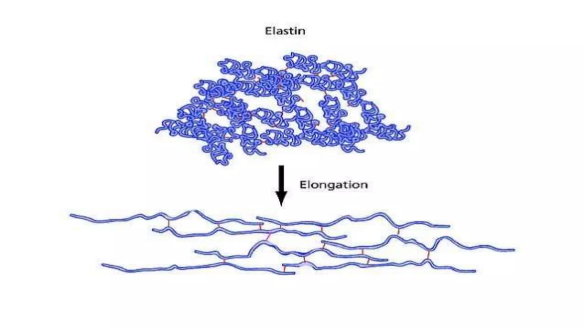
Proteoglycans are protein chains that are covalently bonded at multiple sites to a class of polysaccharides, known as glycosaminoglycans.Glycosaminoglycans constitute 95% of proteins.Proteoglycans are synthesised in RE and transported to GA where they are modified in to various forms.Proteoglycans are major component of ECM and their role is depended on the type of GAGs they associate with.
What is Glycoprotein ?:
Glycoproteins are proteins that contain oligosaccharide chains (glycans) covalently attached to polypeptide side-chains.
This process is known as glycosylation.
The carbohydrate is attached to the protein during the following modifications: Co-translational modification & Post-translational modification.
In proteins that have segments extending extracellularly, the extracellular segments are often glycosylated.
This explains the complex carbohydrates and chemistry of heterpolysaccharides. composition, distribution and its function is explained for each GAGs. brief notes on blood group ag is available. difference between proteoglycan and glycoprotein is explained in a essay way to understand. clinical importance is also added.
Extra cellular matrix is recently being explored in connection with cancer , metastases and autoimmune disorders. It is prepared for the benefit of both UG and PG medical and dental students.
The Indian Dental Academy is the Leader in continuing dental education , training dentists in all aspects of dentistry and
offering a wide range of dental certified courses in different formats.for more details please visit
www.indiandentalacademy.com
Communication between cells and their environment.pptxArunPatel134845
This is a ppt about cells communicate to each other and their environment .This ppt includes basically cell adhesion molecules(CAMs) and Extracellular matrix(ECM)
Collagen is the most abundant protein in your body. It is the major component of connective tissues that make up several body parts, including tendons, ligaments, skin and muscles (1). Collagen has many important functions, including providing your skin with structure and strengthening your bones (
RECOMBINATION MOLECULAR BIOLOGY PPT UPDATED new.pptxSabahat Ali
This ppt is about recombination and where it occurs. Types of recombination and models of recombination along with many factors in prokaryotic and eukaryotic recombination
Folding depends upon sequence of Amino Acids not the Composition. Folding starts with the secondary structure and ends at quaternary structure.
Denaturation occur at secondary, tertiary & quaternary level but not at primary level.
Tertiary Structure basically of Hydrophobic interactions, (interactions in side chains), hydrogen bonding, salt bridges, Vander Waals interactions.
e.g. Globular proteins & Fibrous Proteins
Earliest Galaxies in the JADES Origins Field: Luminosity Function and Cosmic ...Sérgio Sacani
We characterize the earliest galaxy population in the JADES Origins Field (JOF), the deepest
imaging field observed with JWST. We make use of the ancillary Hubble optical images (5 filters
spanning 0.4−0.9µm) and novel JWST images with 14 filters spanning 0.8−5µm, including 7 mediumband filters, and reaching total exposure times of up to 46 hours per filter. We combine all our data
at > 2.3µm to construct an ultradeep image, reaching as deep as ≈ 31.4 AB mag in the stack and
30.3-31.0 AB mag (5σ, r = 0.1” circular aperture) in individual filters. We measure photometric
redshifts and use robust selection criteria to identify a sample of eight galaxy candidates at redshifts
z = 11.5 − 15. These objects show compact half-light radii of R1/2 ∼ 50 − 200pc, stellar masses of
M⋆ ∼ 107−108M⊙, and star-formation rates of SFR ∼ 0.1−1 M⊙ yr−1
. Our search finds no candidates
at 15 < z < 20, placing upper limits at these redshifts. We develop a forward modeling approach to
infer the properties of the evolving luminosity function without binning in redshift or luminosity that
marginalizes over the photometric redshift uncertainty of our candidate galaxies and incorporates the
impact of non-detections. We find a z = 12 luminosity function in good agreement with prior results,
and that the luminosity function normalization and UV luminosity density decline by a factor of ∼ 2.5
from z = 12 to z = 14. We discuss the possible implications of our results in the context of theoretical
models for evolution of the dark matter halo mass function.
Observation of Io’s Resurfacing via Plume Deposition Using Ground-based Adapt...Sérgio Sacani
Since volcanic activity was first discovered on Io from Voyager images in 1979, changes
on Io’s surface have been monitored from both spacecraft and ground-based telescopes.
Here, we present the highest spatial resolution images of Io ever obtained from a groundbased telescope. These images, acquired by the SHARK-VIS instrument on the Large
Binocular Telescope, show evidence of a major resurfacing event on Io’s trailing hemisphere. When compared to the most recent spacecraft images, the SHARK-VIS images
show that a plume deposit from a powerful eruption at Pillan Patera has covered part
of the long-lived Pele plume deposit. Although this type of resurfacing event may be common on Io, few have been detected due to the rarity of spacecraft visits and the previously low spatial resolution available from Earth-based telescopes. The SHARK-VIS instrument ushers in a new era of high resolution imaging of Io’s surface using adaptive
optics at visible wavelengths.
Introduction:
RNA interference (RNAi) or Post-Transcriptional Gene Silencing (PTGS) is an important biological process for modulating eukaryotic gene expression.
It is highly conserved process of posttranscriptional gene silencing by which double stranded RNA (dsRNA) causes sequence-specific degradation of mRNA sequences.
dsRNA-induced gene silencing (RNAi) is reported in a wide range of eukaryotes ranging from worms, insects, mammals and plants.
This process mediates resistance to both endogenous parasitic and exogenous pathogenic nucleic acids, and regulates the expression of protein-coding genes.
What are small ncRNAs?
micro RNA (miRNA)
short interfering RNA (siRNA)
Properties of small non-coding RNA:
Involved in silencing mRNA transcripts.
Called “small” because they are usually only about 21-24 nucleotides long.
Synthesized by first cutting up longer precursor sequences (like the 61nt one that Lee discovered).
Silence an mRNA by base pairing with some sequence on the mRNA.
Discovery of siRNA?
The first small RNA:
In 1993 Rosalind Lee (Victor Ambros lab) was studying a non- coding gene in C. elegans, lin-4, that was involved in silencing of another gene, lin-14, at the appropriate time in the
development of the worm C. elegans.
Two small transcripts of lin-4 (22nt and 61nt) were found to be complementary to a sequence in the 3' UTR of lin-14.
Because lin-4 encoded no protein, she deduced that it must be these transcripts that are causing the silencing by RNA-RNA interactions.
Types of RNAi ( non coding RNA)
MiRNA
Length (23-25 nt)
Trans acting
Binds with target MRNA in mismatch
Translation inhibition
Si RNA
Length 21 nt.
Cis acting
Bind with target Mrna in perfect complementary sequence
Piwi-RNA
Length ; 25 to 36 nt.
Expressed in Germ Cells
Regulates trnasposomes activity
MECHANISM OF RNAI:
First the double-stranded RNA teams up with a protein complex named Dicer, which cuts the long RNA into short pieces.
Then another protein complex called RISC (RNA-induced silencing complex) discards one of the two RNA strands.
The RISC-docked, single-stranded RNA then pairs with the homologous mRNA and destroys it.
THE RISC COMPLEX:
RISC is large(>500kD) RNA multi- protein Binding complex which triggers MRNA degradation in response to MRNA
Unwinding of double stranded Si RNA by ATP independent Helicase
Active component of RISC is Ago proteins( ENDONUCLEASE) which cleave target MRNA.
DICER: endonuclease (RNase Family III)
Argonaute: Central Component of the RNA-Induced Silencing Complex (RISC)
One strand of the dsRNA produced by Dicer is retained in the RISC complex in association with Argonaute
ARGONAUTE PROTEIN :
1.PAZ(PIWI/Argonaute/ Zwille)- Recognition of target MRNA
2.PIWI (p-element induced wimpy Testis)- breaks Phosphodiester bond of mRNA.)RNAse H activity.
MiRNA:
The Double-stranded RNAs are naturally produced in eukaryotic cells during development, and they have a key role in regulating gene expression .
This pdf is about the Schizophrenia.
For more details visit on YouTube; @SELF-EXPLANATORY;
https://www.youtube.com/channel/UCAiarMZDNhe1A3Rnpr_WkzA/videos
Thanks...!
Multi-source connectivity as the driver of solar wind variability in the heli...Sérgio Sacani
The ambient solar wind that flls the heliosphere originates from multiple
sources in the solar corona and is highly structured. It is often described
as high-speed, relatively homogeneous, plasma streams from coronal
holes and slow-speed, highly variable, streams whose source regions are
under debate. A key goal of ESA/NASA’s Solar Orbiter mission is to identify
solar wind sources and understand what drives the complexity seen in the
heliosphere. By combining magnetic feld modelling and spectroscopic
techniques with high-resolution observations and measurements, we show
that the solar wind variability detected in situ by Solar Orbiter in March
2022 is driven by spatio-temporal changes in the magnetic connectivity to
multiple sources in the solar atmosphere. The magnetic feld footpoints
connected to the spacecraft moved from the boundaries of a coronal hole
to one active region (12961) and then across to another region (12957). This
is refected in the in situ measurements, which show the transition from fast
to highly Alfvénic then to slow solar wind that is disrupted by the arrival of
a coronal mass ejection. Our results describe solar wind variability at 0.5 au
but are applicable to near-Earth observatories.
(May 29th, 2024) Advancements in Intravital Microscopy- Insights for Preclini...Scintica Instrumentation
Intravital microscopy (IVM) is a powerful tool utilized to study cellular behavior over time and space in vivo. Much of our understanding of cell biology has been accomplished using various in vitro and ex vivo methods; however, these studies do not necessarily reflect the natural dynamics of biological processes. Unlike traditional cell culture or fixed tissue imaging, IVM allows for the ultra-fast high-resolution imaging of cellular processes over time and space and were studied in its natural environment. Real-time visualization of biological processes in the context of an intact organism helps maintain physiological relevance and provide insights into the progression of disease, response to treatments or developmental processes.
In this webinar we give an overview of advanced applications of the IVM system in preclinical research. IVIM technology is a provider of all-in-one intravital microscopy systems and solutions optimized for in vivo imaging of live animal models at sub-micron resolution. The system’s unique features and user-friendly software enables researchers to probe fast dynamic biological processes such as immune cell tracking, cell-cell interaction as well as vascularization and tumor metastasis with exceptional detail. This webinar will also give an overview of IVM being utilized in drug development, offering a view into the intricate interaction between drugs/nanoparticles and tissues in vivo and allows for the evaluation of therapeutic intervention in a variety of tissues and organs. This interdisciplinary collaboration continues to drive the advancements of novel therapeutic strategies.
Cancer cell metabolism: special Reference to Lactate PathwayAADYARAJPANDEY1
Normal Cell Metabolism:
Cellular respiration describes the series of steps that cells use to break down sugar and other chemicals to get the energy we need to function.
Energy is stored in the bonds of glucose and when glucose is broken down, much of that energy is released.
Cell utilize energy in the form of ATP.
The first step of respiration is called glycolysis. In a series of steps, glycolysis breaks glucose into two smaller molecules - a chemical called pyruvate. A small amount of ATP is formed during this process.
Most healthy cells continue the breakdown in a second process, called the Kreb's cycle. The Kreb's cycle allows cells to “burn” the pyruvates made in glycolysis to get more ATP.
The last step in the breakdown of glucose is called oxidative phosphorylation (Ox-Phos).
It takes place in specialized cell structures called mitochondria. This process produces a large amount of ATP. Importantly, cells need oxygen to complete oxidative phosphorylation.
If a cell completes only glycolysis, only 2 molecules of ATP are made per glucose. However, if the cell completes the entire respiration process (glycolysis - Kreb's - oxidative phosphorylation), about 36 molecules of ATP are created, giving it much more energy to use.
IN CANCER CELL:
Unlike healthy cells that "burn" the entire molecule of sugar to capture a large amount of energy as ATP, cancer cells are wasteful.
Cancer cells only partially break down sugar molecules. They overuse the first step of respiration, glycolysis. They frequently do not complete the second step, oxidative phosphorylation.
This results in only 2 molecules of ATP per each glucose molecule instead of the 36 or so ATPs healthy cells gain. As a result, cancer cells need to use a lot more sugar molecules to get enough energy to survive.
introduction to WARBERG PHENOMENA:
WARBURG EFFECT Usually, cancer cells are highly glycolytic (glucose addiction) and take up more glucose than do normal cells from outside.
Otto Heinrich Warburg (; 8 October 1883 – 1 August 1970) In 1931 was awarded the Nobel Prize in Physiology for his "discovery of the nature and mode of action of the respiratory enzyme.
WARNBURG EFFECT : cancer cells under aerobic (well-oxygenated) conditions to metabolize glucose to lactate (aerobic glycolysis) is known as the Warburg effect. Warburg made the observation that tumor slices consume glucose and secrete lactate at a higher rate than normal tissues.
2. Definition:
Fibrous protein are the group of protein that are specifically
associated with the “rod” or “wire” shape.
They are usually inert structural or storage protein.
They are generally water insoluble proteins.
They are usually used to construct connective tissues,
tendons, bones and muscle fiber
4. History:
In the early 1950s the basic structure of the fibrous
proteins were determined.
It was found that long protein chains were composed
of strings of amino acid, could be folded up in a
systematic manner to generate a limited number of
structures that are
5. consistent with the X-ray data.
Most important of these structures are
alpha-helix, beta-sheets and the collagen
triple helix.
6. Characteristics:
Following are the some major functional
characteristics of the fibrous proteins.
Biological function
Water Solubility
Amino Acid Composition
Secondary Structure
Tertiary structure
Examples
7. contd.,
Biological Functions:
Fibrous protein often have storage or structural roles such as
the collagen that is found in bone and cartilage.
Water Solubility:
Fibrous Proteins are insoluble in water.
8. Amino Acid Composition:
Fibrous protein often have repeated sequence of
amino acid.
Comparatively stronger intermolecular interactions
occur amongst the amino acid of the fibrous protein.
9. Secondary Structure:
Alpha-helixes and the beta-sheets.
Tertiary Structure:
Yes. The tertiary structure of fibrous
protein result in long fiber like structure.
12. Introduction
Collagen is recognized as a complex , interactive compilation of
protiens in dynamic equilibrium that can regulate the genes
expression of cells
It is most abundant protein in mammals . Accounts for 25-30%
of their protein content .
Many fibrous component of skin , bone , tendon and
cartilage.
13. When colagen heated in water , it gradually breaks down to
produce solube derived i.e . Gelatin or animals glue.
Denatures at a high temperature , remains stable at body
temperature
Collagen molecule is rigid and rod like structure that resists
stretching .
It influences cell shape , differentiation and other cellular
activities.
14. Basic structure of collagen:
Composed of three polypeptide alpha chains coiled around
each other to form triple helix configuration
Depending upon the type of collagen, the molecule may be
homometric (3 identical alpha chains)
Hetrometric ( 2 or 3 different alpha chains)
15. Conti…
Alpha chains that are left handed helices wrap around each
other to form right handed rope like triple helical Rod
Each alpha helix is 1.4nm in diameter and 300nm in
length
3 amino acid per turn (Gly-X-Y) repeat,,, frequently 30%
being X=proline & Y=hydroxyproline
Glycine occupies every third position in the repeating
amino acid sequence
16.
17. Types of collagen:
19 types of collagen found Variation occur due to
1) Differences in the assembly of basic polypeptide chains
2) different length of the helix
3) Various interruption in the helix
4) Differences in the termination of the helical domain
18.
19. Functions of Collagen Proteins
Collagen fibers support body tissues, plus collagen is a major
component of the extracellular matrix that supports cells.
Collagen give the skin its strength, waterproofing, and elasticity
. Loss of collagen is a cause of wrinkles.
20. CONTINUED
Connective tissue consists primarily of collagen
Collagen forms fibrils that provide the structure for fibrous
tissue, such as ligaments, tendons, and skin.
Collagen also is found in cartilage, bone, , the cornea of the eye,
intervertebral discs, muscles, and the gastrointestinal tract.
Collagen is used in cosmetic treatments and burn surgery.
Collagen is used to produce gelatin.
21. Synthesis Of Collagen Protein
First, a three-dimensional stranded structure is assembled, with
the amino acids glycine and proline as its principal components.
This is not yet collagen but its precursor, procollagen.
Procollagen is then modified by the addition of hydroxyl; groups
to the amino acids proline and lysine. This step is important for
later glycosylation and the formation of the triple helix structure of
collagen
These hydroxylation reactions are catalyzed by two different
enzymes: prolyl-4-hydroxylase and lysyl-hydroxylase.
Vitamin C also serves with them in inducing these reactions
22. Continued
• Transcription of mRNA
• Pre-pro-peptide formation
• Pre-pro-peptide to pro-
collagen
• Golgi apparatus modification
• Formation of tropocollagen
• Formation of the collagen
fibril
The
synthesis
of collagen
occurs
inside and
outside of
the cell.
Steps
25. Introduction
Keratin is one of a family of fibrous structural proteins
It is the key structural material making
up hair, horns, claws, hooves, and the outer layer of
human skin.
Keratin is extremely insoluble in water and organic solvents.
26. Occurance
Keratin filaments are abundant in keratinocytes in the cornified layer of
the epidermis
keratin filaments are present in epithelial cells in general.
the α-keratins are found in all vertebrates
the harder β-keratins are found only in the sauropsids, that is all living
reptiles and birds.
They are found in the nails, scales, and claws of reptiles, some reptile
shells (Testudines, such as tortoise, turtle, terrapin), and in the feathers,
beaks, and claws of birds.
27. Horns such as those of the impala are made
up of keratin covering a core of live bone.
28. Genes
The human genome encodes 54 functional keratin genes which
are located in two clusters on chromosomes 12 and 17.
The neutral-basic keratins are found on chromosome 12
(12q13.13).
33. Functions of Keratin Proteins
The Role Of Keratin. Keratin is a vital protein for the production of
human skin, hair, and nails. In fact, about 95% of the makeup of hair is
simply keratin
Keratin is also used in keratin treatments, which are designed to smooth
and shine hair. ... In a salon keratin treatment, keratins are mixed with
formaldehyde and applied to the hair with a flat iron and sealing the
protein into your hair.
. Keratin is also the protein that protects epithelial cells from damage or
stress
34. In The Skin
keratin has two main functions in the skin:
1. To hold skin cells together to
form a barrier
2. To form the outermost layer of our skin,
that protects us from the environment
35. Diseases
Diseases caused by mutations in the keratin genes include:
Epidermolysis bullosa simplex
Ichthyosis bullosa of Siemens
Epidermolytic hyperkeratosis
Steatocystoma multiplex
Keratosis pharyngis
Rhabdoid cell formation in Large cell lung carcinoma with rhabdoid
phenotype
36. Intermediate Filaments
Keratin filaments are intermediate filaments. Like all
intermediate filaments, keratin proteins form
filamentous polymers in a series of assembly steps
beginning with dimerization;
dimers assemble into tetramers and octamers and
eventually, if the current hypothesis holds, into unit-
length-filaments (ULF) capable of annealing end-to-
end into long filaments.
37.
38. Elastin
A highly elastic protein in connective tissue
Allows many tissues in body to resume their
shape after stretching or contracting.
Elastin helps the skin to return to the original
position when it is poked or pinched.
39.
40. Occurrence & its importance:
Elastin is found in skin & body tissues
It is a protein that helps to keep our skin flexible & tight.
It also keeps the skin smooth as it stretches to accommodate normal activities like
flexing muscles or opening & closing the mouth to talk or to eat.
Elastin is an imp load bearing tissue in the bodies of vertebrates & used in places
where mechanical energy is required to be stored.
In human Elastin is encoded by ELN gene.
41. Function & structure of elastin:
ELN gene codes for a protein that is one of two component of elastic
fibers
Encoded protein is rich in hydrophobic amino acids such as
glycine and proline which form mobile hydrophobic regions
bound by crosslinks between lysine residues.
Composed primarily of small non polar amino acids residues (G
, A , V).
ALSO rich in proline & lysine but little contains hydroxyproline &
hydroxylysine.
interchain cross-links form desmosine residues.