Downloaded 138 times

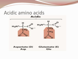
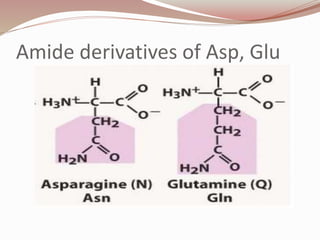
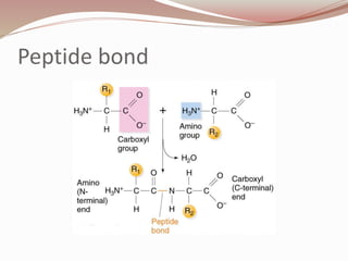
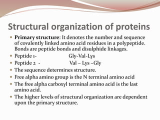
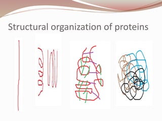
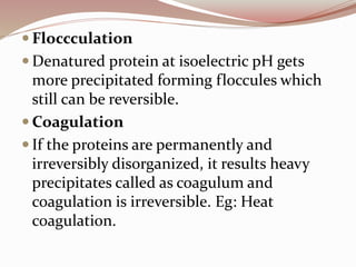
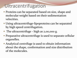


The document discusses the structure and properties of proteins and amino acids. Some key points: - Proteins are polymers of amino acids linked by peptide bonds. There are 20 standard amino acids that vary in properties based on their side chains. - Proteins have primary, secondary, tertiary, and quaternary levels of structure determined by amino acid sequence and interactions. - Amino acids form peptides and proteins through condensation reactions between carboxyl and amino groups, forming peptide bonds. - Proteins can be classified based on function, shape, nutritional value, and composition. Structural proteins like collagen resist digestion while globular proteins are soluble.


![PERI-PROSTHETIC FRACTURE NAIL-PLATE CONSTRUCT [NPC].pptx](https://cdn.slidesharecdn.com/ss_thumbnails/drarunkumardrmohamedashrafperiprostheticfrasturenail-plateconstructnpc-260209164459-7e9d15a1-thumbnail.jpg?width=640&height=640&fit=bounds)

