
Recommended
More Related Content
What's hot
What's hot (20)
Viewers also liked
Viewers also liked (20)
Similar to Amino acids and protein
Similar to Amino acids and protein (20)
Recently uploaded
Recently uploaded (20)
Amino acids and protein
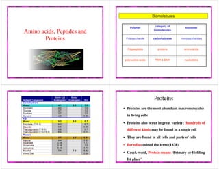
- 1. Amino acids, Peptides and Proteins Biomolecules Polymer category of biomolecules monomer Polysaccharide carbohydrates monosaccharides Polypeptides proteins amino acids polynucleic acids RNA & DNA nucleotides Proteins • Proteins are the most abundant macromolecules in living cells • Proteins also occur in great variety: hundreds of different kinds may be found in a single cell • They are found in all cells and parts of cells • Berzelius coined the term (1838), • Greek word, Protein means ‘Primary or Holding Ist place’
- 2. • Proteins are linear polymers built of monomer units called amino acids • Proteins contain a wide range of• Proteins contain a wide range of functional groups • Some proteins are quite rigid whereas others display considerable flexibility • Proteins are naturally occurring , nitrogen containing bio molecules • They yield amino acids on hydrolysis • They act as building blocks of cells and tissues • They yield 4.0 kcal/g energy on breakdown Amino acid • Amino Acids are the building units of proteins. Proteins are polymers of amino acids linked together by what is called “ Peptide bond”. amino acids linked together by what is called “ Peptide bond”. • There are about 300 amino acids occur in nature. Only 20 of them occur in proteins. Structure of amino acids • Each amino acid has 4 different groups attached to α- carbon ( which is C-atom next to COOH). These 4 groups are : amino group,COOH). These 4 groups are : amino group, COOH gp, • Hydrogen atom and • Side Chain (R)
- 3. Biomedical Importance 1. Proteins play a central role in cell functions & cell structure. It constitutes 17% of body weight. 2. Necessary membrane formation, muscle & connective tissue.& connective tissue. 3. All enzymes are proteins (except Ribozymes, RNA in nature). 4. Many hormones are proteins. 5. Antibodies which confer immunity against viral & bacterial infections are proteins. 7. Proteins function as buffers to maintain pH of the cell. 8. Transport proteins, storage proteins, respiratory pigments ,osmotic pressure & cell signalling. 9. Catabolized to supply energy. Classification 20 amino acids in protein encoded by DNA; three letter abbreviation and one letter symbol. Based on a. Polarity and charge on R groups. b. Structure of side chains. c. Metabolic fate of the amino acid. d. Nutritional importance.
- 4. Based on polarity and charge on R groups. 1. Non-polar R Groups. Aliphatic Hydrocarbon side chains. - Alanine, Valine, Leucine, Isoleucine. Aromatic side chains. -Phenylalanine (Phenyl moiety).-Phenylalanine (Phenyl moiety). -Tryptophan (indole group). - Tyrosine (Phenol gp) 2. Polar – a)uncharged - b)charged(positively charged, negatively charged) a. Uncharged polar R Groups:-OH,-SH,-NH-C=O , hydrogen bond to water. Hydroxylic group: Serine and Theronine. H-Group(Hydrogen, Amide bearing) : Asparagine, Glutamine SH (Thiol gp): Cysteine, Cystine. b. Charged polar R Groups:b. Charged polar R Groups: Basic amino acids: Positive charge on R group at physiological pH (Lysine, Arginine, Histidine). Acidic amino acids: negative charge on R group at physiological pH (Aspartate, Glutamate). Based on structure of side chain 1. Aliphatic amino acid: a. Mono amino mono carboxylic acids. Simple Amino acids: Glycine, Alanine. Branched chain Amino acids: Valine, Leucine, Isoleucine. Hydroxy Amino acids: Serine, Threonine, Tyrosine. Sulphur containing Amino acids: Methionine, Cysteine. Amide Group containing: Asparagine, Glutamine.Amide Group containing: Asparagine, Glutamine. b. Mono amino dicarboxylic acids: Aspartic acid, Glutamic acid c. Di basic monocarboxylic acids: Lysine, Arginine
- 5. 2. Aromatic amino acid: Phenylalanine, Tyrosine. 3.Heterocyclic amino acids: Tryptophan, Histidine . 4.Imino acid: Proline.Proline. Special group in Amino acid. Arginine - guanidinium group. Phenylalanine- benzene. Tyrosine-phenol. Tryptophan-Indole. Histidine- Imidazole Proline- Pyrrolidine, has secondary amino group hence it is Imino acid. Based on Metabolic fate: Glucogenic : Give rise to Glucose. Ketogenic: Give rise to Ketone Bodies. Glucogenic and ketogenic: Give rise to Glucose & Ketone Bodies.Bodies. Based on nutritionally importance Essential amino acid: can not be endogenously synthesized; required for growth. • Isoleucine, Leucine, Threonine, Lysine, Methionine, Phenylalanine, Tryptophan & Valine. Semi essential amino acid: Growing children require them in food but not essential for adult individual. • Histidine , Arginine. Non-essential Amino acid: endogenously synthesized to meet body’s requirement.
- 6. Arginine is listed as an essential amino acid because humans require arginine in their diet to support rapid growth during childhood and pregnancy, even though it is made by urea cycle. 21st Amino acid: Selenocysteine It is present in some enzymes. Instead of SH gp in cysteine selenium is present in selenocysteine. 22nd Amino acid: Pyrrolysine Lysine in amide linkage to substitute-pyrroline-5-Lysine in amide linkage to substitute-pyrroline-5- carboxylate. Present in Methyl transferase enzymes of bacteria. Both are encoded by stop codons. Properties of amino acids 1. Stereoisomerism: D-amino acid and L-amino acid. If NH2 is on right hand, its D-amino acid. While if its on left ;its L-amino acid. Natural proteins in animals and plants. D-amino acid occur in bacteria.animals and plants. D-amino acid occur in bacteria.
- 7. 2.Amphoteric nature and isoelectric pH: The NH2 & COOH groups of amino acids are ionizable groups. Depending on the pH of solution groups act as proton donors(acids) or proton acceptor(bases).This property is called amphoteric & therefore amino acids are called ampholytes. . At specific pH amino acid act carries both the charges in equal number and exist as dipolar ion or zwitterion. At this point net charge is zero i.e. positive and negative charges on proteins /amino acidnegative charges on proteins /amino acid equalizes. The pH at which it occurs pI or isoelectric pH. On the acidic side of pI amino acids exist as cation by accepting proton & on alkaline as anion by donating proton. Chemical properties. Reactions due to carboxylic –COOH group. 1. Formation of amines by decarboxylation. Histidine → Histamine + CO2 Tyrosine → Tyramine + CO2 Tryptophan → Tryptamine +CO2 Lysine → Cadaverine +CO2 Glutamic acid → Gamma amino butyric acid(GABA) +CO2Glutamic acid → Gamma amino butyric acid(GABA) +CO2 2. Formation of amides. COOH group of Dicarboxylic amino acids combine with NH3 to form corresponding Amide. Aspartic acid + NH3 → Asparagine Glutamic + NH3 → Glutamine
- 8. Reactions due to Amino group. Transamination: alpha amino group of amino acid transferred to alpha keto acid to form new keto acid and new amino acid. Imp reaction for synthesis of non essential amino acid. Oxidative deamination: alpha amino gp is removed from amino acid to form corresponding keto acid and ammonia. Glutamic acid is the most common to undergo oxidative deamination. Formation of carbamino compound: Carbon dioxide adds to alpha amino gp of amino acid to form carbamino compounds. Occurs at alkaline pH & serve as mechanism for transport of carbon dioxide fromserve as mechanism for transport of carbon dioxide from tissues to lungs by Hb. Hb-NH2 + CO2 Hb-NH-COOH (Carbamino-Hb) Reactions due to side chains Transmethylation: Methyl group of methionine transferred to acceptor. Methionine + Acceptor → Methylated Acceptor+Homocysteine Ester formation by OH group: Hydroxy amino acid form esters with phosphoric acid- Phosphoprotein. Hydroxyl gp can also form O-glycosidic bonds with carbohydrate residues –Glycoprotein.
- 9. Reactions of amide group: Amide group of glutamine and asparagine can form N-Glycosidic bond-Glycoproteins. Reactions of SH group: Cysteine has SH gp & can form disulphide(S-S) bond with another cysteineform disulphide(S-S) bond with another cysteine residue to form Cystine. Helpful for make interchain disulphide bonds between two polypeptide chains Specific role of some side chains: 1. Hydroxyl groups of serine and theronine exist at catalytic site of certain kinds of enzyme and phosphorylation of site make the enzyme active. 2. Thiol group of cysteine combines with cysteine to form2. Thiol group of cysteine combines with cysteine to form cystine. It is important in linking adjacent polypeptide. 3. Amide bearing side chain of asparagine forms covalent link with oligosaccharide unit in glycoproteins (N-glycosidic linkage). OH-gp of serine and theronine forms linkage of reducing end of oligosaccharide (O-glycosidic linkage). 4. Aromatic side chains are responsible for ultraviolet absorption. 5. Imidazole gp of Histidine is important in the buffering activities of proteins. 6. Cyclic pyrrolidine gp of proline introduces bend in6. Cyclic pyrrolidine gp of proline introduces bend in the peptide chain. 7. R gp of Glycine provides little stearic hindrance due to its small size, so that proteins can bend or rotate easily . Important derivatives of amino acids. 1.GABA-derivative of glutamic acid; Dopamine-Tyrosine precursor(Neurotransmitters) 2.Histamine-synthesized from histidine-mediator of allergic reactions. 3.Thyroxine-from tyrosine-thyroid hormones.3.Thyroxine-from tyrosine-thyroid hormones. 4.Cycloserine-derived from serine-antitubercular drug. 5.Histidine-buffering activity of proteins. 6.Ornithine and citrulline-Arginine-Urea synthesis.
- 10. Biochemical Methods to separate amino acid and Proteins • Electrophoresis • Chromatography: Gel filtration, ion exchange, affinityGel filtration, ion exchange, affinity • Mass Spectrometry, • X-ray Crystallography, Peptides and Proteins 20 amino acids are commonly found in protein. These 20 amino acids are linked together through “peptide bond forming peptides and proteins (what’s the difference?). - The chains containing less than 50 amino acids are called “peptides”, while those containing greater than 50 amino acids are called “proteins”. Peptide bond formation:Peptide bond formation: α-carboxyl group of one amino acid (with side chain R1) forms a covalent peptide bond with α-amino group of another amino acid ( with the side chain R2) by removal of a molecule of water. The result is : Dipeptide ( i.e. Two amino acids linked by one peptide bond). By the same way, the dipeptide can then forms a second peptide bond with a third amino acid (with side chain R3) to give Tripeptide. Repetition of this process generates a polypeptide or protein of specific amino acid sequence. Peptide bond formation: - Each polypeptide chain starts on the left side by free amino group of the first amino acid enter in chain formation . It is termed (N- terminus). - Each polypeptide chain ends on the right side by free COOH group of the last amino acid and termed (C-terminus). Examples on Peptides: 1- Dipeptide ( two amino acids joined by one peptide bond): Example: Aspartame which acts as sweetening agent being used in replacement of cane sugar. It is composed of aspartic acid and phenyl alanine.
- 11. Examples on Peptides: 2- Tripeptides ( 3 amino acids linked by two peptide bonds). Example: GSH which is formed from 3 amino acids: glutamic acid, cysteine and glycine. It helps in absorption of amino acids, protects against hemolysis of RBC by breaking H2O2 which causes cell damage. 3- octapeptides: (8 amino acids)3- octapeptides: (8 amino acids) Examples: Two hormones; oxytocine and vasopressin (ADH). 4- polypeptides: 10- 50 amino acids: e.g. Insulin hormone Classification of proteins I- Simple proteins: i.e. on hydrolysis gives only amino acids Examples: 1- Albumin and globulins: present in egg, milk and blood They are proteins of high biological value i.e. contain all essential amino acids and easily digested. Types of globulins: α1 globulin: e.g. antitrypsin: see later α2 globulin: e.g. hepatoglobin: protein that binds hemoglobin to prevent its excretion by the kidney β-globulin: e.g. transferrin: protein that transport iron γ-globulins = Immunoglobulins (antibodies) : responsible for immunity. 2- Globins (Histones): They are basic proteins rich in histidine amino acid. They are present in : a - combined with DNA b - combined with heme to form hemoglobin of RBCs. 3- Gliadines are the proteins present in cereals. 4- Scleroproteins: They are structural proteins, not digested. include: keratin, collagen and elastin.include: keratin, collagen and elastin. a- α-keratin: protein found in hair, nails, enamel of teeth and outer layer of skin. • It is α-helical polypeptide chain, rich in cysteine and hydrophobic (non polar) amino acids so it is water insoluble. b- collagens: protein of connective tissues found in bone, teeth, cartilage, tendons, skin and blood vessels. • Collagen may be present as gel e.g. in extracellular matrix or in vitreous humor of the eye. • Collagens are the most important protein in mammals. They form about 30% of total body proteins. • There are more than 20 types of collagens, the most common type is collagen I which constitutes about 90% of cell collagens. • Structure of collagen: three helical polypeptide chains (trimeric) twisted around each other forming triplet-helix(trimeric) twisted around each other forming triplet-helix molecule. • ⅓ of structure is glycine, 10% proline, 10% hydroxyproline and 1% hydroxylysine. Glycine is found in every third position of the chain. The repeating sequence –Gly-X-Y-, where X is frequently proline and Y is often hydroxyproline and can be hydroxylysine.
- 12. Solubility: collagen is insoluble in all solvents and not digested. • When collagen is heated with water or dil. HCl it will be converted into gelatin which is soluble , digestible and used as diet ( as jelly). Gelatin is classified as derived protein. Some collagen diseases: 1- Scurvy: disease due to deficiency of vitamin C which is important coenzyme for conversion of proline into hydroxyproline and lysine intocoenzyme for conversion of proline into hydroxyproline and lysine into hydroxylysine. Thus, synthesis of collagen is decreased leading to abnormal bone development, bleeding, loosing of teeth and swollen gum. 2- Osteogenesis Imperfecta (OI): Inherited disease resulting from genetic deficiency or mutation in gene that synthesizes collagen type I leading to abnormal bone formation in babies and frequent bone fracture in children. It may be lethal. C- Elastin: present in walls of large blood vessels (such as aorta). It is very important in lungs, elastic ligaments, skin, cartilage, .. It is elastic fiber that can be stretched to several times as its normal length. Structure: composed of 4 polypeptide chains (tetramer), similar to collagen being having 33% glycine and rich in proline but in that it has low hydroxyproline and absence of hydroxy lysine.lysine. Emphysema: is a chronic obstructive lung disease (obstruction of air ways) resulting from deficiency of α1-antitrypsin particularly in cigarette smokers. Role of α1-antitrypsin: Elastin is a lung protein. Smoke stimulate enzyme called elastase to be secreted form neutrophils (in lung). Elastase cause destruction of elastin of lung. α1-antitrypsin is an enzyme (secreted from liver) and inhibit elastase and prevent destruction of elastin. So deficiency of α1-antitrypsin especially in smokers leads to degradation of lung and destruction of lung ( loss of elasticity of lung, a disease called emphysema. Conjugated proteins i.e. On hydrolysis, give protein part and non protein part and subclassified into:subclassified into: 1- Phosphoproteins: These are proteins conjugated with phosphate group. Phosphorus is attached to OH group of serine or threonine. e.g. Casein of milk and vitellin of yolk. 2- Lipoproteins: These are proteins conjugated with lipids. Functions: a- help lipids to transport in blood b- Enter in cell membrane structure helping lipid soluble substances to pass through cell membranes. 3- Glycoproteins: proteins conjugated with sugar (carbohydrate) e.g. – Mucin - Some hormones such as erythropoeitin- Some hormones such as erythropoeitin - present in cell membrane structure - blood groups. 4- Nucleoproteins: These are basic proteins ( e.g. histones) conjugated with nucleic acid (DNA or RNA). e.g. a- chromosomes: are proteins conjugated with DNA b- Ribosomes: are proteins conjugated with RNA
- 13. 5- Metalloproteins: These are proteins conjugated with metal like iron, copper, zinc, …… a- Iron-containing proteins: Iron may present in heme such as in - hemoglobin (Hb) - myoglobin ( protein of skeletal muscles and cardiacmuscle), - cytochromes, - catalase, peroxidases (destroy H2O2) - tryptophan pyrrolase (desrtroy indole ring of tryptophan).- tryptophan pyrrolase (desrtroy indole ring of tryptophan). Iron may be present in free state ( not in heme) as in: - Ferritin: Main store of iron in the body. ferritin is present in liver, spleen and bone marrow. - Hemosidrin: another iron store. - Transferrin: is the iron carrier protein in plasma. b- Copper containing proteins: e.g. - Ceruloplasmin which oxidizes ferrous ions into ferric ions. - Oxidase enzymes such as cytochrome oxidase. c- Zn containing proteins: e.g. Insulin and carbonic anhydrase d- Mg containing proteins:e.g. Kinases and phosphatases. 6-Chromoproteins: These are proteins conjugated with pigment. e.g. - All proteins containing heme (Hb, myoglobin, ………..)- All proteins containing heme (Hb, myoglobin, ………..) - Melanoprotein:e.g proteins of hair or iris which contain melanin. Derived proteins Produced from hydrolysis of simple proteins. e.g. - Gelatin: from hydrolysis of collagen - Peptone: from hydrolysis of albumin Levels of Protein Structure Protein structure: There are four levels of protein structure (primary, secondary, tertiary and quaternary) Primary structure: • The primary structure of a protein is its unique sequence of amino acids. – Lysozyme, an enzyme that attacks bacteria, consists of a polypeptide chain of 129consists of a polypeptide chain of 129 amino acids. – The precise primary structure of a protein is determined by inherited genetic information. – At one end is an amino acid with a free amino group the (the N-terminus) and at the other is an amino acid with a free carboxyl group the (the C-terminus).
- 14. Protein Primary Structure 1’ structure = ordered sequence of amino acids ProteinPrimaryStructureProteinPrimaryStructure Note the Polarity of the sequence (amino carboxy) Note also the disulfide linkages (cys-cys S- S bonds; actually considered a component of tertiary structure) 2- Secondary structure: Results from hydrogen bond formation between hydrogen of –NH group of peptide bond and the carbonyl oxygen of another peptide bond. According to H-bonding there are two main forms of secondary structure: α-helix: It is a spiral structure resulting from hydrogen bondingresulting from hydrogen bonding between one peptide bond and the fourth one β-sheets: is another form of secondary structure in which two or more polypeptides (or segments of the same peptide chain) are linked together by hydrogen bond between H- of NH- of one chain and carbonyl oxygen of adjacent chain (or segment). 2’Structure:H-Bonds2’Structure:H
- 15. Hydrogen bonding in α-helix: In the α-helix CO of the one amino acid residue forms H-bond with NH of the forth one. Supersecondary structure or Motifs : occurs by combining secondary structure. The combination may be: α-helix- turn- α-helix- turn…..etc Or: β-sheet -turn- β-sheet-turn………etc Or: α-helix- turn- β-sheet-turn- α-helix Turn (or bend): is short segment of polypeptides (3-4 amino acids) that connects successive secondary structures. e.g. β-turn: is small polypeptide that connects successive strands of β- sheets. • Tertiary structure is determined by a variety of interactions (bond formation) among R groups and between R groups and the polypeptide backbone. a. The weak interactions include: Hydrogen bonds among polar side chains Ionic bonds betweenIonic bonds between charged R groups ( basic and acidic amino acids) Hydrophobic interactions among hydrophobic ( non polar) R groups. b. Strong covalent bonds include disulfide bridges, that form between the sulfhydryl groups (SH) of cysteine monomers, stabilize the structure. • Quaternary structure: results from the aggregation (combination) of two or more polypeptide subunits held together by non-covalent interaction like H- bonds, ionic or hydrophobic interactions. • Examples on protein having quaternary structure: – Collagen is a fibrous protein of three polypeptides (trimeric) that are supercoiled like a rope. • This provides the structural strength for their role in connective tissue. – Hemoglobin is a globular protein with four polypeptide chains (tetrameric) – Insulin : two polypeptide chains (dimeric)– Insulin : two polypeptide chains (dimeric)
- 16. CellDiseaseSickle-CellDisease ProteinDenaturationProteinDenaturation Destruction of Conformation = Loss of Function Some simple proteins can spontaneously renature Chaparonins Chaparonins Assist in Protein Folding They segregate protein folding from “bad influences” in the cell
- 17. Chaperones • a large group of proteins, also termed heat shock proteins and chaperones, • During the folding process, they function to prevent unfavorable protein interactions with other potentially complementary surfaces (like other proteins, carbohydrates, lipids, nucleic acids, etc.) • Many of these proteins are ATPases (use hydrolysis of ATP as an energy source). Nitrogen Cycle Sources • Lightning • Inorganic fertilizers • Nitrogen Fixation• Nitrogen Fixation • Animal Residues • Crop residues • Organic fertilizers
- 18. Forms of Nitrogen • Urea CO(NH2)2 • Ammonia NH3 (gaseous) • Ammonium NH4• Ammonium NH4 • Nitrate NO3 • Nitrite NO2 • Atmospheric Dinitrogen N2 • Organic N Microorganisms fixing • Azobacter • Azospirillum • Require the enzyme nitrogenase • Inhibited by oxygen• Azospirillum • Clostridium • Cyanobacteria • Inhibited by ammonia (end product) Rates of Nitrogen Fixation N2 fixing system Nitrogen Fixation (kg N/hect/year) Rhizobium-legume 200-300 Cyanobacteria- moss 30-40