The central nervous system (CNS) is the part of the nervous system consisting of the brain and spinal cord. The central nervous system is so named because it integrates information it receives from, and coordinates and influences the activity of, all parts of the bodies
The central nervous system (CNS) is the part of the nervous system consisting of the brain and spinal cord. The central nervous system is so named because it integrates information it receives from, and coordinates and influences the activity of, all parts of the bodies
You can watch the video on my you tube channel: https://youtu.be/I0FaX-iQfa0
Medulla oblongata or more simply medulla is part of brain stem which forms base of the brain stem. It contains pyramid, olive and above pyramidal structure, there is decussation of pyramids which explains why each part of brain controls opposite part of body. Adding to that medulla also has several nuclei which controls activity of cardiovascular system and respiratory system. Medulla also has nuclei for controlling reflexes of vomiting, swallowing, hiccuping, coughing and sneezing. It has also nuclei for test, hearing and balance. Medulla also contains nuclei of cranial nerve number VIII, IX, X, XI and XII.
Description :
The Indian Dental Academy is the Leader in continuing dental education , training dentists in all aspects of dentistry and
offering a wide range of dental certified courses in different formats.for more details please visit
www.indiandentalacademy.com
The nervous system is the part of an animal's body that coordinates its behavior and transmits signals between different body areas. In vertebrates it consists of two main parts, called the central nervous system (CNS) and the peripheral nervous system (PNS).
The science of yoga is the scientific basis of modern yoga as exercise in human sciences such as anatomy, physiology, and psychology. Yoga's effects are to some extent shared with other forms of exercise,[O 1] though it differs in the amount of stretching involved, and because of its frequent use of long holds and relaxation, in its ability to reduce stress. Yoga is here treated separately from meditation, which has effects of its own, though yoga and meditation are combined in some schools of yoga.
Review of Nervous System, Unconsciousness, and CVA. The Nursing Core FunctionsAyinla Kazeem
This presentation was made at several sessions of Mandatory Continuing Professional Development Programme for Nigerian Nurses in Kwara State, and have undergone series of editing till date. While still working on the final editing to totally conform with global standard of practice, I deemed it necessary to share it in this forum.
THIS PRESENTATION IS UPLOADED TO HELP THE EDUCATOR OF MEDICAL, NURSING & ALLIE HEALTH SCIENCES TO TEACH THEIR STUDENTS ABOUT THE NERVOUS SYSTEM. IT WILL ALSO CREATE AWARENESS AMONG THE COMMON PEOPLE REGARDING NERVOUS SYSTEM.
Briefly explain the ventral and dorsal body cavities and the membran.pdfarchanenterprises
Briefly explain the ventral and dorsal body cavities and the membranes which line the ventral
cavities. What is a mesentery?
Solution
The ventral body cavity is a body cavity that is in the anterior (front) aspect of the body. It is
made up of the thoracic cavity, and the abdominopelvic cavity. The abdominopelvic cavity is
further divided into the abdominal cavity and pelvic cavity, but there is no physical barrier
between the two. The abdominal cavity contains digestive organs, the pelvic cavity contains the
urinary bladder, internal reproductive organs, and rectum.
The thoracic cavity is separated from the abdominopelvic cavity by the diaphragm. The thoracic
cavity is further separated into the pleural cavity which contains the lungs and the superior
mediastinum which includes the pericardial (heart) cavity.
The organs within the ventral body cavity are called the viscera.
The walls of the ventral body cavity and outer covering of its organs contain a thin covering
called the serosa (also called serous membrane). It is a double-layered membrane made up of two
parts called the “parietal serosa” (lines the cavity walls) and “visceral serosa” (covers organs in
the cavity). The serous membranes are separated by a thin layer of fluid called “serous fluid“.
Serous fluid is secreted by both membranes and acts as a lubricant, allowing organs to slide in
the cavity without causing friction.
The dorsal body cavity is located along the dorsal (posterior) surface of the body, where it is
subdivided into the cranial cavity housing the brain and the spinal cavity housing the spinal cord.
The two cavities are continuous with one another.
The cranial cavity is a large, bean-shaped cavity filling most of the upper skull where the brain is
located.The vertebral cavity is a very narrow, thread-like cavity running from the cranial cavity
down the entire length of the spinal cord. Just as the brain and spinal cord make up a continuous,
uninterrupted structure, the cranial and spinal cavities that house them are also continuous. The
brain and spinal cord are protected by the bones of the skull and vertebral column and by
cerebrospinal fluid, a colorless fluid produced by the brain, which cushions the brain and spinal
cord within the posterior (dorsal) cavity.
The covering and protective membranes for the dorsal body cavity are called meninges..
cerebrovascular accidents - types, causes and its managementVarunMahajani
This PowerPoint presentation provides in-depth knowledge regarding cerebrovascular accidents types, stages of management, medical management, surgical management, nursing management, complications and their management
Ozempic: Preoperative Management of Patients on GLP-1 Receptor Agonists Saeid Safari
Preoperative Management of Patients on GLP-1 Receptor Agonists like Ozempic and Semiglutide
ASA GUIDELINE
NYSORA Guideline
2 Case Reports of Gastric Ultrasound
Recomendações da OMS sobre cuidados maternos e neonatais para uma experiência pós-natal positiva.
Em consonância com os ODS – Objetivos do Desenvolvimento Sustentável e a Estratégia Global para a Saúde das Mulheres, Crianças e Adolescentes, e aplicando uma abordagem baseada nos direitos humanos, os esforços de cuidados pós-natais devem expandir-se para além da cobertura e da simples sobrevivência, de modo a incluir cuidados de qualidade.
Estas diretrizes visam melhorar a qualidade dos cuidados pós-natais essenciais e de rotina prestados às mulheres e aos recém-nascidos, com o objetivo final de melhorar a saúde e o bem-estar materno e neonatal.
Uma “experiência pós-natal positiva” é um resultado importante para todas as mulheres que dão à luz e para os seus recém-nascidos, estabelecendo as bases para a melhoria da saúde e do bem-estar a curto e longo prazo. Uma experiência pós-natal positiva é definida como aquela em que as mulheres, pessoas que gestam, os recém-nascidos, os casais, os pais, os cuidadores e as famílias recebem informação consistente, garantia e apoio de profissionais de saúde motivados; e onde um sistema de saúde flexível e com recursos reconheça as necessidades das mulheres e dos bebês e respeite o seu contexto cultural.
Estas diretrizes consolidadas apresentam algumas recomendações novas e já bem fundamentadas sobre cuidados pós-natais de rotina para mulheres e neonatos que recebem cuidados no pós-parto em unidades de saúde ou na comunidade, independentemente dos recursos disponíveis.
É fornecido um conjunto abrangente de recomendações para cuidados durante o período puerperal, com ênfase nos cuidados essenciais que todas as mulheres e recém-nascidos devem receber, e com a devida atenção à qualidade dos cuidados; isto é, a entrega e a experiência do cuidado recebido. Estas diretrizes atualizam e ampliam as recomendações da OMS de 2014 sobre cuidados pós-natais da mãe e do recém-nascido e complementam as atuais diretrizes da OMS sobre a gestão de complicações pós-natais.
O estabelecimento da amamentação e o manejo das principais intercorrências é contemplada.
Recomendamos muito.
Vamos discutir essas recomendações no nosso curso de pós-graduação em Aleitamento no Instituto Ciclos.
Esta publicação só está disponível em inglês até o momento.
Prof. Marcus Renato de Carvalho
www.agostodourado.com
micro teaching on communication m.sc nursing.pdfAnurag Sharma
Microteaching is a unique model of practice teaching. It is a viable instrument for the. desired change in the teaching behavior or the behavior potential which, in specified types of real. classroom situations, tends to facilitate the achievement of specified types of objectives.
These lecture slides, by Dr Sidra Arshad, offer a quick overview of the physiological basis of a normal electrocardiogram.
Learning objectives:
1. Define an electrocardiogram (ECG) and electrocardiography
2. Describe how dipoles generated by the heart produce the waveforms of the ECG
3. Describe the components of a normal electrocardiogram of a typical bipolar lead (limb II)
4. Differentiate between intervals and segments
5. Enlist some common indications for obtaining an ECG
6. Describe the flow of current around the heart during the cardiac cycle
7. Discuss the placement and polarity of the leads of electrocardiograph
8. Describe the normal electrocardiograms recorded from the limb leads and explain the physiological basis of the different records that are obtained
9. Define mean electrical vector (axis) of the heart and give the normal range
10. Define the mean QRS vector
11. Describe the axes of leads (hexagonal reference system)
12. Comprehend the vectorial analysis of the normal ECG
13. Determine the mean electrical axis of the ventricular QRS and appreciate the mean axis deviation
14. Explain the concepts of current of injury, J point, and their significance
Study Resources:
1. Chapter 11, Guyton and Hall Textbook of Medical Physiology, 14th edition
2. Chapter 9, Human Physiology - From Cells to Systems, Lauralee Sherwood, 9th edition
3. Chapter 29, Ganong’s Review of Medical Physiology, 26th edition
4. Electrocardiogram, StatPearls - https://www.ncbi.nlm.nih.gov/books/NBK549803/
5. ECG in Medical Practice by ABM Abdullah, 4th edition
6. Chapter 3, Cardiology Explained, https://www.ncbi.nlm.nih.gov/books/NBK2214/
7. ECG Basics, http://www.nataliescasebook.com/tag/e-c-g-basics
Flu Vaccine Alert in Bangalore Karnatakaaddon Scans
As flu season approaches, health officials in Bangalore, Karnataka, are urging residents to get their flu vaccinations. The seasonal flu, while common, can lead to severe health complications, particularly for vulnerable populations such as young children, the elderly, and those with underlying health conditions.
Dr. Vidisha Kumari, a leading epidemiologist in Bangalore, emphasizes the importance of getting vaccinated. "The flu vaccine is our best defense against the influenza virus. It not only protects individuals but also helps prevent the spread of the virus in our communities," he says.
This year, the flu season is expected to coincide with a potential increase in other respiratory illnesses. The Karnataka Health Department has launched an awareness campaign highlighting the significance of flu vaccinations. They have set up multiple vaccination centers across Bangalore, making it convenient for residents to receive their shots.
To encourage widespread vaccination, the government is also collaborating with local schools, workplaces, and community centers to facilitate vaccination drives. Special attention is being given to ensuring that the vaccine is accessible to all, including marginalized communities who may have limited access to healthcare.
Residents are reminded that the flu vaccine is safe and effective. Common side effects are mild and may include soreness at the injection site, mild fever, or muscle aches. These side effects are generally short-lived and far less severe than the flu itself.
Healthcare providers are also stressing the importance of continuing COVID-19 precautions. Wearing masks, practicing good hand hygiene, and maintaining social distancing are still crucial, especially in crowded places.
Protect yourself and your loved ones by getting vaccinated. Together, we can help keep Bangalore healthy and safe this flu season. For more information on vaccination centers and schedules, residents can visit the Karnataka Health Department’s official website or follow their social media pages.
Stay informed, stay safe, and get your flu shot today!
Title: Sense of Taste
Presenter: Dr. Faiza, Assistant Professor of Physiology
Qualifications:
MBBS (Best Graduate, AIMC Lahore)
FCPS Physiology
ICMT, CHPE, DHPE (STMU)
MPH (GC University, Faisalabad)
MBA (Virtual University of Pakistan)
Learning Objectives:
Describe the structure and function of taste buds.
Describe the relationship between the taste threshold and taste index of common substances.
Explain the chemical basis and signal transduction of taste perception for each type of primary taste sensation.
Recognize different abnormalities of taste perception and their causes.
Key Topics:
Significance of Taste Sensation:
Differentiation between pleasant and harmful food
Influence on behavior
Selection of food based on metabolic needs
Receptors of Taste:
Taste buds on the tongue
Influence of sense of smell, texture of food, and pain stimulation (e.g., by pepper)
Primary and Secondary Taste Sensations:
Primary taste sensations: Sweet, Sour, Salty, Bitter, Umami
Chemical basis and signal transduction mechanisms for each taste
Taste Threshold and Index:
Taste threshold values for Sweet (sucrose), Salty (NaCl), Sour (HCl), and Bitter (Quinine)
Taste index relationship: Inversely proportional to taste threshold
Taste Blindness:
Inability to taste certain substances, particularly thiourea compounds
Example: Phenylthiocarbamide
Structure and Function of Taste Buds:
Composition: Epithelial cells, Sustentacular/Supporting cells, Taste cells, Basal cells
Features: Taste pores, Taste hairs/microvilli, and Taste nerve fibers
Location of Taste Buds:
Found in papillae of the tongue (Fungiform, Circumvallate, Foliate)
Also present on the palate, tonsillar pillars, epiglottis, and proximal esophagus
Mechanism of Taste Stimulation:
Interaction of taste substances with receptors on microvilli
Signal transduction pathways for Umami, Sweet, Bitter, Sour, and Salty tastes
Taste Sensitivity and Adaptation:
Decrease in sensitivity with age
Rapid adaptation of taste sensation
Role of Saliva in Taste:
Dissolution of tastants to reach receptors
Washing away the stimulus
Taste Preferences and Aversions:
Mechanisms behind taste preference and aversion
Influence of receptors and neural pathways
Impact of Sensory Nerve Damage:
Degeneration of taste buds if the sensory nerve fiber is cut
Abnormalities of Taste Detection:
Conditions: Ageusia, Hypogeusia, Dysgeusia (parageusia)
Causes: Nerve damage, neurological disorders, infections, poor oral hygiene, adverse drug effects, deficiencies, aging, tobacco use, altered neurotransmitter levels
Neurotransmitters and Taste Threshold:
Effects of serotonin (5-HT) and norepinephrine (NE) on taste sensitivity
Supertasters:
25% of the population with heightened sensitivity to taste, especially bitterness
Increased number of fungiform papillae
NVBDCP.pptx Nation vector borne disease control programSapna Thakur
NVBDCP was launched in 2003-2004 . Vector-Borne Disease: Disease that results from an infection transmitted to humans and other animals by blood-feeding arthropods, such as mosquitoes, ticks, and fleas. Examples of vector-borne diseases include Dengue fever, West Nile Virus, Lyme disease, and malaria.
Title: Sense of Smell
Presenter: Dr. Faiza, Assistant Professor of Physiology
Qualifications:
MBBS (Best Graduate, AIMC Lahore)
FCPS Physiology
ICMT, CHPE, DHPE (STMU)
MPH (GC University, Faisalabad)
MBA (Virtual University of Pakistan)
Learning Objectives:
Describe the primary categories of smells and the concept of odor blindness.
Explain the structure and location of the olfactory membrane and mucosa, including the types and roles of cells involved in olfaction.
Describe the pathway and mechanisms of olfactory signal transmission from the olfactory receptors to the brain.
Illustrate the biochemical cascade triggered by odorant binding to olfactory receptors, including the role of G-proteins and second messengers in generating an action potential.
Identify different types of olfactory disorders such as anosmia, hyposmia, hyperosmia, and dysosmia, including their potential causes.
Key Topics:
Olfactory Genes:
3% of the human genome accounts for olfactory genes.
400 genes for odorant receptors.
Olfactory Membrane:
Located in the superior part of the nasal cavity.
Medially: Folds downward along the superior septum.
Laterally: Folds over the superior turbinate and upper surface of the middle turbinate.
Total surface area: 5-10 square centimeters.
Olfactory Mucosa:
Olfactory Cells: Bipolar nerve cells derived from the CNS (100 million), with 4-25 olfactory cilia per cell.
Sustentacular Cells: Produce mucus and maintain ionic and molecular environment.
Basal Cells: Replace worn-out olfactory cells with an average lifespan of 1-2 months.
Bowman’s Gland: Secretes mucus.
Stimulation of Olfactory Cells:
Odorant dissolves in mucus and attaches to receptors on olfactory cilia.
Involves a cascade effect through G-proteins and second messengers, leading to depolarization and action potential generation in the olfactory nerve.
Quality of a Good Odorant:
Small (3-20 Carbon atoms), volatile, water-soluble, and lipid-soluble.
Facilitated by odorant-binding proteins in mucus.
Membrane Potential and Action Potential:
Resting membrane potential: -55mV.
Action potential frequency in the olfactory nerve increases with odorant strength.
Adaptation Towards the Sense of Smell:
Rapid adaptation within the first second, with further slow adaptation.
Psychological adaptation greater than receptor adaptation, involving feedback inhibition from the central nervous system.
Primary Sensations of Smell:
Camphoraceous, Musky, Floral, Pepperminty, Ethereal, Pungent, Putrid.
Odor Detection Threshold:
Examples: Hydrogen sulfide (0.0005 ppm), Methyl-mercaptan (0.002 ppm).
Some toxic substances are odorless at lethal concentrations.
Characteristics of Smell:
Odor blindness for single substances due to lack of appropriate receptor protein.
Behavioral and emotional influences of smell.
Transmission of Olfactory Signals:
From olfactory cells to glomeruli in the olfactory bulb, involving lateral inhibition.
Primitive, less old, and new olfactory systems with different path
These simplified slides by Dr. Sidra Arshad present an overview of the non-respiratory functions of the respiratory tract.
Learning objectives:
1. Enlist the non-respiratory functions of the respiratory tract
2. Briefly explain how these functions are carried out
3. Discuss the significance of dead space
4. Differentiate between minute ventilation and alveolar ventilation
5. Describe the cough and sneeze reflexes
Study Resources:
1. Chapter 39, Guyton and Hall Textbook of Medical Physiology, 14th edition
2. Chapter 34, Ganong’s Review of Medical Physiology, 26th edition
3. Chapter 17, Human Physiology by Lauralee Sherwood, 9th edition
4. Non-respiratory functions of the lungs https://academic.oup.com/bjaed/article/13/3/98/278874
Muktapishti is a traditional Ayurvedic preparation made from Shoditha Mukta (Purified Pearl), is believed to help regulate thyroid function and reduce symptoms of hyperthyroidism due to its cooling and balancing properties. Clinical evidence on its efficacy remains limited, necessitating further research to validate its therapeutic benefits.
- Video recording of this lecture in English language: https://youtu.be/kqbnxVAZs-0
- Video recording of this lecture in Arabic language: https://youtu.be/SINlygW1Mpc
- Link to download the book free: https://nephrotube.blogspot.com/p/nephrotube-nephrology-books.html
- Link to NephroTube website: www.NephroTube.com
- Link to NephroTube social media accounts: https://nephrotube.blogspot.com/p/join-nephrotube-on-social-media.html
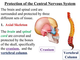
1. The brain and spinal cord are covered and protected by the bones of the skull, specifically the cranium , and the vertebral column . Protection of the Central Nervous System 1. Axial Skeleton Vertebral Column The brain and spinal cord are surrounded and protected by three different sets of tissue. Cranium
2. Protection of the Central Nervous System The brain has hollow, fluid-filled cavities called ventricles . Inside the ventricles is a structure called the choroid plexus (see Neuroglia ; slide 5; ependymal cells) that makes a clear, colorless fluid called cerebrospinal fluid (CSF) . CSF circulates within and around the brain and spinal cord to help cushion it from injury. 2. Ventricles and cerebrospinal fluid
3. CSF is constantly being absorbed and replenished. A “perfect” balance is maintained between absorption and replenishment. A disruption or blockage in the system can cause a build up of CSF, which can cause enlargement of the ventricles, called hydrocephalus, or cause a collection of fluid in the spinal cord, called syringomyelia. Protection of the Central Nervous System
4. Protection of the Central Nervous System 3. The Meningies The brain and spinal cord are covered and protected by three layers of tough, connective tissue called meninges . From the outermost layer inward they are: the dura mater , arachnoid mater , and pia mater . White matter Gray matter
5. The dura mater is a strong, thick membrane that closely lines the inside of the skull. It has two layers, the periosteal and meningeal dura , which are mostly fused, but do separate to form venous sinuses. Protection of the Central Nervous System 3. The Meningies White matter Gray matter
6. The dura creates little folds or compartments. There are two special dural folds, the falx and the tentorium . The falx separates the right and left hemispheres of the brain and the tentorium separates the cerebrum from the cerebellum . Protection of the Central Nervous System White matter Gray matter
7. The arachnoid mater is a thin, web-like membrane that covers the entire brain. The arachnoid is made of elastic tissue . The space between the dura and arachnoid membranes is called the subdural space . Protection of the Central Nervous System 3. The Meningies White matter Gray matter
8. The pia mater hugs the surface of the brain following its folds and grooves. The pia mater has many blood vessels that reach deep into the brain. Protection of the Central Nervous System 3. The Meningies White matter Gray matter
9. The space between the arachnoid and pia is called the subarachnoid space . It is here where the cerebrospinal fluid bathes and cushions the brain . Protection of the Central Nervous System White matter Gray matter
10. Blood is carried to the brain by two paired arteries, the internal carotid arteries and the vertebral arteries . The internal carotid arteries supply most of the cerebrum . The vertebral arteries supply the cerebellum , brainstem , and the underside of the cerebrum . After passing through the skull, the right and left vertebral arteries join together to form the basilar artery. Arterial Blood Supply of the Brain
11. The basilar artery and the internal carotid arteries “communicate” with each other at the base of the brain called the Circle of Willis. The communication between the internal carotid and vertebral-basilar systems is an important safety feature of the brain. If one of the major vessels becomes blocked, it is possible for collateral blood flow to come across the Circle of Willis and prevent brain damage.
12. The venous circulation of the brain is very different than the rest of the body. Usually arteries and veins run together as they supply and drain specific areas of the body. In the brain, the major vein collectors are integrated into the dura to form venous sinuses - not to be confused with the air sinuses in the face and nasal region. Venous Blood Drainage of the Brain
13. The venous sinuses collect the blood from the brain and pass it to the internal jugular veins. The superior and inferior sagittal sinuses drain the cerebrum, the cavernous sinuses drains the anterior skull base. All sinuses eventually drain to the sigmoid sinuses, which exit the skull and form the jugular veins. These two jugular veins are essentially the only drainage of the brain.
14. The Blood Brain Barrier is a physiological mechanism that alters the permeability of brain capillaries so that some substances are prevented from entering brain tissue, while other substances are allowed to enter freely. The Blood Brain Barrier
15. A key aspect of the blood-brain barrier are the thin, flat cells known as endothelial cells which form the walls of capillaries. In most parts of the body, the endothelial cells in the capillaries overlap at what are called junctions . These junctions are leaky enough to let a lot of different materials move through the wall of the blood vessel into the tissue and back again.
16. However, in the brain there's a different arrangement. The endothelial cells meet at what are called tight junctions . These tight junctions block the passage of most things except for small, hydrophobic molecules like O 2 , CO 2 , & hormones. Cells of the barrier also actively transport metabolic products such as glucose molecules.
17. In addition to tight junctions , the "end feet" of astrocytes (see Neuroglia; slide 3; Astrocytes) surround the outside of capillary endothelial cells . The reason for this endothelial-glial connection is unclear, but may reflect an influence of astrocytes on the formation and maintenance of the blood-brain barrier. Astrocyte Capillary “ End Foot” of Astrocyte