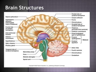
The document describes several structures of the brain and ventricular system. It discusses:
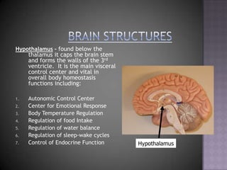
- The hypothalamus, located below the thalamus, which controls autonomic and endocrine functions.
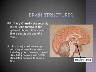
- The pituitary gland, located in the sella turcica bone, which secretes 9 hormones including growth hormone.
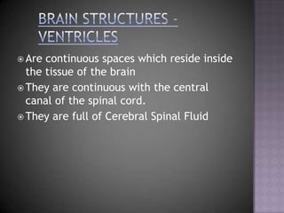
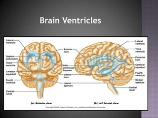
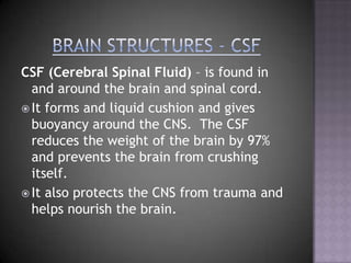
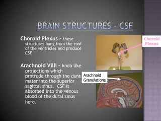
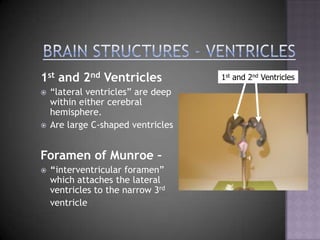
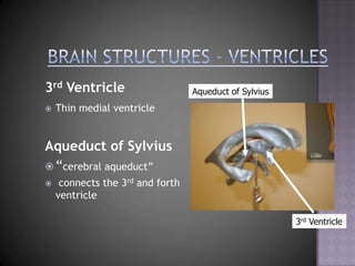
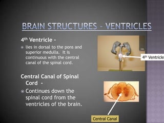
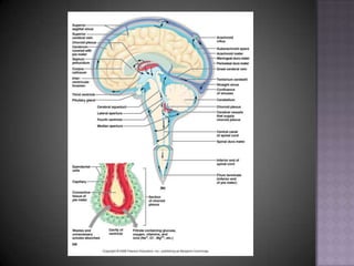
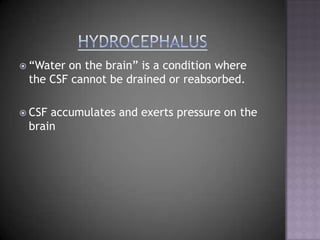
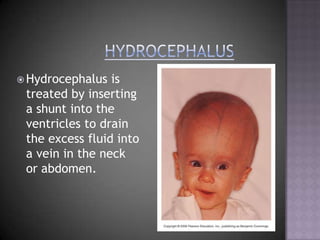
- The ventricles which are continuous spaces within the brain that contain cerebrospinal fluid (CSF). The CSF cushions the brain and is produced in the choroid plexus.
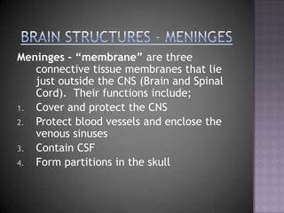
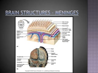
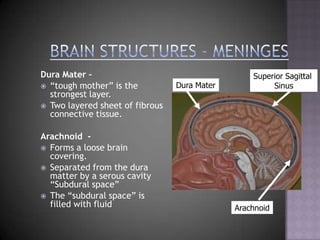
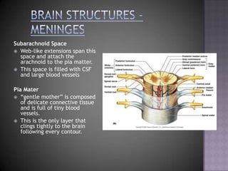
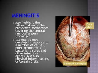
- Meningitis, which is inflammation of the meninges layers covering the brain and spinal cord caused by bacteria, viruses or physical injury.