The science of yoga is the scientific basis of modern yoga as exercise in human sciences such as anatomy, physiology, and psychology. Yoga's effects are to some extent shared with other forms of exercise,[O 1] though it differs in the amount of stretching involved, and because of its frequent use of long holds and relaxation, in its ability to reduce stress. Yoga is here treated separately from meditation, which has effects of its own, though yoga and meditation are combined in some schools of yoga.
The nervous system controls and coordinates the activities of the body. It has both voluntary and involuntary functions. The autonomic nervous system regulates involuntary functions like heart rate and digestion. It has two divisions - the sympathetic and parasympathetic systems which generally have opposing effects on organs. The sympathetic system prepares the body for fight or flight while the parasympathetic maintains normal functions. Dysfunctions of the autonomic nervous system can cause issues like high blood pressure, digestive problems, and more.
The document summarizes key aspects of respiratory anatomy and physiology. It describes the structure and function of the lungs, including details on lobes and fissures, vascular and lymphatic supply, innervation and development. It also discusses common respiratory diseases and clinical relevance of respiratory anatomy, such as in bronchoscopy, pneumonia, tuberculosis, asthma and lung cancer.
The document provides an overview of human anatomy and physiology, with a focus on the respiratory and circulatory systems. It defines anatomy and physiology, describes anatomical terms and positions, and reviews the key structures and functions of the skeletal, respiratory, and circulatory systems. The respiratory system section examines the upper airway, lower airway, gas exchange, and respiratory evaluation. The circulatory system section states its overall function of nutrient/oxygen transport and waste removal.
This document provides an overview of the nervous system's general and special senses. It discusses the different types of receptors, including mechanoreceptors, chemoreceptors, thermoreceptors, and nociceptors. It describes the pathways for different senses such as olfaction, gustation, equilibrium, hearing, and vision. Key structures are highlighted, including the cochlea, semicircular canals, maculae, and organ of Corti. Diagrams and figures illustrate the anatomical structures associated with different sensory systems.
The document discusses the anatomy of the thorax as seen on MRI. It describes the various components of the thorax including the thoracic wall with its skeleton and joints, the thoracic cavities containing the mediastinum and pleural cavities, the neurovasculature including thoracic arteries and veins, and internal organs such as the heart, lungs, and trachea. The thoracic wall forms a protective cage around the thoracic contents. Within the wall are openings that connect the thorax superiorly to the neck and inferiorly to the abdomen.
The thoracic cavity contains the lungs, heart, esophagus, and other structures. It is bounded by the thoracic wall anteriorly and the vertebral column posteriorly. The mediastinum divides the cavity into left and right pleural cavities which contain the lungs. The diaphragm separates the thoracic and abdominal cavities. Key structures in the thoracic cavity include the lungs, trachea, bronchi, blood vessels, esophagus, thymus gland, and diaphragm.
The chest cavity contains the lungs, heart, major blood vessels, and other structures. It is bounded by the ribs, sternum, vertebral column, and diaphragm. The chest cavity is further divided into the pleural cavities and mediastinum. The mediastinum is the median partition between the lungs and contains the esophagus, trachea, thymus, and major blood vessels. Each lung is surrounded by a pleural membrane made of visceral and parietal layers that create a pleural cavity containing fluid.
The thorax refers to the region which forms a major part of the appendicular skeleton. Knowledge of its surface anatomy is essential for surgical techniques, to say the least.
This document provides an overview of lung anatomy including:
1. The upper respiratory system including the nose, nasal cavity, paranasal sinuses, and pharynx.
2. The structure and lobes of the lungs including fissures, surfaces, and impressions.
3. Details of the airways including the trachea, bronchi, bronchioles and their branching patterns.
4. Other aspects such as the muscles involved in respiration, circulation, innervation, and lymphatics of the lungs.
The respiratory system functions to provide oxygen to tissues and remove carbon dioxide through gas exchange that occurs in the alveoli of the lungs. It includes both conducting zones that transport air, and respiratory zones involved in gas exchange. Key structures are the nasal cavity, pharynx, larynx, trachea, bronchi, lungs, and pleurae. Breathing occurs through changes in pressure between the atmosphere and lungs, driven by the diaphragm and intercostal muscles, with inspiration occurring when the thoracic cavity volume increases and expiration when it decreases.
The document summarizes the main components and functions of the central nervous system. It discusses the brain regions including the cerebrum, diencephalon, brain stem, and cerebellum. It also covers the spinal cord, protective coverings, blood supply, common disorders like strokes, and the cranial nerves.
The thorax contains the organs of the respiratory and cardiovascular systems. It is bounded by the sternum, vertebrae, and ribs. Within the thoracic cavity are two pleural cavities housing the lungs and a central mediastinum containing the heart, great vessels, trachea and esophagus. The lungs, pleura, chest wall and mediastinal structures were described in detail.
Segmental anatomy of lungs , anatomy of mediastinum and secondary lobuleGamal Agmy
The document discusses the segmental anatomy of the lungs and secondary lobule. It notes that there are approximately 23 generations of dichotomous branching from the trachea to the alveolar sacs. The secondary lobule is described as the basic anatomic unit of pulmonary structure and function, measuring 1-2 cm and containing 5-15 pulmonary acini. It is supplied by a terminal bronchiole in the center and surrounded by connective tissue septa and two lymphatic systems. Diseases typically manifest in either the centrilobular or perilymphatic areas based on how they enter the lungs.
Anatomy and physiology of cerebral circulation and csfSeri Lollen
The document discusses the anatomy and physiology of the cerebral circulation and cerebrospinal fluid (CSF). It describes the major arteries and veins that make up the brain's blood supply, including the internal carotid arteries, vertebral arteries, and circle of Willis. It explains how blood flows into the anterior and posterior circulations to supply different brain regions. It also outlines the ventricles and flow of CSF within the brain and spinal cord.
The document discusses the pleura, which forms the pleural cavity surrounding the lungs. It describes the two layers of pleura - the visceral pleura adhering to the lungs and the parietal pleura lining the thoracic wall. It then discusses pleural reflections, recesses, nerve supply, and various pleural diseases and conditions like pleurisy, effusions, pneumothorax, and haemothorax. It also shows images related to pleural anatomy, demonstrations of tension pneumothorax, and labels parts on a chest X-ray.
The urinary system develops through three successive phases - the pronephros, mesonephros, and metanephros. The pronephros is nonfunctional in mammals. The mesonephros allows for filtration of blood into mesonephric tubules. During the fifth week of gestation, the ureteric bud induces nearby mesoderm to form the metanephros, or permanent kidneys. The kidneys filter waste from the blood to form urine, which drains via the ureters into the bladder and is then excreted through the urethra. The basic functional unit of the kidneys is the nephron, which filters the blood and reabsorbs necessary molecules
The document provides an overview of the anatomy, internal structure, blood supply, and functional organization of the spinal cord. Key points include:
- The spinal cord extends from the foramen magnum to L1-L2 and is segmented into cervical, thoracic, lumbar, and sacral regions.
- It has a butterfly-shaped gray matter H-shape surrounded by white matter tracts that contain ascending and descending nerve fibers.
- The principal blood supply comes from the anterior and posterior spinal arteries along with segmental radicular arteries like the artery of Adamkiewicz.
- The spinal cord has a segmental organization corresponding to 31 pairs of spinal nerves that mediate motor and sensory functions.
Unit-III, chapter-2- Lymphatic System,
Functions of Lymphatic System,
Major Parts of Lymphatic System,
Composition of Lymph,
Lymph and Lymphatic Capillaries,
Structure of lymph node,
Mechanisms of Lymph Flow,
Functions of Lymph Node,
Mucosa-Associated Lymphoid Tissue (MALT),
As per PCI syllabus,
B. Pharm. First Year,
Human Anatomy and Physiology-I.
The pleura is a membrane that surrounds the lungs and lines the chest cavity. It has two layers - the parietal pleura lining the chest wall and diaphragm, and the visceral pleura attached directly to the lungs. Pleurisy is inflammation of the pleura that causes sharp chest pain worsened by breathing. It is usually caused by viral infection but can result from other lung and heart conditions. Diagnosis involves listening for pleural friction rubs and tests like chest x-rays. Treatment focuses on pain relief, draining excess fluid in the pleural space, and addressing the underlying cause.
The document summarizes the anatomy of the thorax. It contains:
- The thorax is bounded superiorly by the root of the neck and inferiorly by the diaphragm. It contains the heart, lungs, and major blood vessels.
- The mediastinum is the thick soft tissue partition that separates the two pleural cavities and contains the heart and other structures.
- The lungs are enclosed in pleural cavities and separated from each other by the mediastinum.
This document provides an overview of the anatomy of the lungs, pleura, and diaphragm. It describes the structure and function of the lungs including lobes and fissures. It discusses the pleura, its layers and reflections. It also details the origins, parts and functions of the diaphragm. Throughout it includes labeling of diagrams to enhance understanding of respiratory structures.
This document provides an overview of the pleura and lung. It begins by introducing the lung and its parts, including lobes and borders. It then describes the pleural layers in detail, including the parietal pleura layers of cervical, costal, mediastinal and diaphragmatic pleura. Relations and blood supply of the pleura are discussed. Bronchopulmonary segments and clinical significance are summarized. Common pleural conditions like pleurisy, pleural effusion and pneumothorax are briefly mentioned.
This document provides information about anatomy of the chest wall, thoracic cavity, lungs, heart and related structures. It covers topics such as layers of the thoracic wall, bones and muscles of the rib cage, structures passing through thoracic inlets and outlets, anatomy of breathing including the pleural membranes and diaphragm, vascular and lymphatic drainage of the chest, lobes and fissures of the lungs, structures within the mediastinum, and basic cardiac anatomy including the pericardium and internal structures of the atria and ventricles. The document is in a question and answer format to test knowledge of these anatomical structures and relationships.
This document describes the anatomy of the lungs and pleura. It discusses the borders and lobes of the lungs, noting key landmarks like the apex and angles of Louis. It also details the fissures that divide the lungs into lobes, such as the oblique and transverse fissures. The document outlines the segmental bronchi and notes their anterior and posterior divisions. Furthermore, it describes the layers of the pleura, including the parietal and visceral pleura. Several important surface anatomy landmarks are indicated, such as Traube's area, Kronig's isthmus, and the bare area of the heart. In closing, the upper border of the liver is delineated starting at the 6th rib.
The mediastinum is the space within the thorax bounded by the sternum anteriorly, the thoracic vertebral column posteriorly, and the mediastinal pleura on either side. It is divided into superior and inferior portions by a line passing through the sternal angle and lower border of T4 vertebra. The inferior mediastinum is further divided into the anterior, middle, and posterior mediastinum. The superior mediastinum contains structures such as the trachea, esophagus, thymus, thoracic duct, and arch of the aorta. The middle mediastinum includes the heart within the pericardium as well as the bifurcation of the trachea and principal bronchi.
What is the art of yoga?
Yoga is a portable art form, free from costly equipment and supplies. All it requires is a sense of adventure and a willingness to explore the vastness of the Self. ... The art of life is life as art. Yoga means joining together what we do with who we are: Love itself.
SCOPE OF HUMAN ANATOMY AND PHYSIOLOGY.pptxAvneshSingh3
This document provides an introduction to human anatomy and physiology. It defines anatomy and physiology, and describes the subdivisions of anatomy including gross (macroscopic) anatomy, microscopic anatomy, and clinical anatomy. It also discusses the levels of organization in the human body from the chemical level to the tissue, organ, and system levels. Key body cavities like the cranial, thoracic, abdominal, and pelvic cavities are outlined. Major body organs in each cavity are identified. Anatomical terminology, planes, quadrants, and medical imaging techniques are also summarized.
This document provides an overview of human anatomy, including:
1) It describes the different branches of anatomy such as gross anatomy, microscopic anatomy, developmental anatomy, and comparative anatomy.
2) It explains the hierarchical structural organization of the human body from simplest to most complex, including chemical, cellular, tissue, organ, and organ system levels.
3) It lists and briefly describes the main organ systems in the human body.
The document provides an overview of 11 human body systems: digestive, urinary, respiratory, circulatory, skeletal, muscular, nervous, integumentary, immune, endocrine, and reproductive. For each system, the key organs and their functions are described. Common diseases associated with each system are also listed. The document emphasizes how body systems work interdependently, with the circulatory system connecting many organs and the nervous system coordinating responses across systems.
The document defines various medical terms related to the study of disease causation (etiology, idiology), tissue (histology), the heart (systole, diastole), fluid accumulation (edema), inflammation (-itis), and blood clots (thrombus, embolus). It also provides trivia questions about anatomy and physiology, including the parts of organs like the kidney, small intestine, brain, and skeletal system. Finally, it defines some common medical conditions like Parkinson's disease, epilepsy, stroke, Bell's palsy, and sciatica.
The document summarizes key information about the structure and function of the brain and cerebrospinal fluid (CSF). It describes the four ventricles that produce CSF and how it circulates through the brain and spinal cord. It discusses the formation of CSF by the choroid plexus and its absorption. The structure and functions of structures like the basal ganglia, thalamus, and blood-brain barrier are also summarized briefly.
The document provides information about the skeletal, ear, eye, and lung systems. It describes the main bones and parts of the skeletal, ear, eye and lung anatomy. It explains key functions such as support, protection, movement, hearing, vision, respiration and gas exchange. The skeletal system has 206 bones and provides structure, while the ear detects sounds, the eye allows vision, and the lungs enable breathing and oxygen/carbon dioxide exchange.
1. The document provides an outline for a presentation on nursing assessment of the neck, including objectives, introduction to neck anatomy, and procedures for physical examination of the neck and lymphatic system.
2. Key steps in physical examination of the neck involve inspection, palpation, auscultation, and assessment of lymph nodes at different locations in the neck.
3. The lymphatic system acts as a drainage system and filters for lymph fluid, and enlarged or tender lymph nodes may indicate various inflammatory or infectious conditions.
The hippocampus is located deep in the brain and plays an important role in memory formation and spatial navigation. It is involved in fear conditioning and damage to the hippocampus has been linked to anxiety disorders like PTSD. The hippocampus contains anxiety cells that respond to unfamiliar places, and suppressing these cells reduces anxiety in mice. CBD may help reduce activity in the amygdala and aid signaling in the endocannabinoid system involving the hippocampus to help treat anxiety symptoms.
The brain and spinal cord are protected and cushioned by three layers of tissue (the meninges) and cerebrospinal fluid. The meninges include the dura mater, arachnoid mater, and pia mater. Cerebrospinal fluid is produced by the choroid plexus within the ventricles and circulates around the brain and spinal cord. Blood enters the brain through the internal carotid and vertebral arteries, and drains into venous sinuses which form the jugular veins. The blood brain barrier regulates what substances can enter the brain by restricting passage through tight junctions between endothelial cells.
The document lists various important points about anatomy and physiology:
- Motor nerves carry impulses from the brain to sense organs, while afferent nerves carry impulses from sense organs to the brain. Blood plasma and cerebrospinal fluid are examples of extracellular fluids.
- The human body develops from a single cell called a zygote, and cells are the smallest functional units of the body. The brain and spinal cord are surrounded by three layers of tissue.
- Gases are exchanged between the lungs, blood, and body tissues through diffusion. External respiration is the exchange between the lungs and blood, while internal respiration is the exchange between blood and tissues.
This document provides a review of key terms and concepts for an anatomy final exam. It defines anatomical directions like anterior and posterior. It describes the four main tissue types - epithelial, connective, muscle and neural tissue. It discusses the skeletal system including bones and joints. Finally, it reviews topics related to the nervous system like neurons, the brain and spinal cord, and the autonomic nervous system.
This document provides an overview of anatomy and physiology. It defines anatomy as the structure of body parts and physiology as the function of body parts. It notes that structure and function are interrelated. Examples are provided of how hand and foot structure relates to grasping ability. It also discusses how heart valve function impacts heart structure over time. Characteristics of life and requirements for maintaining life like water, food, oxygen, heat, and pressure are outlined. The concept of homeostasis and examples of maintaining core temperature and blood pressure and glucose levels are described. Feedback systems and levels of organization from atoms to organisms are defined. The major body cavities, portions, tissues, organs and systems are identified. Finally, membranes in the thoracic and abdominal cav
This document provides an introduction and overview of anatomy and physiology. It begins by defining anatomy as the study of the body's structure and physiology as the study of how the body's parts work and cooperate. It then discusses the different levels of human organization from chemical to cellular to tissue to organ to organ systems. Key concepts covered include homeostasis, positive and negative feedback systems, and common anatomical terminology prefixes. The document provides a high-level introduction to the scope and goals of studying anatomy and physiology.
The pituitary gland, also called the hypophysis, is a small gland located at the base of the brain. It is divided into three lobes - the anterior, intermediate, and posterior lobes. The anterior lobe secretes hormones that regulate other endocrine glands like the thyroid, while the posterior lobe stores and releases hormones important for water balance and childbirth. The pituitary is attached to the hypothalamus and receives signals that stimulate or inhibit hormone release to maintain homeostasis. Diseases of the pituitary can disrupt hormone levels throughout the body.
The document summarizes the human movement, respiratory, circulatory, and digestive systems. It describes the key organs in each system, such as muscles, bones, and joints in the movement system. It explains how each system functions, for example how gas exchange occurs in the lungs and oxygenated blood is transported by the circulatory system. It also lists some common abnormalities and disorders that can affect each bodily system.
The nervous system is composed of nerve cells called neurons that communicate with each other and other cells. It has two main divisions - the central nervous system consisting of the brain and spinal cord, and the peripheral nervous system consisting of nerves linking the body to the CNS. The brain is protected by membranes and bone and has four main regions - the cerebrum, cerebellum, diencephalons, and brain stem. The cerebrum controls intellectual functions, the cerebellum controls muscle coordination, and the diencephalons relay sensory information and regulate body functions. Cerebrospinal fluid cushions and nourishes the brain and spinal cord. Blood circulates to the brain through arteries and veins, protected by the blood
Review of Nervous System, Unconsciousness, and CVA. The Nursing Core FunctionsAyinla Kazeem
This presentation was made at several sessions of Mandatory Continuing Professional Development Programme for Nigerian Nurses in Kwara State, and have undergone series of editing till date. While still working on the final editing to totally conform with global standard of practice, I deemed it necessary to share it in this forum.
This presentation contains the explanation of different systems in our body. For example : respiratory,circulatory etc.I hope you will enjoy the presentation :)
Photography is the art, application, and practice of creating durable images by recording light, either electronically by means of an image sensor, or chemically by means of a light-sensitive material such as photographic film.
This is the real secret of the yoga practice. When we learn not only to breathe with the postures, but actually breathe them, we can enter into a place where our focus and attention is so unified that there’s nowhere else to be but inside our bodies. This is the root meaning of yoga in Sanskrit: “union.” This can be such a sweet experience that a friend of mine once described it as “the feeling of someone brushing your fur in the right direction.” That might sound nuts—until you try it. Here’s how:
First you must understand that breath already is movement. Take a deep breath and notice what happens. On the inhale, you get a little bigger, and more expansive: your belly and chest rise. As you exhale, you get a little smaller, a little denser: your belly and chest contract. The inhale has an upward flowing energy, called prana. The exhale is a downward flowing energy called apana.
The Yoga Sūtra of Patañjali is a collection of Sanskrit sutras (aphorisms) on the theory and practice of yoga - 195 sutras (according to Vyāsa and Krishnamacharya) and 196 sutras (according to other scholars including BKS Iyengar). The Yoga Sutra was compiled sometime between 500 BC and AD400 by the sage Patanjali in India who synthesized and organized knowledge about yoga from much older traditions.[1][2][3] The Yoga Sūtra of Patañjali was the most translated ancient Indian text in the medieval era, having been translated into about forty Indian languages and two non-Indian languages: Old Javanese and Arabic.[4] The text fell into relative obscurity for nearly 700 years from the 12th to 19th century, and made a comeback in late 19th century due to the efforts of Swami Vivekananda, the Theosophical Society and others. It gained prominence again as a comeback classic in the 20th century.[5]
Swami Vivekananda introduced yoga to Western audiences in 1893 at the World's Parliament of Religions in Chicago. However, yoga was not well known at the time and Vivekananda presented it as a philosophical practice rather than the posture-based practice it is known as today. Yoga started gaining more popularity in the US in the 1920s-1930s through Indian immigrants. The physical aspects of hatha yoga were merged with Western exercise in the early 1900s. Yoga truly became mainstream in the US starting in the 1960s through its adoption by the hippie and New Age movements and its presentation on television and in fitness studios.
Students and Learners can participate in the national-level quiz competition by logging onto Diksha App until 20th July 2020. Alternatively, students and learners can also click on the link given below to get more information about the quiz and register for the competition directly.
Rishikesh in India is considered the birthplace of yoga. The practice of yoga began over 5,000 years ago in northern India. Though practiced earlier, the great sage Patanjali systematized yoga around 500 BC by compiling existing practices into his Yoga Sutras text. Yoga aims for self-realization and liberation through harmonizing the mind and body.
The document provides information on an individual's work experience and qualifications as a yoga instructor. It lists multiple roles as a yoga instructor from 2005 to present at various locations. It also lists educational and skill qualifications including a Bachelor's Degree in Product Knowledge and over 200 hours of yoga training and certification. The individual has extensive experience teaching yoga and fitness classes and maintaining safety and cleanliness standards.
This document provides information about Sharadha Yoga Center and Ayurveda International Academy, including its affiliations, courses offered, values, mission, and vision. It discusses the importance of certification for yoga teachers and the objectives of yoga teacher training courses. The document also outlines the course content, including fundamentals of yoga education, yoga practices, yoga therapy, and practical lessons.
This document provides biographical information about Yogacharya Dr. S. Yegneshwaran. It details his educational background and qualifications in fields including yoga, Ayurveda, medicine and counseling. It also outlines his professional experience as the founder and director of Sharadha Yoga Center and Ayurveda International Academy, and lists his publications, productions and awards related to yoga education, practice and therapy.
The document provides a history of Ashtanga Yoga, distinguishing between Ashtanga Yoga as Patanjali's eight limbed path and Ashtanga Vinyasa Yoga as the physical practice developed by Pattabhi Jois. It describes how the physical yoga postures were introduced later to prepare the body for meditation. It then discusses the origins and development of Ashtanga Vinyasa Yoga, including how Pattabhi Jois and Krishnamacharya were said to have discovered the ancient Yoga Korunta text describing its six series of postures. However, the original text was later destroyed by ants.
Yoga is a group of physical, mental, and spiritual practices or disciplines which originated in ancient India. Yoga is one of the six Āstika schools of Indian philosophical traditions. There is a broad variety of yoga schools, practices, and goals in Hinduism, Buddhism, and Jainism.
Strategies for Effective Upskilling is a presentation by Chinwendu Peace in a Your Skill Boost Masterclass organisation by the Excellence Foundation for South Sudan on 08th and 09th June 2024 from 1 PM to 3 PM on each day.
বাংলাদেশের অর্থনৈতিক সমীক্ষা ২০২৪ [Bangladesh Economic Review 2024 Bangla.pdf] কম্পিউটার , ট্যাব ও স্মার্ট ফোন ভার্সন সহ সম্পূর্ণ বাংলা ই-বুক বা pdf বই " সুচিপত্র ...বুকমার্ক মেনু 🔖 ও হাইপার লিংক মেনু 📝👆 যুক্ত ..
আমাদের সবার জন্য খুব খুব গুরুত্বপূর্ণ একটি বই ..বিসিএস, ব্যাংক, ইউনিভার্সিটি ভর্তি ও যে কোন প্রতিযোগিতা মূলক পরীক্ষার জন্য এর খুব ইম্পরট্যান্ট একটি বিষয় ...তাছাড়া বাংলাদেশের সাম্প্রতিক যে কোন ডাটা বা তথ্য এই বইতে পাবেন ...
তাই একজন নাগরিক হিসাবে এই তথ্য গুলো আপনার জানা প্রয়োজন ...।
বিসিএস ও ব্যাংক এর লিখিত পরীক্ষা ...+এছাড়া মাধ্যমিক ও উচ্চমাধ্যমিকের স্টুডেন্টদের জন্য অনেক কাজে আসবে ...
A workshop hosted by the South African Journal of Science aimed at postgraduate students and early career researchers with little or no experience in writing and publishing journal articles.
The simplified electron and muon model, Oscillating Spacetime: The Foundation...RitikBhardwaj56
Discover the Simplified Electron and Muon Model: A New Wave-Based Approach to Understanding Particles delves into a groundbreaking theory that presents electrons and muons as rotating soliton waves within oscillating spacetime. Geared towards students, researchers, and science buffs, this book breaks down complex ideas into simple explanations. It covers topics such as electron waves, temporal dynamics, and the implications of this model on particle physics. With clear illustrations and easy-to-follow explanations, readers will gain a new outlook on the universe's fundamental nature.
This presentation includes basic of PCOS their pathology and treatment and also Ayurveda correlation of PCOS and Ayurvedic line of treatment mentioned in classics.
This presentation was provided by Steph Pollock of The American Psychological Association’s Journals Program, and Damita Snow, of The American Society of Civil Engineers (ASCE), for the initial session of NISO's 2024 Training Series "DEIA in the Scholarly Landscape." Session One: 'Setting Expectations: a DEIA Primer,' was held June 6, 2024.
ISO/IEC 27001, ISO/IEC 42001, and GDPR: Best Practices for Implementation and...PECB
Denis is a dynamic and results-driven Chief Information Officer (CIO) with a distinguished career spanning information systems analysis and technical project management. With a proven track record of spearheading the design and delivery of cutting-edge Information Management solutions, he has consistently elevated business operations, streamlined reporting functions, and maximized process efficiency.
Certified as an ISO/IEC 27001: Information Security Management Systems (ISMS) Lead Implementer, Data Protection Officer, and Cyber Risks Analyst, Denis brings a heightened focus on data security, privacy, and cyber resilience to every endeavor.
His expertise extends across a diverse spectrum of reporting, database, and web development applications, underpinned by an exceptional grasp of data storage and virtualization technologies. His proficiency in application testing, database administration, and data cleansing ensures seamless execution of complex projects.
What sets Denis apart is his comprehensive understanding of Business and Systems Analysis technologies, honed through involvement in all phases of the Software Development Lifecycle (SDLC). From meticulous requirements gathering to precise analysis, innovative design, rigorous development, thorough testing, and successful implementation, he has consistently delivered exceptional results.
Throughout his career, he has taken on multifaceted roles, from leading technical project management teams to owning solutions that drive operational excellence. His conscientious and proactive approach is unwavering, whether he is working independently or collaboratively within a team. His ability to connect with colleagues on a personal level underscores his commitment to fostering a harmonious and productive workplace environment.
Date: May 29, 2024
Tags: Information Security, ISO/IEC 27001, ISO/IEC 42001, Artificial Intelligence, GDPR
-------------------------------------------------------------------------------
Find out more about ISO training and certification services
Training: ISO/IEC 27001 Information Security Management System - EN | PECB
ISO/IEC 42001 Artificial Intelligence Management System - EN | PECB
General Data Protection Regulation (GDPR) - Training Courses - EN | PECB
Webinars: https://pecb.com/webinars
Article: https://pecb.com/article
-------------------------------------------------------------------------------
For more information about PECB:
Website: https://pecb.com/
LinkedIn: https://www.linkedin.com/company/pecb/
Facebook: https://www.facebook.com/PECBInternational/
Slideshare: http://www.slideshare.net/PECBCERTIFICATION
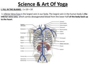
1. Science & Art Of Yoga
I. FILL IN THE BLANKS : 1 x 10 = 10
1. inferior Vena Cava is the largest vein in our body. The largest vein in the human body is the
inferior vena cava, which carries deoxygenated blood from the lower half of the body back up
to the heart.
2. CSF is secreted by ……………
choroid plexus
The epithelial cells of the choroid plexus secrete cerebrospinal fluid (CSF), by a process that
involves the movement of Na(+), Cl(-) and HCO(3)(-) from the blood to the ventricles of the
brain. This creates the osmotic gradient, which drives the secretion of H(2)O.
3. A disease in which the hyposecretion of thyroid glands in adult is called…… For example,
insufficient production (hyposecretion) of thyroid stimulating hormone (TSH) in the pituitary
gland will cause hypothyroidism, while overproduction (hypersecretion) of TSH will cause
hyperthyroidism. ……
4. Q4.the shortest rib in human body is called
A.twelfth rib
The twelfth rib is an atypical rib. It is the shortest rib, and one of two floating ribs.
1. There are seven pairs of true ribs. ... They differ from false and floating ribs because they
directly articulate with the sternum by means of their costal cartilages. The
shortest true rib is rib 1 and their length increases all the way to rib 7. Also, the radius of their
curvature increases progressing inferiorly.
5. The human skeleton consists of … 206 …….. bones
The adult human skeleton usually consists of 206 named bones. These bones can be grouped in
two divisions: axial skeleton and appendicular skeleton. The 80 bones of the axial skeleton form
the vertical axis of the body. They include the bones of the head, vertebral column, ribs and
breastbone or sternum.
6. The study of bone is called ……..
Orthopaedics is the study of the musculoskeletal system. ... The
musculoskeletal system provides form, stability, and movement to
the human body. It is made up of the body's bones (the skeleton),
muscles, cartilage, tendons, ligaments, joints, and other connective
tissue.
7. arterioles is a small artery
The aorta branches into a network of smaller arteries that extend throughout the body.
The arteries' smaller branches are called arterioles and capillaries. The pulmonary arteries carry
oxygen-poor blood from the heart to the lungs under low pressure, making
these arteries unique.
8. The first vertebra is known as Atlas (C1)
The first cervical vertebra is a bony ring with a thin anterior arch and posterior laminae, which
are joined by lateral masses having articular facets that articulate with the occipital condyles
superiorly and the lateral masses of C2 inferiorly.
Atlas (anatomy)
9. The contraction of the ventricles produces…
first heart sound, S1 ……….
The ventricles begin to contract (ventricular systole), raising pressure within the ventricles.
When ventricular pressure rises above the pressure in the atria, blood flows toward the
atria, producing the first heart sound, S1 or lub.
Cardiac Cycle | Anatomy and Physiology
10. Ovaries in the females which secrete
…………….and…………… oestrogen and progesterone.
The ovaries produce and release eggs (oocytes) into the female reproductive tract at the mid-
point of each menstrual cycle. They also produce the female
hormones oestrogen and progesterone.
11. II. DEFINE THE FOLLOWING : 2 x 10 = 20
1) Thyroid gland
The thyroid, or thyroid gland, is an
endocrine gland in the neck consisting of two
connected lobes. The lower two thirds of the
lobes are connected by a thin band of tissue
called the thyroid isthmus. The thyroid is
located at the front of the neck, below the
Adam's apple.
12. Digestion
Digestion is the breakdown of large insoluble food molecules into small water-soluble food
molecules so that they can be absorbed into the watery blood plasma. In certain organisms,
these smaller substances are absorbed through the small intestine into the blood stream.
13. Pericardium
The pericardium, also called pericardial sac, is
a double-walled sac containing the heart and
the roots of the great vessels. It has two layers,
an outer layer made of strong connective
tissue (fibrous pericardium), and an inner layer
made of serous membrane
(serous pericardium).
Location: A sac around the heart
Nerve: Phrenic nerve
14. Liver cirrhosis
Cirrhosis is a late stage of scarring (fibrosis) of
the liver caused by many forms
of liver diseases and conditions, such as
hepatitis and chronic alcoholism. Each time
your liver is injured — whether by disease,
excessive alcohol consumption or another
cause — it tries to repair itself.06-Feb-2021
Risk Factors: Non-alcoholic fatty liver disease
15. Write about mid brain?
Midbrain, also called mesencephalon, region
of the developing vertebrate brain that is
composed of the tectum and tegmentum. ...
The midbrain serves important functions in
motor movement, particularly movements of
the eye, and in auditory and visual processing.
16. Explain pulmonary circulation.
The pulmonary circulation is the portion of the
circulatory system which carries deoxygenated
blood away from the right ventricle, to the
lungs, and returns oxygenated blood to the left
atrium and ventricle of the heart. The term
pulmonary circulation is readily paired and
contrasted with the systemic circulation.
17. Define Pancha Kosha
Panchakoshas, are the layers of body that
seemingly cover the Atman. The Tvam
padartha of the Mahavakya Tat Tvam Asi is
determined by the analysis of Panchakoshas
that are not the atman.
18. Define Pathya.
What is Pathya in Ayurveda?
Pathya refers to that which gives relief to the
person by the use of diet, regimen and
medicine. On the contrary, Apathya aggravates
the disease. The Pathya and Apathya are
effective tools in Ayurveda for diagnosis as
well as management of diseases.
19. Hormones
Hormones are your body's chemical
messengers. They travel in your bloodstream
to tissues or organs. They work slowly, over
time, and affect many different processes,
including. Growth and development.
Metabolism - how your body gets energy from
the foods you eat.
20. atlas
The atlas is one of the two upper cervical
vertebrae, also known as C1, which is the
topmost vertebra of the spinal column. ... It is
the vertebra that is in contact with the
occipital bone, a flat bone located at the back
portion of the head.
what is atlas anatomy
21. III. WRITE BRIEF ANSWER FOR ANY 5
QUESTIONS : 5 x 5 = 25
Brain ?
The human brain is the central organ of the human nervous system, and with the spinal
cord makes up the central nervous system. The brain consists of the cerebrum,
the brainstem and the cerebellum. It controls most of the activities of the body, processing,
integrating, and coordinating the information it receives from the sense organs, and making
decisions as to the instructions sent to the rest of the body. The brain is contained in, and
protected by, the skull bones of the head.
The cerebrum is the largest part of the human brain. It is divided into two cerebral hemispheres.
The cerebral cortex is an outer layer of grey matter, covering the core of white matter. The
cortex is split into the neocortex and the much smaller allocortex. The neocortex is made up of
six neuronal layers, while the allocortex has three or four. Each hemisphere is conventionally
divided into four lobes – the frontal, temporal, parietal, and occipital lobes. The frontal lobe is
associated with executive functions including self-control, planning, reasoning, and abstract
thought, while the occipital lobe is dedicated to vision. Within each lobe, cortical areas are
associated with specific functions, such as the sensory, motor and association regions. Although
the left and right hemispheres are broadly similar in shape and function, some functions
are associated with one side, such as language in the left and visual-spatial ability in the right.
The hemispheres are connected by commissural nerve tracts, the largest being the corpus
callosum.
22. Pneumonia
Pneumonia is an infection that inflames the air sacs in one or both lungs. The air sacs may fill
with fluid or pus (purulent material), causing cough with phlegm or pus, fever, chills, and
difficulty breathing. A variety of organisms, including bacteria, viruses and fungi, can
cause pneumonia.
Infection that inflames air sacs in one or both lungs, which may fill with fluid.
With pneumonia, the air sacs may fill with fluid or pus. The infection can be life-threatening to
anyone, but particularly to infants, children and people over 65.
Symptoms include a cough with phlegm or pus, fever, chills and difficulty breathing.
Antibiotics can treat many forms of pneumonia. Some forms of pneumonia can be prevented by
vaccines.
Very common
More than 10 million cases per year (India)
Some types preventable by vaccine
Treatable by a medical professional
Requires a medical diagnosis
Lab tests or imaging always required
Spreads by airborne droplets
Short-term: resolves within days to weeks
23. Tumors in pituitary gland
Pituitary tumor
Pituitary tumors are abnormal growths that develop in your pituitary gland. Some pituitary
tumors result in too much of the hormones that regulate important functions of your body.
Some pituitary tumors can cause your pituitary gland to produce lower levels of hormones.
A pituitary tumor is a tumor that forms in the pituitary gland near the brain that can cause
changes in hormone levels in the body. This illustration shows a
smaller tumor (microadenoma). Pituitary tumors are abnormal growths that develop in
your pituitary gland.
24. Femur
The femur is the only bone located within the human thigh. It is both the longest and the
strongest bone in the human body, extending from the hip to the knee.
25. Explain the structure of eye
The outer covering of the eyeball consists of a relatively tough, white layer called the sclera (or
white of the eye). Near the front of the eye, in the area protected by the eyelids, the sclera is
covered by a thin, transparent membrane (conjunctiva), which runs to the edge of the cornea.
26. Functions of skin
Functions of the skin
Provides a protective barrier against mechanical, thermal and physical injury and hazardous
substances.
Prevents loss of moisture.
Reduces harmful effects of UV radiation.
Acts as a sensory organ (touch, detects temperature).
Helps regulate temperature.
An immune organ to detect infections etc.
Production of vitamin D.
27. Autonomic nervous system
The autonomic nervous system is a control system that acts largely unconsciously and regulates
bodily functions, such as the heart rate, digestion, respiratory rate, pupillary response, urination,
and sexual arousal. This system is the primary mechanism in control of the fight-or-flight
response.
28. Kidneys and their functions
Your kidneys remove wastes and extra fluid from your body. Your kidneys also remove acid that
is produced by the cells of your body and maintain a healthy balance of water, salts,
and minerals—such as sodium, calcium, phosphorus, and potassium—in your blood.
29. IV. WRITE LONG ANSWER FOR ANY 3
QUESTIONS : 15 x 3 = 45
Explain the reproductive system
The reproductive system of an organism, also known as the genital system, is the biological
system made up of all the anatomical organs involved in sexual reproduction. Many non-living
substances such as fluids, hormones, and pheromones are also important accessories to the
reproductive system.
30. Name the clotting factors. Describe
the mechanism of clotting
The coagulation factors are generally serine proteases (enzymes), which act by
cleaving downstream proteins. ... The coagulation cascade is therefore
classically divided into three pathways. The tissue factor and contact activation
pathways both activate the "final common pathway" of factor X, thrombin and
fibrin.
31. Describe the spinal cord
The spinal cord is a long, thin, tubular structure made up of
nervous tissue, which extends from the medulla oblongata in
the brainstem to the lumbar region of the vertebral column. It
encloses the central canal of the spinal cord, which contains
cerebrospinal fluid.
32. Describe the actions of skeletal
muscle?
Skeletal muscles are attached to bones by tendons, and
they produce all the movements of body parts in relation to
each other. Unlike smooth muscle and
cardiac muscle, skeletal muscle is under voluntary control.
... For more information on the structure and function
of skeletal muscle, see muscle and muscle system, human.
33. Name the clotting factors. Describe
the mechanism of clotting?
The coagulation factors are generally serine proteases (enzymes), which act by
cleaving downstream proteins. ... The coagulation cascade is therefore
classically divided into three pathways. The tissue factor and contact activation
pathways both activate the "final common pathway" of factor X, thrombin and
fibrin.
34. Different Types Of Asanas
Techniques
YNS001-03 :
TIME: 3 Hours
Marks: 100
I. FILL IN THE BLANKS : 1 x 10 = 10
35. Meditation acts on ……. nervous
system.
A. Autonomic Nervous
System (ANS)
36. Meditation can change beta
waves to …… in brain.
Meditation or Exercise
Regular meditation has been shown to increase alpha waves –
your relaxation brain waves — and reduce beta waves – the brain
waves of active thought and learning. That's why it's most
commonly recommended for reducing stress.
Right Answer is Alpha Waves.
37. ….. posture should be maintain
to practice meditation
To get in the right position to meditate, sit in your chair
with a straight back and with your feet flat on the floor.
They should form a 90-degree angle with your knees. You
may need to scoot to the edge of the chair. Sit up straight,
so that your head and neck are in line with your spine
Right Answer Is Padmasana
38. According to Rishi Patanjali
meditation is the…… Stage to attain
Samadhi?
Patanjali lists Samadhi as the eighth and final step on
the path of yoga. Samadhi is often achieved
through meditation. In this state, the three aspects
of meditation — meditator, an act of meditation, the
object of meditation known as God — are finally
united.
39. Heart rate can be reduced
through …… Practice?
Through Pranayama Can be reduced
the Heart Rate.