Meningitis
•Download as PPSX, PDF•
5 likes•1,576 views

Meningitis refers to an inflammatory process of the membranes (meninges) surrounding the brain and spinal cord. There are different types including bacterial, viral, and fungal meningitis. Symptoms include fever, headache, stiff neck, nausea, confusion, and photophobia. Diagnosis involves physical exam, imaging tests, and analyzing cerebrospinal fluid obtained via lumbar puncture. Complications can include seizures, brain damage, and hearing loss. Treatment depends on the cause but may include antibiotics, antivirals, or antifungals to treat the infection as well as medications to manage symptoms and complications.

Recommended
More Related Content
What's hot
What's hot (20)
Viewers also liked
Similar to Meningitis
Similar to Meningitis (20)
Recently uploaded
Recently uploaded (20)
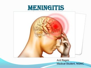
Meningitis
- 2. Meningitis refers an inflammatory process of leptomeninges and csf within subarachnoid space. Clinically, fever with chills, severe headache, nausea, vomiting photophobia altered mental sensation coma, death
- 3. Anatomy The central nervous system (CNS) is surrounded by several layers of tissue, with several outer layers not directly related to the CNS and three membranes that directly envelope the CNS. The outer layers are the skin and then a bone layer with associated periosteum. This layer includes the skull and the vertebrae. Below the periosteum is the Epidural Space which lies between periosteum and dura in the vertebral canal. The epidural space contains adipose tissue, loose connective tissue, veins and lymphatics.
- 4. Dura mater The Dura mater is the outer menix and is made up of a dense fibrous connective tissue. In the cranium, the dura layer is fused with the periosteum and therefore is in effect single layer without an epidural space. The dura contains a number of folds throughout its coverage of the brain including the Falx cerebri, a midline fold between cerebral hemispheres, the Tentorium cerebelli, an oblique fold between the cerebrum and cerebellum and the Diaphragma sellae which forms a collar around the neck of the pituitary and forms the roof of the hypophyseal fossa. This layer and these associated folds all provide structural support to the brain and prevent the brain from undergoing excess movement within the skull. Where the dura mater folds between brain tissues it splits into two distinct layers that are separated by large blood filled spaces called venous sinuses. Venous sinuses are directly connected to the venous system and venous blood from vessels supplying the brain return to the heart via these sinuses. Subdural space The subdural space lies between the dura and the next meningial layer, the arachnoid mater (not fused). The subdural space is thought to contain only lymph-like fluid. This space can also be the site of a subdural hematoma.
- 5. Arachnoid mater This is the middle meningial layer and lies between the dura mater and the pia mater, the innermost meningeal layer. The arachnoid mater is a delicate structure and is constructed with non-vascular connective tissue. This layer also has small protrusions through the dura mater into the previously mentioned venous sinuses called Arachnoid villus and these allow cerebrospinal fluid (CSF) to enter and exit the blood stream. These protrusions adhere to the inner surface of the skull via calvaria processes. Subarachnoid Space The subarachnoid space lies between the arachnoid mater and pia mater. Both meninges are connected via a fine network of connective tissue filaments (spider-like) which run through the space, originating from the arachnoid mater. This space also contains CSF from ventricular system. The largest part of this space are called the cisterns which are used for the collection of CSF. For example there is a cerebellomedullary cistern around the foramen magnum. The lumbar cistern used for lumbar puncture in man.
- 6. Pia Mater This is the innermost layer and is firmly bound to the underlying neural tissue of the brain and spinal cord. The inner surface of the brain facing this meningial layer is lined with ependymal cells. The pia mater is highly vascular and is formed from connective tissue.
- 7. meninges
- 8. Blood-Brain Barrier Since the brain is such a delicate organ, nature has taken extra measures to protect the brain by creating the blood-brain barrier to limit the diffusion of substances from the bloodstream into brain tissue selectively. The blood-brain barrier mainly consists of tight junctions, which seals the endothelial cells that line the brain capillaries. Astrocytes, a type of neuroglia from the brain, closely attached to the endothelial cells and release chemicals to regulate the permeabilities of the tight junctions. The major sites of the blood brain barrier are the arachnoid membrane, choroid plexus epithelium, and the cerebral microvascular endothelium. Only a few kinds water-soluble substance can move across the blood-brain barrier, such as glucose by active transport, urea, creatinine, and ions move across by slow diffusion. On the other hand, lipid-soluble substances can easily cross the blood-brain barrier, such as oxygen, carbon dioxide, alcohol, and most anesthetic agents.
- 10. Cerebrospinal Fluid Cerebrospinal fluid (CSF) is a colorless, transparent liquid that continuously circulates through the cavities of the brain and spinal cord, and as such, it acts as an internal circulation system to transport nutrients and wastes between the bloodstream and the brain and spinal cord. This reducdant circulation protects the brain and spinal cord from chemical injuries similar to the function of the blood-brain barrier. The CSF also protects the brain and spinal cord from physical injuries by acting as a shock absorber between the brain and spinal cord from the skeletal structures (cranium and vertebrae).
- 11. Circulation of csf CSF is produced in the choroid plexuses, which are networks of capillaries in the ventricles. The choroid plexuses filter out blood plasma from the bloodstream, which is the main component of CSF. The choroid plexuses are covered by ependymal cells that are sealed together with tight junctions. These tight junctions forces the blood plasma to pass through these ependymal cells, which further filter out the blood plasma, producing CSF. From the choroid plexuses of each lateral ventricle, CSF flows into the third ventricle through the interventricular foramina, which are two narrow oval openings. The choroid plexuses in the third ventricle adds more CSF. Then, CSF flows into the fourth ventricle throught the cerebral aqueduct. Again, the choroid plexuses in the fourth ventricle adds more CSF. The fluid then enters the subarachnoid spacethrough the three openings in the roof of the fourth ventricle. These three openings are a median aperture and a pair of lateral apertures. Then, CSF circultates in the central canal of the spinal cord and in the subarachnoid space around the surface of the brain and spinal cord
- 12. Classification of meningitis 1. acute pyogenic (bacterial) meningitis 2.acute aseptic (viral) meningitis 3.acute focal suppurative infection (brain abscess,subdural and extradural empyema) 4.chronic bacterial infection (tuberculosis).
- 13. Signs and Symptoms of Meningitis Symptoms of bacterial meningitis are usually acute, developing within a few hours and last 2 to 3 weeks. It is important to seek immediate medical attention when symptoms occur, because acute bacterial meningitis can be fatal within hours. Viral meningitis may develop suddenly or within days or weeks, depending on the virus and the overall health of the patient. Characteristic symptoms of both viral and bacterial meningitis are stiff neck, headache, and fever. Symptoms may develop over the course a few hours (acute bacterial meningitis) or a few days. Some patients experience cough, runny nose, and congestion prior to developing other symptoms.
- 14. Other signs and symptoms of include: Confusion Drowsiness Joint pain Lethargy Nausea and vomiting Seizures Sensitivity to light (photophobia) Skin rash (commonly near the armpits and on the hands and feet) Symptoms of meningitis in infants may be difficult to detect and include the following: Bulging of the soft spots (fontanels) in the head caused by increased intracranial pressure Decreased activity Difficulty nursing or eating Excessive sleeping High-pitched cry Increased crying and irritability Vomiting
- 15. Diagnosis of Meningitis A diagnosis of meningitis depends primarily on a thorough physical examination and cerebrospinal fluid (CSF) analysis. In the physical examination stiff neck, severe headache, and fever indicate meningitis. It may be extremely painful to move the neck forward. The neck may be so stiff that attempting to move it causes the entire body to move. Other signs the physician may look for include swelling in the eyes, which indicates elevated intracranial pressure, and skin rash. Computed tomography (CT scan) or magnetic resonance imaging (MRI scan) of the brain may be used to evaluate possible swelling (edema) and bleeding (hemorrhage) and to rule out other neurological disorders. Laboratory tests that may be performed include complete blood count (CBC), blood culture, and spinal tap. CBC will show elevated levels of white blood cells if there is an active infection in the body. Blood is cultured to identify bacteria in the blood. Spinal tap, or lumbar puncture, is essential in diagnosing and selecting appropriate treatment for meningitis. About 2 tablespoons of cerebrospinal fluid is drawn into a needle inserted between two lumbar vertebrae. Lab analysis looks for elevated levels of white blood cells and blood. The fluid also is cultured to identify the organism causing meningitis.
- 16. Meningitis Complications Complications such as the following can develop during the course of meningitis: Disseminated intravascular coagulation (DIC; blood-clotting disorder) Encephalitis Persistent fever Seizures Syndrome of inappropriate antidiuretic hormone (SIADH; causes fluid build-up) Prompt medical treatment decreases the risk for brain damage and long-term complications, including these: Behavioral and personality changes Vision loss (partial or total) Cerebral palsy Hearing loss (partial or total) Learning disabilities or mental retardation Paralysis (partial or total) Speech loss (partial or total) Severe bacterial meningitis also may cause the head and heels to bend backward and the body to bow forward (called opisthotonos), coma, and death. Newborns and young children may develop heart, liver, intestinal problems, or malformed limbs.
- 17. Treatment for Meningitis Treatment is determined by the type of meningitis and the organism causing the disease. Viral meningitis usually requires only symptom relief (palliative care). Palliative care may include bed rest, increased fluid intake to prevent dehydration, and analgesics (e.g., aspirin, acetaminophen) to reduce fever and relieve body aches. Meningitis caused by herpesvirus can be treated using antiviral medication such as acyclovir or ribavirin. Side effects of these medications include nausea, vomiting, and headache. Suspected bacterial meningitis requires prompt intravenous (IV) antibiotic treatment in the hospital to prevent serious complications and neurological damage. If symptoms are severe, IV treatment may be initiated before the lumbar puncture is performed. Severly ill patients are treated immediately with a combination of antibiotics. Penicillin combined with a cephalosporin (e.g., ceftriaxone cefotaxime ) is commonly used. Because some bacteria are resistant to these drugs, vancomycin, with or without rifampin, ampicillin, and gentamicin may be added to cover resistant pneumococcal strains of bacteria and Listeria monocytogenes. Side effects include abdominal pain, nausea, vomiting, and diarrhea. Once the CSF culture has revealed the disease-causing organism (pathogen), antibiotic treatment is adjusted accordingly. Amphotericin B and fluconazole are effective against most disease-causing fungi and are the drugs of choice for treatment of fungal meningitis. They may be administered singly or as combined therapy. Both drugs are well tolerated in most patients. Possible side effects of fluconazole include nausea and vomiting, diarrhea, headache, skin rash, and abdominal pain. Intravenously administered amphotericin B may produce the same side effects, as well as shaking chills and fever, slowed heart rate, low blood pressure (hypotension), body ache, and weight loss. Parasitic meningitis usually is treated with a benzimidazole derivative or other antihelminthic agent. Complications that develop also must be treated. Corticosteroids (e.g., dexamethasone) may be administered to reduce the risk for hearing loss. Increased intracranial pressure may be reduced with diuretics (e.g., mannitol) and a surgically placed shunt that drains excess fluid.
- 18. Its only for my practice, so please do not depend upon it fully